

Paper Menu >>
Journal Menu >>
 J. Biomedical Science and Engineering, 2011, 4, 506-510 doi:10.4236/jbise.2011.47064 Published Online July 2011 (http://www.SciRP.org/journal/jbise/ JBiSE ). Published Online July 2011 in SciRes. http://www.scirp.org/journal/JBiSE Uncoupling protein 2 regulates myocardial apoptosis via the diabetogenic action of streptozotocin Xiu-Zhen Li1, Ruo-Yun Tan2, Xiang Lu1* 1The First Clinical College, NJMU, Nanjing, China; 2The Second Clinical College, NJMU, Nanjing, China. Email: [email protected], [email protected] Received 10 January 2010; revised 26 April 2010; accepted 24 May 2011. ABSTRACT Objective: Determine the role of uncoupling protein 2 (UCP2) in the myocardial apoptosis of diabetic melli- tus(DM). Methods: DM animal models were induced by streptozotocinon (STZ) on UCP2 knock-out mice (UCP2KO) and wild-type mice (WT), which were reared for 7 and 28 days after successful modeling, respectively. The expressions of relative protein for myocardial apoptosis, pro-caspase-9, were investigated using western blot. However, the terminal deoxynu- cleotidyltransferase-mediated dUTP-biotin nick end labeling (TUNEL) was used to explain apoptosis at the DNA level. Results: Image analysis showed that the expression of pro-caspase-9 protein levels increased slightly in UCP-/- + DM-7-day group comparing with DM-7-day group (P > 0.05). The expression of pro-caspase-9 protein levels increased significantly (P < 0.05)in UC P-/- + DM-28- day group comparin g with DM-28-day group. TUNEL analysis indicated that UCP2 reduced the number of apoptotic myocytes in the DM-28-day group by 70% in comparison to DM-7-day group by 30% (P < 0.05). Conclusion UCP2 may be one of the most important factors that con- tribute to the myocardial apoptosis of DM. Keywords: Uncoupling Protein 2; Diabetes; Myocardium; Apoptosis 1. INTRODUCTION An inner mitochondrial protein known as uncoupling pro- tein 2 (UCP2) is a member of the uncoupling protein fam- ily and belongs to the inner mitochondrial membrane an- ion-carrier superfamily. UCP2 can be detected in the heart [1], brain, lung, spleen, kidney, liver, and adipose tissue. UCP2 is a regulator of reactive oxygen species during electron transport in the mitochondrial inner membrane, plays an active role in the prevention of atherosclerosis [2], is one of the etiologies of type 2 diabetes [3], participates in inflammation [4], and regulates cell apoptosis [5]and aging [6]. The purpose of this study was to investigate the relationship between UCP2 and the myocardial apoptosis of diabetic mellitus, demonstrate the important role of UCP2 in diabetic cardiomyopathy (DC), and provide ad- ditional theoretical foundation for DC therapy. 2. MATERIALS AND METHODS 2.1 Animals Male, 8- to 12-week old WT and UCP2KO littermate mice were used in this study. UCP2KO and WT mice were generated on a C57B/6 mixed background. The WT mice were purchased from the Shanghai Silaike Laboratory Animal Company. UCP2KO mice were pre- viously generated by homologous recombination in em- bryonic stem cells on a C57BL/6 mixed background in the Laboratory Animal Center of Nanjing University, wherein their genomic loci were analyzed using PCR, as previously described [7]. The WT and UCP2KO mice were raised in a colony at the Nanjing Medical Univer- sity, wherein all of the animal protocols were approved by the Animal Care Committee, and the animals were handled according to the guidelines of the Chinese Council of Animal Care. 2.2. Chemicals and Reagents STZ (N-(methylnitrosocarbamoyl)-α-D-glucosamine), anti-caspase9 antibody, and a TUNEL Apoptosis Assay Kit was obtained from Bioworld. 2.3. STZ-Induced Diabetes Each mouse received a single daily injection of STZ (60 mg/kg) for three consecutive days. Animals in the nega- tive control group (NC) and blank control group (BC) were identically handled, but were injected with only vehicle (citrate buffer). All STZ-Injected animals were assessed for the development of diabetes as described previously [8] and sacrificed on days 7 or 28, and their tissues were harvested for further analyses. 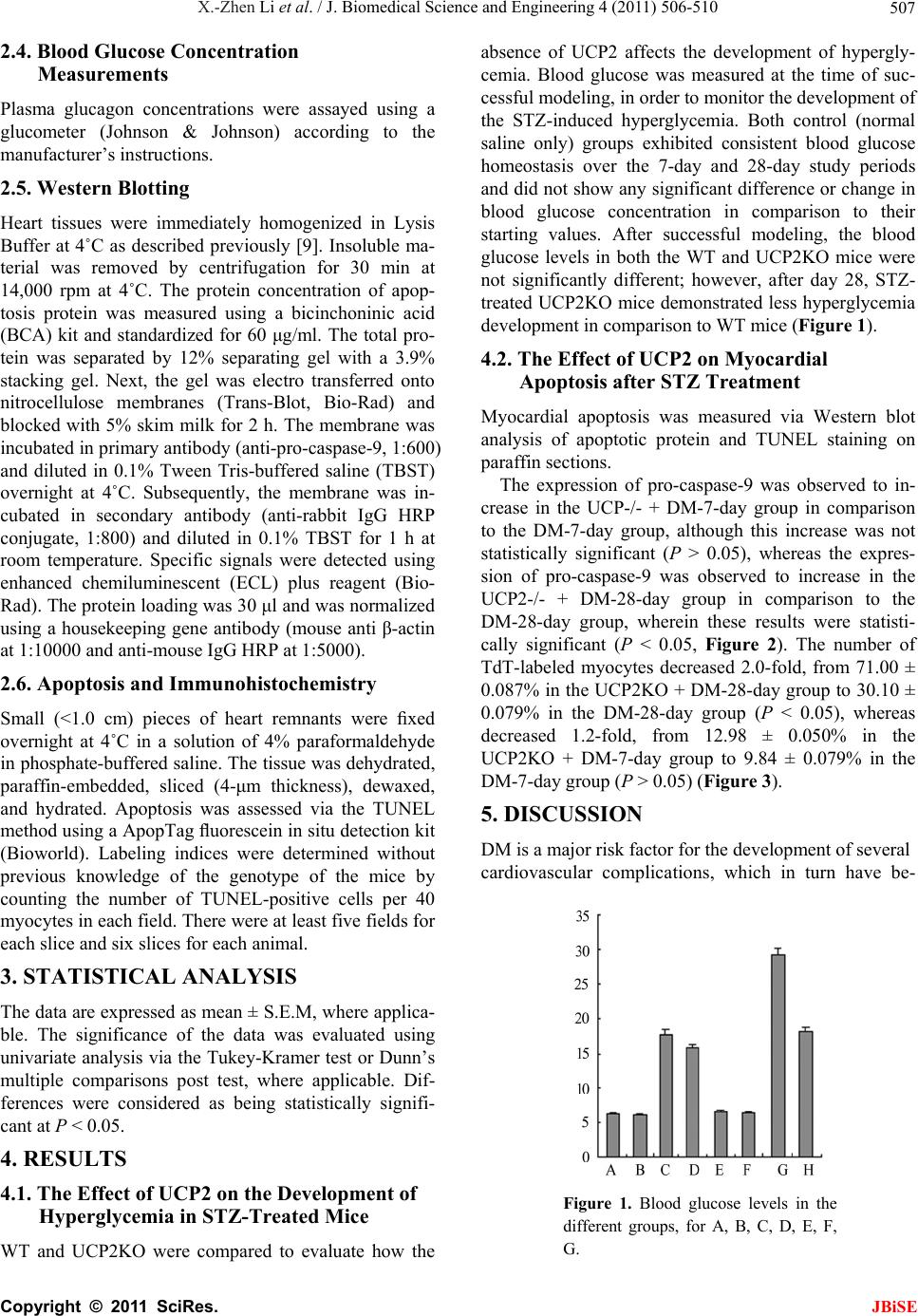 X.-Zhen Li et al. / J. Biomedical Science and Engineering 4 (2011) 506-510 507 2.4. Blood Glucose Concentration Measurements Plasma glucagon concentrations were assayed using a glucometer (Johnson & Johnson) according to the manufacturer’s instructions. 2.5. Western Blotting Heart tissues were immediately homogenized in Lysis Buffer at 4˚C as described previously [9]. Insoluble ma- terial was removed by centrifugation for 30 min at 14,000 rpm at 4˚C. The protein concentration of apop- tosis protein was measured using a bicinchoninic acid (BCA) kit and standardized for 60 μg/ml. The total pro- tein was separated by 12% separating gel with a 3.9% stacking gel. Next, the gel was electro transferred onto nitrocellulose membranes (Trans-Blot, Bio-Rad) and blocked with 5% skim milk for 2 h. The membrane was incubated in primary antibody (anti-pro-caspase-9, 1:600) and diluted in 0.1% Tween Tris-buffered saline (TBST) overnight at 4˚C. Subsequently, the membrane was in- cubated in secondary antibody (anti-rabbit IgG HRP conjugate, 1:800) and diluted in 0.1% TBST for 1 h at room temperature. Specific signals were detected using enhanced chemiluminescent (ECL) plus reagent (Bio- Rad). The protein loading was 30 μl and was normalized using a housekeeping gene antibody (mouse anti β-actin at 1:10000 and anti-mouse IgG HRP at 1:5000). 2.6. Apoptosis and Immunohistochemistry Small (<1.0 cm) pieces of heart remnants were fixed overnight at 4˚C in a solution of 4% paraformaldehyde in phosphate-buffered saline. The tissue was dehydrated, paraffin-embedded, sliced (4-μm thickness), dewaxed, and hydrated. Apoptosis was assessed via the TUNEL method using a ApopTag fluorescein in situ detection kit (Bioworld). Labeling indices were determined without previous knowledge of the genotype of the mice by counting the number of TUNEL-positive cells per 40 myocytes in each field. There were at least five fields for each slice and six slices for each animal. 3. STATISTICAL ANALYSIS The data are expressed as mean ± S.E.M, where applica- ble. The significance of the data was evaluated using univariate analysis via the Tukey-Kramer test or Dunn’s multiple comparisons post test, where applicable. Dif- ferences were considered as being statistically signifi- cant at P < 0.05. 4. RESULTS 4.1. The Effect of UCP2 on the Development of Hyperglycemia in STZ-Treated Mice WT and UCP2KO were compared to evaluate how the absence of UCP2 affects the development of hypergly- cemia. Blood glucose was measured at the time of suc- cessful modeling, in order to monitor the development of the STZ-induced hyperglycemia. Both control (normal saline only) groups exhibited consistent blood glucose homeostasis over the 7-day and 28-day study periods and did not show any significant difference or change in blood glucose concentration in comparison to their starting values. After successful modeling, the blood glucose levels in both the WT and UCP2KO mice were not significantly different; however, after day 28, STZ- treated UCP2KO mice demonstrated less hyperglycemia development in comparison to WT mice (Figure 1). 4.2. The Effect of UCP2 on Myocardial Apoptosis after STZ Treatment Myocardial apoptosis was measured via Western blot analysis of apoptotic protein and TUNEL staining on paraffin sections. The expression of pro-caspase-9 was observed to in- crease in the UCP-/- + DM-7-day group in comparison to the DM-7-day group, although this increase was not statistically significant (P > 0.05), whereas the expres- sion of pro-caspase-9 was observed to increase in the UCP2-/- + DM-28-day group in comparison to the DM-28-day group, wherein these results were statisti- cally significant (P < 0.05, Figure 2). The number of TdT-labeled myocytes decreased 2.0-fold, from 71.00 ± 0.087% in the UCP2KO + DM-28-day group to 30.10 ± 0.079% in the DM-28-day group (P < 0.05), whereas decreased 1.2-fold, from 12.98 ± 0.050% in the UCP2KO + DM-7-day group to 9.84 ± 0.079% in the DM-7-day group (P > 0.05) (Figure 3). 5. DISCUSSION DM is a major risk factor for the development of several cardiovascular complications, which in turn have be- Figure 1. Blood glucose levels in the different groups, for A, B, C, D, E, F, G. C opyright © 2011 SciRes. JBiSE 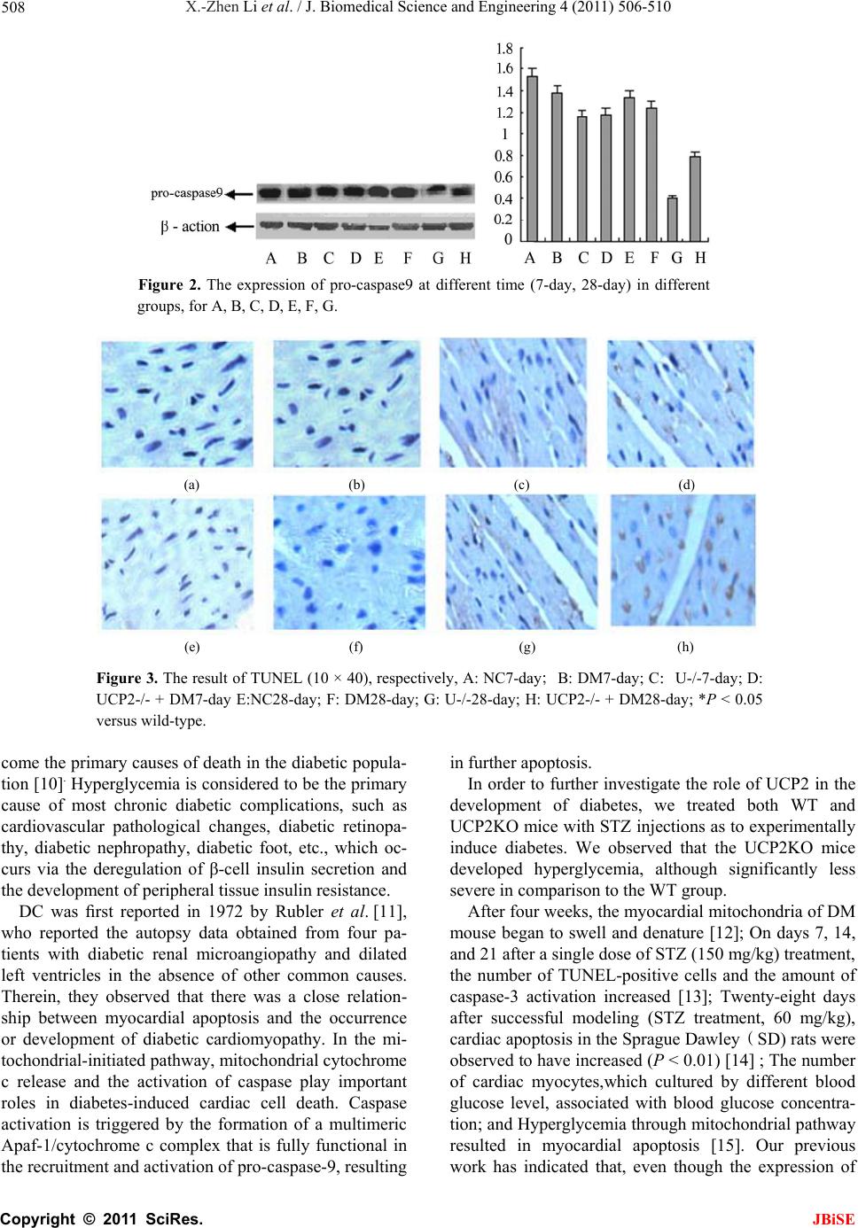 X.-Zhen Li et al. / J. Biomedical Science and Engineering 4 (2011) 506-510 508 Figure 2. The expression of pro-caspase9 at different time (7-day, 28-day) in different groups, for A, B, C, D, E, F, G. (a) (b) (c) (d) (e) (f) (g) (h) Figure 3. The result of TUNEL (10 × 40), respectively, A: NC7-day;B: DM7-day; C:U-/-7-day; D: UCP2-/- + DM7-day E:NC28-day; F: DM28-day; G: U-/-28-day; H: UCP2-/- + DM28-day; *P < 0.05 versus wild-type. come the primary causes of death in the diabetic popula- tion [10]. Hyperglycemia is considered to be the primary cause of most chronic diabetic complications, such as cardiovascular pathological changes, diabetic retinopa- thy, diabetic nephropathy, diabetic foot, etc., which oc- curs via the deregulation of β-cell insulin secretion and the development of peripheral tissue insulin resistance. DC was first reported in 1972 by Rubler et al. [11], who reported the autopsy data obtained from four pa- tients with diabetic renal microangiopathy and dilated left ventricles in the absence of other common causes. Therein, they observed that there was a close relation- ship between myocardial apoptosis and the occurrence or development of diabetic cardiomyopathy. In the mi- tochondrial-initiated pathway, mitochondrial cytochrome c release and the activation of caspase play important roles in diabetes-induced cardiac cell death. Caspase activation is triggered by the formation of a multimeric Apaf-1/cytochrome c complex that is fully functional in the recruitment and activation of pro-caspase-9, resulting in further apoptosis. In order to further investigate the role of UCP2 in the development of diabetes, we treated both WT and UCP2KO mice with STZ injections as to experimentally induce diabetes. We observed that the UCP2KO mice developed hyperglycemia, although significantly less severe in comparison to the WT group. After four weeks, the myocardial mitochondria of DM mouse began to swell and denature [12]; On days 7, 14, and 21 after a single dose of STZ (150 mg/kg) treatment, the number of TUNEL-positive cells and the amount of caspase-3 activation increased [13]; Twenty-eight days after successful modeling (STZ treatment, 60 mg/kg), cardiac apoptosis in the Sprague Dawley(SD) rats were observed to have increased (P < 0.01) [14] ; The number of cardiac myocytes,which cultured by different blood glucose level, associated with blood glucose concentra- tion; and Hyperglycemia through mitochondrial pathway resulted in myocardial apoptosis [15]. Our previous work has indicated that, even though the expression of C opyright © 2011 SciRes. JBiSE 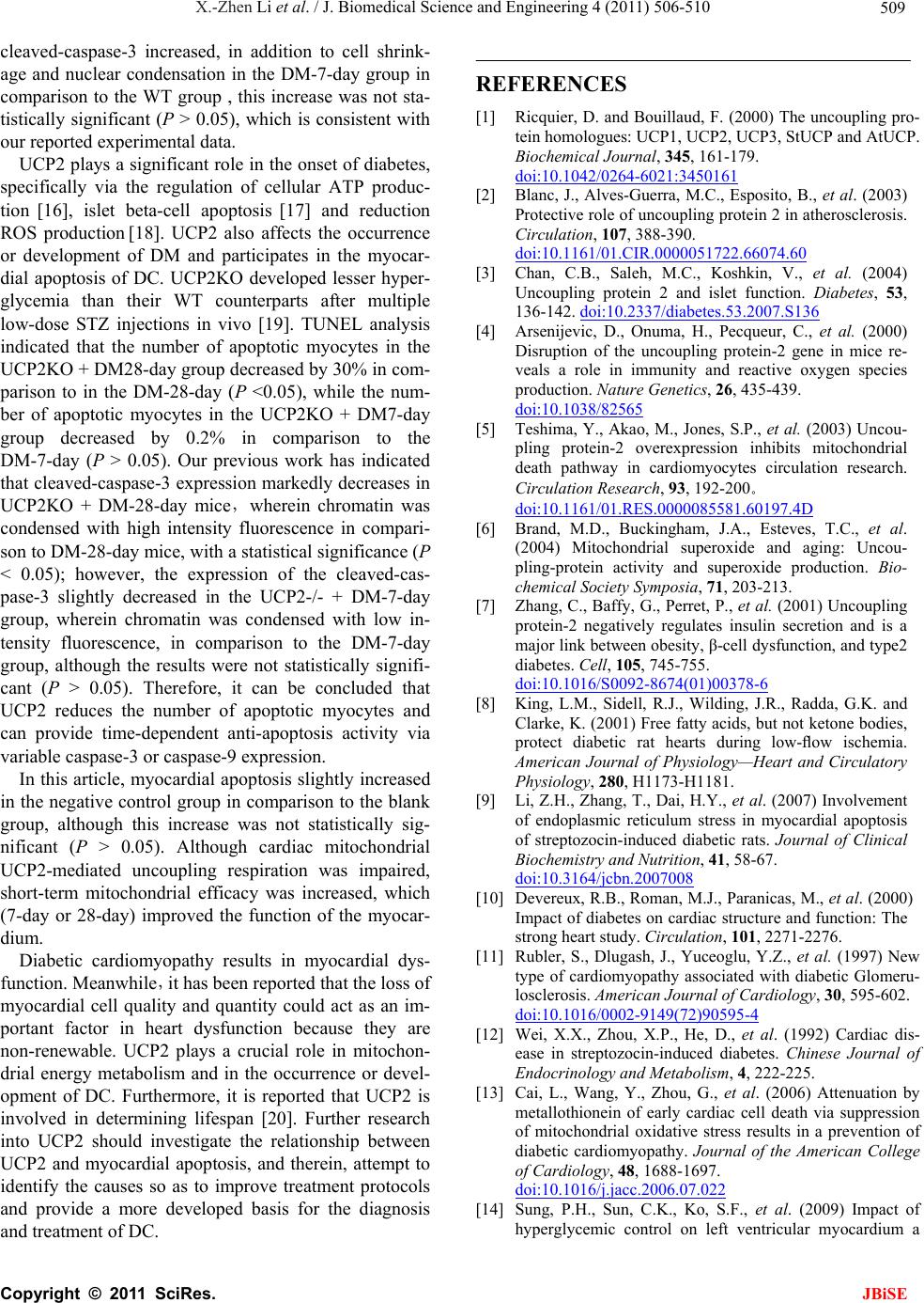 X.-Zhen Li et al. / J. Biomedical Science and Engineering 4 (2011) 506-510 509 cleaved-caspase-3 increased, in addition to cell shrink- age and nuclear condensation in the DM-7-day group in comparison to the WT group , this increase was not sta- tistically significant (P > 0.05), which is consistent with our reported experimental data. UCP2 plays a significant role in the onset of diabetes, specifically via the regulation of cellular ATP produc- tion [16], islet beta-cell apoptosis [17] and reduction ROS production [18]. UCP2 also affects the occurrence or development of DM and participates in the myocar- dial apoptosis of DC. UCP2KO developed lesser hyper- glycemia than their WT counterparts after multiple low-dose STZ injections in vivo [19]. TUNEL analysis indicated that the number of apoptotic myocytes in the UCP2KO + DM28-day group decreased by 30% in com- parison to in the DM-28-day (P <0.05), while the num- ber of apoptotic myocytes in the UCP2KO + DM7-day group decreased by 0.2% in comparison to the DM-7-day (P > 0.05). Our previous work has indicated that cleaved-caspase-3 expression markedly decreases in UCP2KO + DM-28-day mice,wherein chromatin was condensed with high intensity fluorescence in compari- son to DM-28-day mice, with a statistical significance (P < 0.05); however, the expression of the cleaved-cas- pase-3 slightly decreased in the UCP2-/- + DM-7-day group, wherein chromatin was condensed with low in- tensity fluorescence, in comparison to the DM-7-day group, although the results were not statistically signifi- cant (P > 0.05). Therefore, it can be concluded that UCP2 reduces the number of apoptotic myocytes and can provide time-dependent anti-apoptosis activity via variable caspase-3 or caspase-9 expression. In this article, myocardial apoptosis slightly increased in the negative control group in comparison to the blank group, although this increase was not statistically sig- nificant (P > 0.05). Although cardiac mitochondrial UCP2-mediated uncoupling respiration was impaired, short-term mitochondrial efficacy was increased, which (7-day or 28-day) improved the function of the myocar- dium. Diabetic cardiomyopathy results in myocardial dys- function. Meanwhile,it has been reported that the loss of myocardial cell quality and quantity could act as an im- portant factor in heart dysfunction because they are non-renewable. UCP2 plays a crucial role in mitochon- drial energy metabolism and in the occurrence or devel- opment of DC. Furthermore, it is reported that UCP2 is involved in determining lifespan [20]. Further research into UCP2 should investigate the relationship between UCP2 and myocardial apoptosis, and therein, attempt to identify the causes so as to improve treatment protocols and provide a more developed basis for the diagnosis and treatment of DC. REFERENCES [1] Ricquier, D. and Bouillaud, F. (2000) The uncoupling pro- tein homologues: UCP1, UCP2, UCP3, StUCP and AtUCP. Biochemical Journal, 345, 161-179. doi:10.1042/0264-6021:3450161 [2] Blanc, J., Alves-Guerra, M.C., Esposito, B., et al. (2003) Protective role of uncoupling protein 2 in atherosclerosis. Circulation, 107, 388-390. doi:10.1161/01.CIR.0000051722.66074.60 [3] Chan, C.B., Saleh, M.C., Koshkin, V., et al. (2004) Uncoupling protein 2 and islet function. Diabetes, 53, 136-142. doi:10.2337/diabetes.53.2007.S136 [4] Arsenijevic, D., Onuma, H., Pecqueur, C., et al. (2000) Disruption of the uncoupling protein-2 gene in mice re- veals a role in immunity and reactive oxygen species production. Nature Genetics, 26, 435-439. doi:10.1038/82565 [5] Teshima, Y., Akao, M., Jones, S.P., et al. (2003) Uncou- pling protein-2 overexpression inhibits mitochondrial death pathway in cardiomyocytes circulation research. Circulation Research, 93, 192-200。 doi:10.1161/01.RES.0000085581.60197.4D [6] Brand, M.D., Buckingham, J.A., Esteves, T.C., et al. (2004) Mitochondrial superoxide and aging: Uncou- pling-protein activity and superoxide production. Bio- chemical Society Symposia, 71, 203-213. [7] Zhang, C., Baffy, G., Perret, P., et al. (2001) Uncoupling protein-2 negatively regulates insulin secretion and is a major link between obesity, β-cell dysfunction, and type2 diabetes. Cell, 105, 745-755. doi:10.1016/S0092-8674(01)00378-6 [8] King, L.M., Sidell, R.J., Wilding, J.R., Radda, G.K. and Clarke, K. (2001) Free fatty acids, but not ketone bodies, protect diabetic rat hearts during low-flow ischemia. American Journal of Physiology—Heart and Circulatory Physiology, 280, H1173-H1181. [9] Li, Z.H., Zhang, T., Dai, H.Y., et al. (2007) Involvement of endoplasmic reticulum stress in myocardial apoptosis of streptozocin-induced diabetic rats. Journal of Clinical Biochemistry and Nutrition, 41, 58-67. doi:10.3164/jcbn.2007008 [10] Devereux, R.B., Roman, M.J., Paranicas, M., et al. (2000) Impact of diabetes on cardiac structure and function: The strong heart study. Circulation, 101, 2271-2276. [11] Rubler, S., Dlugash, J., Yuceoglu, Y.Z., et al. (1997) New type of cardiomyopathy associated with diabetic Glomeru- losclerosis. American Journal of Cardiology, 30, 595-602. doi:10.1016/0002-9149(72)90595-4 [12] Wei, X.X., Zhou, X.P., He, D., et al. (1992) Cardiac dis- ease in streptozocin-induced diabetes. Chinese Journal of Endocrinology and Metabolism, 4, 222-225. [13] Cai, L., Wang, Y., Zhou, G., et al. (2006) Attenuation by metallothionein of early cardiac cell death via suppression of mitochondrial oxidative stress results in a prevention of diabetic cardiomyopathy. Journal of the American College of Cardiology, 48, 1688-1697. doi:10.1016/j.jacc.2006.07.022 [14] Sung, P.H., Sun, C.K., Ko, S.F., et al. (2009) Impact of hyperglycemic control on left ventricular myocardium a C opyright © 2011 SciRes. JBiSE  X.-Zhen Li et al. / J. Biomedical Science and Engineering 4 (2011) 506-510 Copyright © 2011 SciRes. 510 JBiSE molecular and cellular basic study in a diabetic rat mode. International Heart Journal, 50, 191-206. doi:10.1536/ihj.50.191 [15] Li, C.Y., Cao, L.S., Zeng, Q.T., et al. (2005) Taurine may prevent diabetic rats from developing cardiomyopathy also by down regulating angiotensintypeⅡ-2 receptor expression. Cardiovascular Drugs and Therapy, 19, 105-112. doi:10.1007/s10557-005-0443-x [16] Joseph, J.W., Koshkin, V., Zhang, C.Y., et al. (2002) Uncoupling protein 2 knockout mice have enhanced in- sulin secretary capacity after a high-fat diet. Diabetes, 51, 3211-3219. doi:10.2337/diabetes.51.11.3211 [17] Joseph, J.W., Koshkin, V., Saleh, M.C., et al. (2004) Free fatty acid-induced beta-cell defects are dependent on un- coupling protein 2 expressions. The Journal of Biological Chemistry, 279, 51049-51056. doi:10.1074/jbc.M409189200 [18] Arsenijevic, D., Onuma, H., Pecqueur, C., et al. (2000) Disruption of the uncoupling protein gene in mice reveals a role in immunity and reactive oxygen species produc- tion. Nature Genetics, 26, 435-439. doi:10.1038/82565 [19] Lee, S.C., Robson-Doucette, C.A. and Wheeler, M.B. (2009) Uncoupling protein 2 regulates reactive oxygen species formation in islets and influences susceptibility to diabetogenic action of streptozotocin. Journal of Endo- crinology, 203, 33-43. doi:10.1677/JOE-09-0117 [20] Andrews, Z.B. and Horvath, T. (2009) Uncoupling pro- tein-2 regulates lifespan in mice. American Journal of Physiology—Endocrinology and Metabolism, 296, E621-E627. doi:10.1152/ajpendo.90903.2008 |