

Paper Menu >>
Journal Menu >>
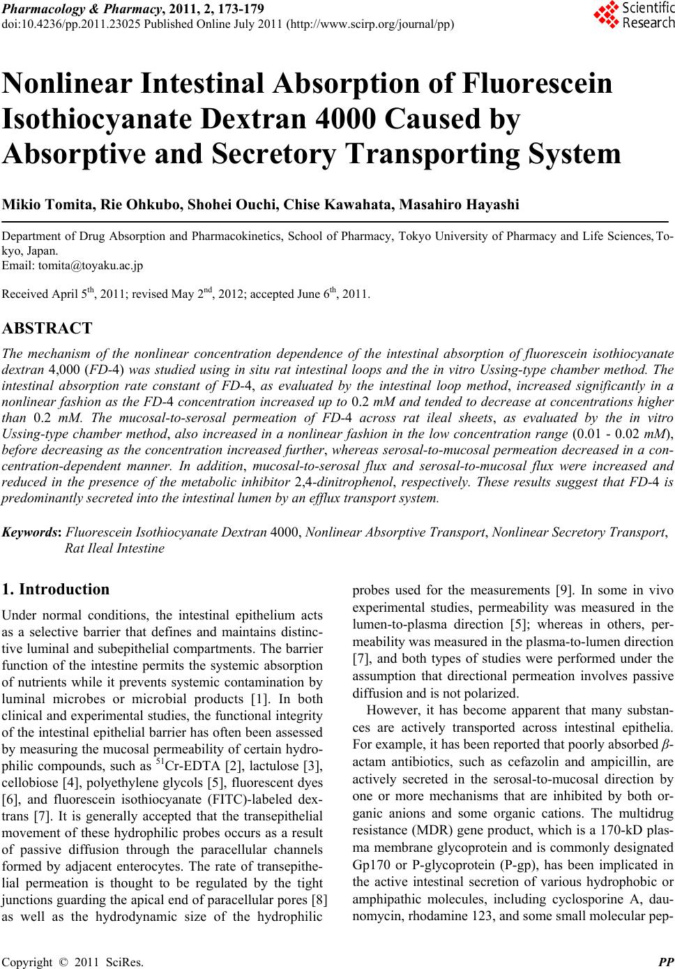 Pharmacology & Pharmacy, 2011, 2, 173-179 doi:10.4236/pp.2011.23025 Published Online July 2011 (http://www.scirp.org/journal/pp) Copyright © 2011 SciRes. PP 173 Nonlinear Intestinal Absorption of Fluorescein Isothiocyanate Dextran 4000 Caused by Absorptive and Secretory Transporting System Mikio Tomita, Rie Ohkubo, Shohei Ouchi, Chise Kawahata, Masahiro Hayashi Department of Drug Absorption and Pharmacokinetics, School of Pharmacy, Tokyo University of Pharmacy and Life Sciences, To- kyo, Japan. Email: [email protected] Received April 5th, 2011; revised May 2nd, 2012; accepted June 6th, 2011. ABSTRACT The mechanism of the nonlinear concentration dependence of the intestinal absorption of fluorescein isothiocyanate dextran 4,000 (FD-4) was studied using in situ rat intestinal loops and the in vitro Ussing-type chamber method. The intestinal absorption rate constant of FD-4, as evaluated by the intestinal loop method, increased significantly in a nonlinear fashion as the FD-4 concentration increased up to 0.2 mM and tended to decrease at concentrations higher than 0.2 mM. The mucosal-to-serosal permeation of FD-4 across rat ileal sheets, as evaluated by the in vitro Ussing-type chamber method, also increased in a nonlinear fashion in the low concentration range (0.01 - 0.02 mM), before decreasing as the concentration increased further, whereas serosal-to-mucosal permeation decreased in a con- centration-dependent manner. In addition, mucosal-to-serosal flux and serosal-to-mucosal flux were increased and reduced in the presence of the metabolic inhibitor 2,4-dinitrophenol, respectively. These results suggest that FD-4 is predominantly secreted into the intestinal lumen by an efflux transport system. Keywords: Fluorescein Isothiocyanate Dextran 4000, Nonlinear Absorptive Transport, Nonlinear Secretory Transport, Rat Ileal Intestine 1. Introduction Under normal conditions, the intestinal epithelium acts as a selective barrier that defines and maintains distinc- tive luminal and subepithelial compartments. The barrier function of the intestine permits the systemic absorption of nutrients while it prevents systemic contamination by luminal microbes or microbial products [1]. In both clinical and experimental studies, the functional integrity of the intestinal epithelial barrier has often been assessed by measuring the mucosal permeability of certain hydro- philic compounds, such as 51Cr-EDTA [2], lactulose [3], cellobiose [4], polyethylene glycols [5], fluorescent dyes [6], and fluorescein isothiocyanate (FITC)-labeled dex- trans [7]. It is generally accepted that the transepithelial movement of these hydrophilic probes occurs as a result of passive diffusion through the paracellular channels formed by adjacent enterocytes. The rate of transepithe- lial permeation is thought to be regulated by the tight junctions guarding the apical end of paracellular pores [8] as well as the hydrodynamic size of the hydrophilic probes used for the measurements [9]. In some in vivo experimental studies, permeability was measured in the lumen-to-plasma direction [5]; whereas in others, per- meability was measured in the plasma-to-lumen direction [7], and both types of studies were performed under the assumption that directional permeation involves passive diffusion and is not polarized. However, it has become apparent that many substan- ces are actively transported across intestinal epithelia. For example, it has been reported that poorly absorbed β- actam antibiotics, such as cefazolin and ampicillin, are actively secreted in the serosal-to-mucosal direction by one or more mechanisms that are inhibited by both or- ganic anions and some organic cations. The multidrug resistance (MDR) gene product, which is a 170-kD plas- ma membrane glycoprotein and is commonly designated Gp170 or P-glycoprotein (P-gp), has been implicated in the active intestinal secretion of various hydrophobic or amphipathic molecules, including cyclosporine A, dau- nomycin, rhodamine 123, and some small molecular pep- 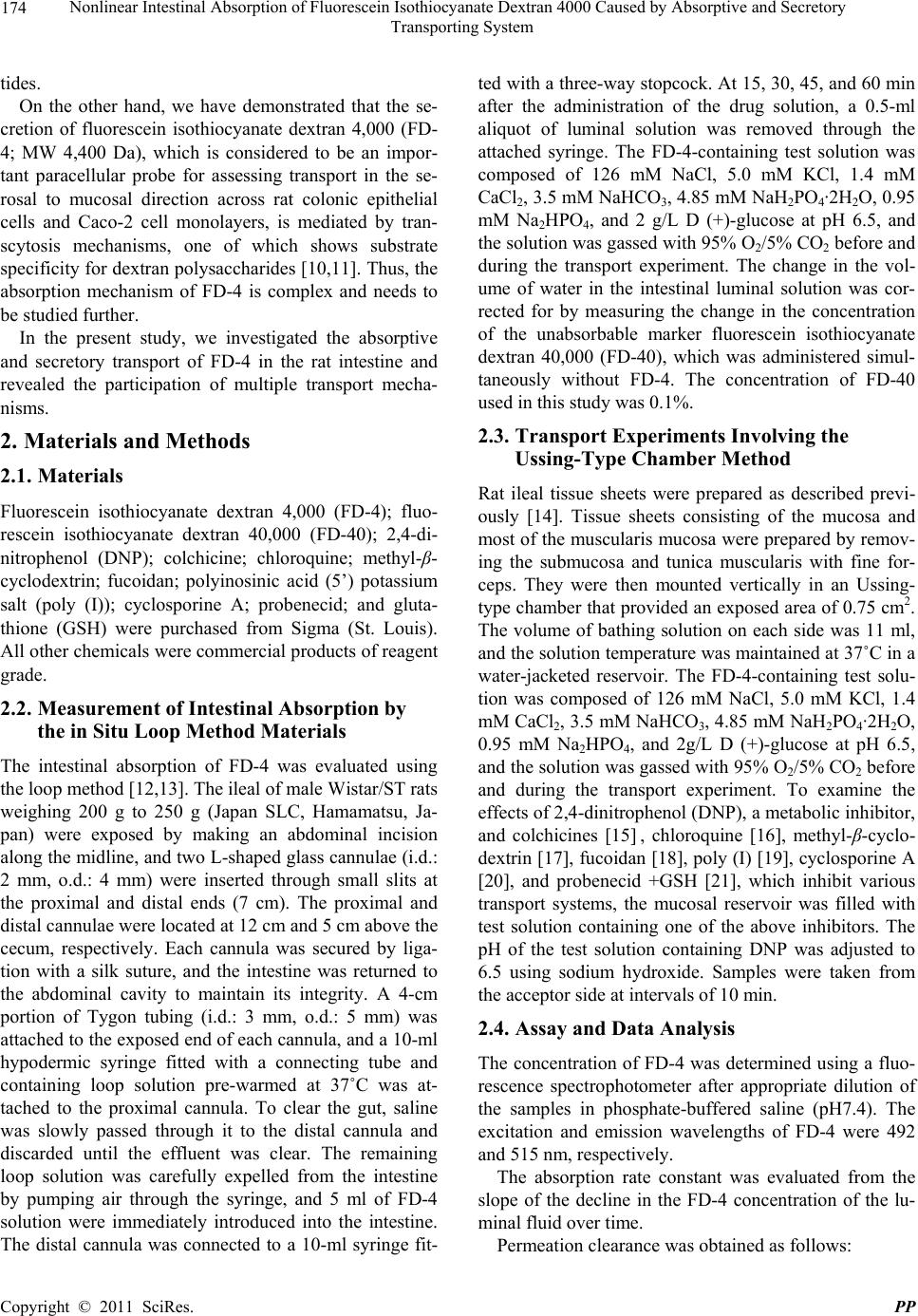 Nonlinear Intestinal Absorption of Fluorescein Isothiocyanate Dextran 4000 Caused by Absorptive and Secretory Transporting System 174 tides. On the other hand, we have demonstrated that the se- cretion of fluorescein isothiocyanate dextran 4,000 (FD- 4; MW 4,400 Da), which is considered to be an impor- tant paracellular probe for assessing transport in the se- rosal to mucosal direction across rat colonic epithelial cells and Caco-2 cell monolayers, is mediated by tran- scytosis mechanisms, one of which shows substrate specificity for dextran polysaccharides [10,11]. Thus, the absorption mechanism of FD-4 is complex and needs to be studied further. In the present study, we investigated the absorptive and secretory transport of FD-4 in the rat intestine and revealed the participation of multiple transport mecha- nisms. 2. Materials and Methods 2.1. Materials Fluorescein isothiocyanate dextran 4,000 (FD-4); fluo- rescein isothiocyanate dextran 40,000 (FD-40); 2,4-di- nitrophenol (DNP); colchicine; chloroquine; methyl-β- cyclodextrin; fucoidan; polyinosinic acid (5’) potassium salt (poly (I)); cyclosporine A; probenecid; and gluta- thione (GSH) were purchased from Sigma (St. Louis). All other chemicals were commercial products of reagent grade. 2.2. Measurement of Intestinal Absorption by the in Situ Loop Method Materials The intestinal absorption of FD-4 was evaluated using the loop method [12,13]. The ileal of male Wistar/ST rats weighing 200 g to 250 g (Japan SLC, Hamamatsu, Ja- pan) were exposed by making an abdominal incision along the midline, and two L-shaped glass cannulae (i.d.: 2 mm, o.d.: 4 mm) were inserted through small slits at the proximal and distal ends (7 cm). The proximal and distal cannulae were located at 12 cm and 5 cm above the cecum, respectively. Each cannula was secured by liga- tion with a silk suture, and the intestine was returned to the abdominal cavity to maintain its integrity. A 4-cm portion of Tygon tubing (i.d.: 3 mm, o.d.: 5 mm) was attached to the exposed end of each cannula, and a 10-ml hypodermic syringe fitted with a connecting tube and containing loop solution pre-warmed at 37˚C was at- tached to the proximal cannula. To clear the gut, saline was slowly passed through it to the distal cannula and discarded until the effluent was clear. The remaining loop solution was carefully expelled from the intestine by pumping air through the syringe, and 5 ml of FD-4 solution were immediately introduced into the intestine. The distal cannula was connected to a 10-ml syringe fit- ted with a three-way stopcock. At 15, 30, 45, and 60 min after the administration of the drug solution, a 0.5-ml aliquot of luminal solution was removed through the attached syringe. The FD-4-containing test solution was composed of 126 mM NaCl, 5.0 mM KCl, 1.4 mM CaCl2, 3.5 mM NaHCO3, 4.85 mM NaH2PO4·2H2O, 0.95 mM Na2HPO4, and 2 g/L D (+)-glucose at pH 6.5, and the solution was gassed with 95% O2/5% CO2 before and during the transport experiment. The change in the vol- ume of water in the intestinal luminal solution was cor- rected for by measuring the change in the concentration of the unabsorbable marker fluorescein isothiocyanate dextran 40,000 (FD-40), which was administered simul- taneously without FD-4. The concentration of FD-40 used in this study was 0.1%. 2.3. Transport Experiments Involving the Ussing-Type Chamber Method Rat ileal tissue sheets were prepared as described previ- ously [14]. Tissue sheets consisting of the mucosa and most of the muscularis mucosa were prepared by remov- ing the submucosa and tunica muscularis with fine for- ceps. They were then mounted vertically in an Ussing- type chamber that provided an exposed area of 0.75 cm2. The volume of bathing solution on each side was 11 ml, and the solution temperature was maintained at 37˚C in a water-jacketed reservoir. The FD-4-containing test solu- tion was composed of 126 mM NaCl, 5.0 mM KCl, 1.4 mM CaCl2, 3.5 mM NaHCO3, 4.85 mM NaH2PO4·2H2O, 0.95 mM Na2HPO4, and 2g/L D (+)-glucose at pH 6.5, and the solution was gassed with 95% O2/5% CO2 before and during the transport experiment. To examine the effects of 2,4-dinitrophenol (DNP), a metabolic inhibitor, and colchicines [15] , chloroquine [16], methyl-β-cyclo- dextrin [17], fucoidan [18], poly (I) [19], cyclosporine A [20], and probenecid +GSH [21], which inhibit various transport systems, the mucosal reservoir was filled with test solution containing one of the above inhibitors. The pH of the test solution containing DNP was adjusted to 6.5 using sodium hydroxide. Samples were taken from the acceptor side at intervals of 10 min. 2.4. Assay and Data Analysis The concentration of FD-4 was determined using a fluo- rescence spectrophotometer after appropriate dilution of the samples in phosphate-buffered saline (pH7.4). The excitation and emission wavelengths of FD-4 were 492 and 515 nm, respectively. The absorption rate constant was evaluated from the slope of the decline in the FD-4 concentration of the lu- minal fluid over time. Permeation clearance was obtained as follows: C opyright © 2011 SciRes. PP  Nonlinear Intestinal Absorption of Fluorescein Isothiocyanate Dextran 4000 Caused by Absorptive and Secretory Transporting System 175 Permeation clearance = (dQ/dt)/(A × C0), where dQ/dt is the transport rate (µg/min) and corre- sponds to the slope of the linear regression line between the amount transported and time. C0 is the initial concen- tration in the donor chamber (µg/mL), and A is the area of the membrane (0.75 cm2). 2.5. Statistical Analysis All results are expressed as the mean value ± standard error (Mean ± S.E.). The statistical significance of dif- ferences between two groups was analyzed using Dun- nett’s test. Differences were considered to be significant at a level of p < 0.05. 3. Results 3.1. Concentration Dependence of the Intestinal Absorption Rate of FD-4 in Rats The relationship between the first-order absorption rate constant and the FD-4 concentration was evaluated by the loop method. When 0.01 to 1 mM FD-4 was admin- istered to the ileal loop, the intestinal absorption rate changed nonlinearly, as shown in Figure 1. The intesti- nal absorption rate increased from 0.01 mM to 0.2 mM, and then decreased from 0.2 mM to 1 mM. The absorp- tion rate constant at 0.2 mM was significantly higher than that at 0.01 or 0.02 mM (p < 0.05). This result sug- gests that at least two nonlinear events are involved in Figure 1. Concentration dependence of the FD-4 ileal ab- sorption rate constant in rats. The intestinal absorption rate constant (ka) of FD-4 (0.01 mM - 1 mM) was evaluated throughout the time course of the decrease in the luminal FD-4 concentration measured by the in situ loop method; Each point represents the mean ±S.E. of seven experiments. ***Significantly different from the absorption rate constant observed at 0.01 µM FD-4 (p < 0.001). N.S. not significantly different from the absorption rate constant measured at 0.2 mM FD-4. the I testinal absorption of FD-4. The transport of FD-4 in rat intestinal tissue was ex- amined further using the diffusion chamber method. The permeation of FD-4 at various concentrations across a rat ileal sheet was measured in both the mucosal-to-serosal and serosal-to-mucosal directions. As shown in Figure 2, the serosal-to-mucosal permeation coefficient signifi- cantly decreased in a concentration-dependent manner, whereas the mucosal-to-serosal permeation coefficient increased up to 0.02 mM, and then tended to decrease at concentrations higher than 0.02 mM. Furthermore, in the presence of 1 mM DNP, the serosal-to-mucosal transport clearance of 0.01 mM FD-4 significantly decreased, whereas transport in the reverse direction increased sig- nificantly (Figure 2). At FD-4 concentrations above 0.02 mM, no vectorial transport of FD-4 across the rat ileal membrane was observed (Figure 2), and the effect of DNP disappeared (data not shown) 3.2. Transcellular Transport of FD-4 across a Rat Ileal Sheet To determine whether the transport of FD-4 across a rat- ileal sheet was temperature dependent, transport fluxes Figure 2. Concentration dependence of FD-4 permeation clearance across rat ileal tissue. The permeation of FD-4 was determined using a rat ileum mounted in an Ussing- type chamber. The closed squares and closed circles re- present the permeation clearance in the serosal-to-mucosal and mucosal-to-serosal directions, respectively. The open squares and open circles represent the serosal-to-mucosal and mucosal-to serosal clearance of FD-4 in the presence of 1 mM 2,4-dinitrophenol. Each point represents the mean ±S.E. of seven experiments. Significantly different from the mucosal-to-serosal clearance in the absence of 2,4-dini- trophenol (p < 0.05). **Significantly different from the se- rosal-to-mucosal clearance in the absence of 2,4-dinitro- phenol (p < 0.01). Copyright © 2011 SciRes. PP  Nonlinear Intestinal Absorption of Fluorescein Isothiocyanate Dextran 4000 Caused by Absorptive and Secretory Transporting System 176 were measured at 4˚C and 37˚C (Figure 3). The permea- tion of FD-4 was linear over 120 min after an initial lag time of a few minutes. The flux in the serosal-to mucosal direction was about 2.0 - 3.3 times larger than the reverse flux (Figure 3), and the flux observed at 4˚C, which is regarded as a measure of passive permeability, was sig- nificantly lower than that seen at 37˚C. Therefore, the transport of FD-4 can be ascribed to active transcellular permeation. To investigate whether FD-4 interacts with receptors or transporters present in rat ileal epithelial cells, the effects of colchicines [15], chloroquine [16], methyl-β- cyclodertin [17], fucoidan [18], poly(I) [19], cyclospo- rine A [20], and probenecid + GSH [21] on the serosal to mucosal transport of FD-4 were examined. When colchi- cines, a typical inhibitor of transcytosis including endo- cytosis and exocytosis, was used at a concentration of 2 mM, the transport of FD-4 in the serosal-to-mucosal di- rection decreased (Figure 4), while that of FITC without dextran was not decreased (Figure 5). When Cyclos- porine A, a typical substrate for P-gp, was added at a concentration that was 2 times higher than that of FD-4, the transport of FD-4 was significantly decreased, and it was also decreased in the presence of ABC transport inhibitors such as a probenecid + GSH and various other endocytosis inhibitors such as chloroquine, methyl-β- cyclodextrin, and fucoidan (Figure 4). However, no marked changes in the serosal-to-mucosal transport of FITC were observed in the presence or absence of pro- Figure 3. Effect of temperature on the polarized permeation of FD-4 across rat ileal tissue. The concentration of FD-4 used in this study was 10 µM. The circles and triangles represent the data obtained at 37˚C and 4˚C, respectively. The white and black symbols represent the data for the serosal-to-mucosal and mucosal-to-serosal directions, re- spectively. Each point represents the mean ±S.E. of seven experiments. Figure 4. Effects of various inhibitors on the serosal to mu- cosal transport of FD-4 across rat ileal tissue. The concen- trations of colchicines, chloroquine, methyl-β-cyclodextrin, fucoidan, poly (I), cyclosporine A, probenecid, and GSH used in the present study were 2 mM, 0.1 mM, 5 mM, 0.2 mg/ml, 50 µg/ml, 20 µM, 1 mM, and 10 mM, respectively. Each point represents the mean ±S.E. of seven experiments; *( p < 0.05); **( p < 0.01); ***( p < 0.001), significantly differ- ent from the data obtained without the inhibitor (no addi- tive). Figure 5. Effects of various inhibitors on the serosal to mu- cosal transport of FITC across rat ileal tissue. The concen- trations of colchicines, chloroquine, methyl-b-cyclodextrin, fucoidan, poly (I), cyclosporine A, probenecid, and GSH used in the present study were 2 mM, 0.1 mM, 5 mM, 0.2 mg/ml, 50 µg/ml, 20 µM, 1 mM, and 10 mM, respectively. Each point represents the mean ±S.E. of seven experi- ments. benecid +GSH (Figure 5). Chloroquine, methyl-β-cy- clodextrin, fucoidan, and cyclosporine A had no signifi- cant effect on FITC transport (Figure 5). 4. Discussion The concept that carrier-mediated intestinal absorption and luminal secretion mechanisms as well as intestinal tissue metabolic activity regulate the bioavailability of various drugs has been established previously [22,23]. Such saturable physiological mechanisms can sometimes produce nonlinear pharmacokinetic phenomena. The re- C opyright © 2011 SciRes. PP  Nonlinear Intestinal Absorption of Fluorescein Isothiocyanate Dextran 4000 Caused by Absorptive and Secretory Transporting System 177 FD-4. sults obtained in the present study represent the first evi- dence that a complex pattern of nonlinear FD-4 bio- availability is generated by the interaction of intestinal absorptive and secretory transport systems when FD-4 is present at a concentration beyond that which induces linear absorbability in the intestinal luminal fluid. The intestinal absorption of FD-4 in rats, as assessed by the in situ loop method, exhibited distinctive nonlin- earity, involving a significant increase of the first-order absorption rate constant at a drug concentration of 0.2 mM compared with those observed at 0.01 and 0.02 mM, followed by a decrease at higher concentrations (1 mM) (Figure 1). As the disappearance of intact FD-4 from the intestinal luminal fluid was measured in this experiment, the above observations can be accounted for by saturable secretory and absorptive transport mechanisms as these phenomena can not be induced by metabolism saturation or a solubility limitation. Absorptive-directed (mucosal- to-serosal) flux of FD-4 across ileal tissue preparations mounted on an Ussing-type chamber increased with the concentration of the drug up to 0.02 mM (Figure 2). This result is consistent with that obtained by the in situ loop method, both of which can be explained by a satur- able secretory mechanism. This hypothesis is further supported by our measurements of serosal-to-mucosal flux using the Ussing-type chamber method, which showed a marked decrease in the permeation coefficient as the FD-4 concentration increased (Figure 2). The break points shown in Figures 1 and 2 occurred at 0.2 mM and 0.02 mM, respectively. This concentration dif- ference can be ascribed to the difference in the thick- ness of the unstirred water layer between these two ex- perimental systems because the loop method is expected to have a thicker unstirred water layer than the isolated ileal sheet chamber method. Conversely, in the in vitro system, the solution in contact with the tissue was stirred at 95% O2/5% CO2 throughout the experiment. The vectorial transport of FD-4 across a rat ileal sheet displayed a higher permeation constant in the serosal-to- mucosal direction than in the reverse direction (Figure 3). Accordingly, concentration-dependent and vectorial transport may operate during rat intestinal absorption. Here, the mucosal-to-serosal flux of FD-4 at 37˚C was significantly higher than that at 4˚C (Figure 3), support- ing the active transport of FD-4 across rat ileal sheets. In a previous study using an Ussing-type chamber, we showed that the permeability of laminaran, a (1→3)-β- D-glucan used as a water soluble and high molecular weight probe, is greater in the serosal-to-mucosal direc- tion than the mucosal-to-serosal direction [24]. Lamina- ran is a dextran polysaccharide that consists of repeating D-glucose units connected by α-glycoside linkages. The polarized serosal-to-mucosal flux of FD-4 but not Luci- fer yellow (a substrate of fluid-phase endocytosis) was inhibited when excess dextran 10,000, a structurally similar polysaccharide, was added to the FD-4 solution [25]. These findings suggest that at least two distinct polarized transport systems exist, one of which shows some degree of substrate specificity for dextrans. We also considered the possibility that the polarized trans- port of FD-4 and laminaran was mediated by membrane traffic such as fluid-phase endocytosis. This notion is not a novel concept. Using primary cultures of canine proximal tubular renal epithelial cells, Goligorsky et al. reported that the flux of LY, which is generally regarded as a marker of fluid-phase endocytosis, was 3-fold greater in the basal-to-apical direction than in the api- cal-to-basal direction [26]. In contrast, Pantzar et al. re- ported that the net rate of fluid-phase transcytosis of [3H] inulin across cultured MDCK epithelial cells was ap- proximately equal in both directions, even though the basolateral endocytotic rate was 6-fold greater than the apical rate [27]. In our study, we observed that the mu- cosa-to-serosal and serosal-to-mucosal fluxes of FD-4 involve nonlinear transport that is dependent on the con- centration of FD-4 in the donor chamber. These findings are consistent with transport in both directions occurring as a result of membrane traffic and paracellular permea- tion, or a combination of both processes. Our results are insufficient to entirely exclude the possibility that polar- ized fluid phase transcytosis (or some other nonsaturable process) contributes to the vectorial transport of In conclusion, fluorescein isothiocyanate dextran 4,000 (FD-4), displayed nonlinear intestinal absorption involv- ing increases at lower concentrations followed by de- creases at higher concentrations. Such nonlinearity can be explained by the operation of absorptive and secretory transporters or other mechanisms in the intestine, al- though no specific secretory or absorptive transporters have been identified yet. Clarification of the mechanistic and kinetic features of the nonlinear intestinal absorption of FD-4 is important to aid our understanding of the sys- tems used to transport high molecular and hydrophilic compounds in humans. 5. Acknowledgments This research was supported partly by a Grant-in-Aid for Scientific Research (No. 21590182) from the Ministry of Education, Science, and Culture of Japan and by grants from the Japan Health Sciences Foundation. 6. References [1] J. S. Aranow and M. P. Fink, “Determinant of Intestinal Barrier Failure in Critical Illness,” British Journal of An- Copyright © 2011 SciRes. PP  Nonlinear Intestinal Absorption of Fluorescein Isothiocyanate Dextran 4000 Caused by Absorptive and Secretory Transporting System 178 esthesia, Vol. 77, 1996, pp. 71-81. [2] D. W. Riddington, B. Venkatesh, C. M. Boivin, et al., “Intestinal Permeability, Gastric Intramucosal pH, and Systemic Endotoxemia in Patients Undergoing Cardio- pulmonaly Bypass,” The Journal of the American Medi- cal Association, Vol. 275, 1996, pp. 1007-1012. doi:10.1001/jama.275.13.1007 [3] P. L. Faries, R. J. Simon, A. T. Martella, et al., “Intestinal Permeability Correlates with Severity of Injury in Trauma Patients,” The Journal of Trauma, Vol. 44, No. 6, 1998, pp. 1031-1036. doi:10.1097/00005373-199806000-00016 [4] H. M. Oudemans van Straaten, P. G. Jansen, F. J. Hoek, et al., “Intestinal Permeability, Circulating Endotoxin, and Postoperative Systemic Responses in Cardiac Sur- gery Patients,” Journal of Cardiothoracic and Vascular Anesthesia, Vol. 10, 1996, pp. 187-194. doi:10.1097/00005373-199806000-00016 [5] R. Schleiffer and F. Raul, “Prophylactic Administration of L-Arginine Improves the Intestinal Barrier Function after Mesenteric Ischemia,” Gut, Vol. 39, No. 2, 1996, pp. 194-198. doi:10.1097/00005373-199806000-00016 [6] A. L. Salzman, P. S. Wollert, H. Wang, et al., “Intralu- minal Oxygenation Ameliorates Ischemia/Reperfusion-in- Duced Gut Mucosal Hyperpermeability in Pigs,” Cir- culatory Shock, Vol. 40, No. 1, 1993, pp. 37-46. [7] A. L. Salzman, H. Wang, P. S. Wollert, et al., “En- dotoxin-Induced Ileal Mucosal Hyperpermeability in Pigs: Role of Tissue Acidosis,” American Journal of Physiol- ogy, Vol. 266, No. 4, 1994, pp. G633-G646. [8] J. L. Madara, “Loosening Tight Junctions: Lessons from the Intestine,” The Journal of Clinical Investgation, Vol. 83, No. 4, 1989, pp. 1089-1094. doi:10.1172/JCI113987 [9] T. Sawada, T. Ogawa, M. Tomita, et al., “Role of Para- cellular Pathway in Nonelectrolyte Permeation across Rat Colon Epithelium Enhanced by Sodium Caprate and Sosium Caprylate,” Pharmaceutical Research, Vol. 8, No. 11, 1991, pp. 1365-1371. doi:10.1023/A:1015840921203 [10] M. Tomita, Y. Hotta, R. Ohkubo and S. Awazu, “Polar- ized Transport Wa Observed not in Hydrophilic Com- pounds but in Dextran in Caco-2 cell Monolayers,” Bio- logical & Pharmaceutical Bulletin, Vol. 22, 1999, pp. 330-331. [11] M. Tomita, M. J. Menconi, R. L. Delude and M. P. Fink., “Polarized Transport of Hydrophilic Compounds across Rat Colonic Mucosa from Serosa to Mucosa is Tempera- ture Dependent,” Gastroenterology, Vol. 118, No. 3, 2000, pp. 535-543. doi:10.1016/S0016-5085(00)70259-9 [12] A. Iida, M. Tomita, Y. Matsuura, et al., “Improvement of Intestinal Absorption of P-Glycoprotein Substrate by D-Tartaric Acid,” Drug Metabolism and Pharmacokinet- ics, Vol. 21, No. 5, 2006, pp. 424-428. doi:10.2133/dmpk.21.424 [13] J. T. Doluisio, N. F. Billups, E. T. Sugita, et al., “Drug Absorption I: An in Situ Rat Gut Technique Yielding Re- alistic Absorption Rates,” Journal of Pharmaceutical Sciences, Vol. 58, No. 10, 1969, pp. 1196-1202. doi:10.1002/jps.2600581006 [14] M. Tomita, R. Ohkubo and M. Hayashi, “Lipopolysac- charide Transport System across Colonic Epithelial Cells in Normal and Infective Rat,” Drug Metabolism and Pharmacokinetics, Vol. 19, No. 1, 2004, pp. 33-40. doi:10.2133/dmpk.19.33 [15] K. Sandvig and B. van Deurs, “Selective Modification of the Endocytic Uptake of Eicin and Fluid Phase Markers without Alteration in Transferring Endocytosis,” The Journal of Biological Chemistry, Vol. 265, 1990, pp. 6382-6388. [16] S. Wu-Pong, T. L. Weiss and C. A. Hunt, “Antisense C- myc Oligodeoxynucleotide Cellular Uptake,” Pharma- ceutical Research, Vol. 9, No. 8, 1992, pp. 1010-1017. doi:10.1023/A:1015846209681 [17] S. K. Rodal, G. Skretting, O. Garred, et al., “Extraction of Cholesterol with Methyl-β-Cyclodextrin Perturbs Forma- tion of Clathrin-Coated Endocytic Vesicles,” Molecular Biology of the Cell, Vol. 10, No. 4, 1999, pp. 961-974. [18] D. J. Falcone, “Heparin Stimulation of Plasminogen Acti- vator Secretion by Macrophage-Like Cell Line RAW264.7: Role of the Scavenger Receptor,” Journal of Cellular Physiology, Vol. 140, No. 2, 1989, pp. 219-226. doi:10.1023/A:1015846209681 [19] L. Rohrer, M. Freeman, T. Kodama, et al., “Coiled Coli fibrous Domains Mediated Ligand Binding by Macro- phage Scavenger Receptor Type II,” Nature, Vol. 343, 1990, pp. 570-572. doi:10.1038/343570a0 [20] B. L. Leu and J. D. Huang, “Inhibition of Intestinal P- Glycoprotein and Effect on Etoposide Absorption,” Can- cer Chemotherapy and Pharmacology, Vol. 35, No. 5, 1995, pp. 432-436. doi:10.1007/s002800050258 [21] G. Rappa, A. Lorico, R. A. Flavell, et al., “Evidence that the Multidrug Resistance Protein (MRP) Function as a Co-transporter of Glutathione and Natural Product Tox- ins,” Cancer Research, Vol. 57, No. 23, 1997, pp. 5232- 5237. [22] I. Tamai, A. Saheki, R. Saitoh, et al., “Nonlinear Intesti- nal Absorption of 5-Hydroxytrip-Tamine Receptor An- tagonist Caused by Absorptive and Secterory Transport- ers,” The Journal of Pharmacology and Experimental Therapeutics, Vol. 283, No. 1, 1997, pp. 108-115. [23] L. Z. Benet, C. Y. Wu, M. F. Hebert, et al., “Intestinal Drug Metabolism and Antiport Processes: A Potential Paradigm I Oral Drug Delivery,” Journal of Controlled Release, Vol. 39, 1996, pp. 139-143. doi:10.1016/0168-3659(95)00147-6 [24] M. Tomita, M. Miwa, S. Ohuchi, et al., “Nonlinear Intes- tinal Absorption of (1→3) β-D-Glucan Caused by Ab- sorptive and Secretory Transporting System,” Biological & Pharmaceutical Bulletin, Vol. 32, No. 7, 2009, pp. 1295-1297. doi:10.1248/bpb.32.1295 [25] M. Tomita, M. J. Menconi, R. L. Dekude, et al., “Polar- ized Transport of Hydrophilic Compounds across Rat Colonic Mucosa from Serosa to Mucosa is Temperature C opyright © 2011 SciRes. PP  Nonlinear Intestinal Absorption of Fluorescein Isothiocyanate Dextran 4000 Caused by Absorptive and Secretory Transporting System Copyright © 2011 SciRes. PP 179 Dependent,” Gastroenterology, Vol. 118, No. 3, 2000, pp. 535-543. doi:10.1016/S0016-5085(00)70259-9 [26] M. S. Goligorsky and K. A. Hruska, “Transcytosis in Cultured Proximal Tubular Cells,” Journal of Membrane Biology, Vol. 93, No. 3, 1986, pp. 237-247. doi:10.1007/BF01871178 [27] N. Pantzar, S. Lundin and B. R. Westrom, “Bidirectional Small-Intestinal Permeability in the Rat to Some Com- mon Marker Molecules in Vitro,” Scandinavian Journal of Gastroenterology, Vol. 29, 1994, pp. 703-709. doi:10.3109/00365529409092497 |