

Paper Menu >>
Journal Menu >>
 Vol.1, No.2, 21-28 (2011) doi:10.4236/scd.2011.12003 C opyright © 2011 SciRes. Openly accessible at http://www.sc irp.org/journal/SCD/ Stem Cell Discovery Establishment and molecular characterization of breast cancer mesenchymal stem cell line derived from human non-metastasis breast cancer tumor Pravin Potdar, Sachin Chaugule Department of Molecular Medicine and Biology, Jaslok Hospital and Research Centre, Mumbai, India; Corresponding Author: [email protected] Received April 29th, 2011; revised May 23rd, 2011; accepted May 31st, 2011. ABSTRACT Breast cancer remains a leading cause of mor- bidity and mort ality in women mainly because o f the propensity of primary breast tumors to me- tastasize. It is composed of heterogeneous cell populations with different biological properties. Breast cancer-initiating c ells have been rec ently identified in breast carcinoma as CD44+/CD24−/low cells, which display stem cell like properties. In the present study, we have isolated breast can- cer stem cells from non-met astasis tumor tissue, which is presently at passage 18 and designated as human Breast Cancer Mesenchymal Stem Cells (hBCMSCs) line. These cells showed spin- dle shaped morphology and formed mammos- pheres as well as pluripotency clones indicating their stem cell nature. Molecular marker study confirmed mesenchymal nature as well as pluripotency, plasticity and oncogenicity of these cells. The hBCMSCs cell line may likely contain a heterogeneous population of malig- nant cells. Interestingly, we also found that these cells exhibit BRCA 2 mutation, which was found in Indian population. Overall, this study revealed that hBCMSCs cell line may represent a suitable in vitro model to study the mecha- nism of breast cancer which further leads to an identification of molecular targets for future breast cancer targeted therapy. Keywords: Breast Cancer Stem Cells; Breast Cancer, BRCA2; Mesenchymal Stem Cells; Molecular Markers 1. INTRODUCTION A fundamental problem in breast cancer research is to identify the cell type capable of sustaining the growth of the neoplastic clone. It is less clear from most of the breast cancers studies that which cells within the tumor clone possess tumor-initiating cell function for main- taining tumor growth. The identification of potential breast cancer stem cells is the most important task for the study due to the characteristics of these stem cells which are resistant to conventional cancer therapy [1]. Pardal et al. [2003] have shown that a small subset of cells within a tumor, termed Cancer Stem Cells (CSCs) or tumor-initiating cells which are responsible for tumor initiation and tumor growth progression [2]. The concept about tumor containing heterogeneous populations of cells was demonstrated firstly by Lapidot and colleagues in leukemia [3]. Fidler and Kripke observed striking heterogeneity of dissociated tumor cells with respect to the clonogenic ability to form metastasis [4]. The exis- tence of “cancer stem cells” or “cancer initiating cells” has been identified in a variety of solid tumors including gliomas, medulloblastoma, lung cancer, prostate cancer, colon cancer and breast cancer by various investigators [5-11]. Recent studies have suggested the existence of stem cells in the human mammary gland similar to those in the rodent and also shown that non-adherent mam- mospheres are highly enriched for cells with stem and progenitor cell properties [12,13]. The isolation and characterization of mammary cancer stem cells also has important implications for understanding mammary car- cinogenesis. Significant progress has already been made in the identification of the stem cell in mammary gland of the mouse, though relatively little work has been car- ried out in humans [1]. It may help to design therapies targeted to the unique properties of the tumor stem cell to get selective killing. There is a need of comparing Breast cancer initiating cells phenotypes with non-breast cancer initiating cells phenotypes. The focus of future studies involves cell signaling pathways, molecular and cellular markers for normal and tumorigenic cells and drug development.  P. Potdar et al. / Stem Cell Discovery 1 (2011) 21-28 Copyright © 2011 SciRes. Openl y accessible at http://www.scirp.org/journal/SCD/ 22 Molecular marker study is one of the important stud- ies to characterize and establish any cell type. Recent studies on stem cell research have identified CD105, CD13 and CD73 as a Mesenchymal Stem Cells (MSCs) markers [14], CD34, CD45 as a Hematopoietic Stem Cells (HSCs) markers [15,16], Oct4, Nanog, SOX2 as a Pluripotency and self renewal capacity markers [17]. It has been established that there is change in expression of molecular markers like DAPK, COX2 and LIF in can- cerous cell types [18-20]. CD44 is a receptor for halu- ronic acid, which has been shown to play a role in tumor migration and metastasis [21]. CD24 has recently been described as a negative regulator of CXCR4, a chemo- kine receptor important in facilitating breast cancer me- tastasis [22]. An understanding of the biology of the cells that drive tumorogenesis has the potential to lead to new therapeutic approaches for breast cancer. In present study, we have mainly focused on isolation, characterization and propagation of in vitro breast cancer stem cells from non-metastasis human breast tumor. Moreover, our research further confined to the isolation of breast cancer stem cells and characterizes these cells for their mesenchymal or hematopoietic phenotypes as well as pluripotency, oncogenic, cytokines and chemoki- nes markers. As our laboratory has already established Breast cancer mutation in BRCA2 gene in Indian popu- lation [23]. We have decided to study this mutation in vivo tumor tissue and in vitro isolated hBCMSCs cell line to confirm the specificity of Breast cancer tumor cells. Overall, this study will develop in vitro model system to understanding mechanism of breast cancer development as well as to help in developing specific targeted therapies for breast cancer. 2. MATERIALS AND METHODS 2.1. Collection and Processing of Human Breast Cancer Tissue Fresh specimen of human breast tumor was received in sterile condition within an hour after surgery with prier consent of patient as per the guidelines of ethical committee of Jaslok Hospital and Research Center, Mumbai, India and it was sent to the stem cell laboratory. The tumor was cut into 2 mm pieces in sterile 1X Phos- phate Buffer Saline (PBS) containing 1% Penstrep (HiMedia, India) and washed 2 - 3 times. The cleaned tissue fragments, then digested in 0.25% Trypsin-EDTA (HiMedia, India) at 37˚C for 40 min. Tumor tissue ex- plants were plated in 65 mm Nunc dishes which were scratched with scarpel blade to adhere these tissue frag- ments. The tissues pieces were fed with freshly prepared DMEM (Dulbecco’s Modified Eagle’s Medium, HiMe- dia, India) supplemented with 10% FBS (Fetal Bovine Serum, Invitrogen, Carlsbad, CA), 1% Penicillin-strep- tomycin, 1l/ml Insulin (Sigma, USA), 2 l/ml L-Gluta- mine (HiMedia, India), 20 ng/ml EGF (Epidermal Growth Factor, Sigma, USA) and dishes were incubated in a CO2 incubator at 37˚C with 5% CO2. Explants cultures were observed daily under a phase contrast microscope for outgrowth of cells from partially digested tissue frag- ments. After 5th day many cells were seen outgrowing from tissue, which were adhered to the bottom of the petridish. These adhered cells were fed with fresh growth media three times in a week. These adhered cells started to multiply and reached confluent within 15 - 20 days. After attending confluency, the cells were passaged using 0.25% Trypsin-EDTA and plated in new 50 mm tissue culture flasks. The same procedure was repeated after every confluency and cells were crypreserved at –85˚C. 2.2. Phase Contrast Microscop y The morphology of cultured cells was observed using phase contrast microscope equipped with TSview soft- ware (Tucsen Imaging, Fuzhou, PR China) for observing and capturing the images. Cells were regularly moni- tored and images were captured for analysis. 2.3. Cryopreservation and Revival Semi-confluent cultures of isolated breast cancer stem cells were trypsinized with 0.25% Trypsin-EDTA for 3 - 5 minutes, centrifuged and washed with 1X PBS. The cell pellet was suspended slowly in ready-made cell freezing medium (Himedia, India) and transferred into the cryovial and store at −85˚C. For revival, vials were thawed and freezing medium was replaced by freshly prepared DMEM medium. These cells maintained 90% viability after revival. 2.4. Molecular Characterization of Breast Cancer Stem Cells Total RNA was extracted from tumor tissue of breast cancer patient (In vivo study) and isolated breast cancer stem cells (In vitro study) by using Trizol Reagent (Invi- trogen, Carlsbad, CA). RNA was transcribed to cDNA by using Applied Biosystems High Capacity cDNA Kit. (Applied Biosystem, USA). The molecular markers study was carried out by RT/PCR for Mesenchymal phenotypes (CD105, CD13, CD73), Hematopoietic phenotypes (CD34, CD45), Pluripotency and differenti- ating markers (Oct4, Nanog, SOX2, LIF, Keratin18 and Actin), Oncogenic markers (c-MYC, BCL2, COX2, DAPK, EGFR and CD44), Make Singular - Cytokine and Chemokine (IL6, TNF, SDF1 and CXCR4) and BRCA2 gene mutation. PCR conditions and primer se- 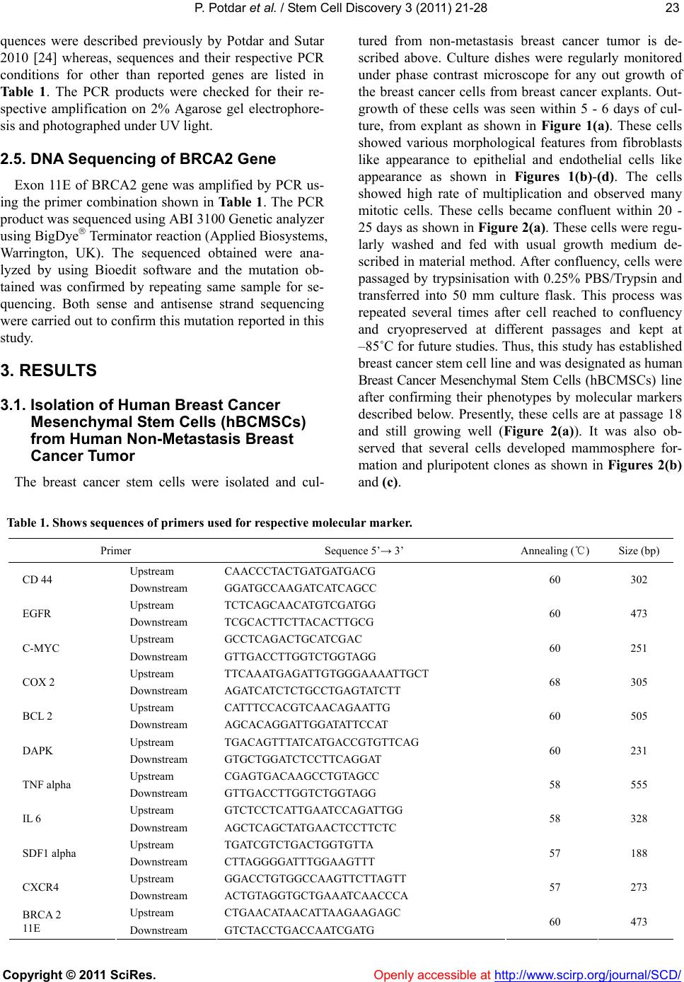 P. Potdar et al. / Stem Cell Discovery 3 (2011) 21-28 Copyright © 2011 SciRes. http://www.scirp.org/journal/SCD/Openly accessible at 2323 quences were described previously by Potdar and Sutar 2010 [24] whereas, sequences and their respective PCR conditions for other than reported genes are listed in Table 1. The PCR products were checked for their re- spective amplification on 2% Agarose gel electrophore- sis and photographed under UV light. 2.5. DNA Se quencing of BRCA2 Gene Exon 11E of BRCA2 gene was amplified by PCR us- ing the primer combination shown in Table 1. The PCR product was sequenced using ABI 3100 Genetic analyzer using BigDye Terminator reaction (Applied Biosystems, Warrington, UK). The sequenced obtained were ana- lyzed by using Bioedit software and the mutation ob- tained was confirmed by repeating same sample for se- quencing. Both sense and antisense strand sequencing were carried out to confirm this mutation reported in this study. 3. RESULTS 3.1. Isolation of Human Breast Cancer Mesenchymal Stem Cells (hBCMSCs) from Human Non-Metastasis Breast Cancer Tumor The breast cancer stem cells were isolated and cul- tured from non-metastasis breast cancer tumor is de- scribed above. Culture dishes were regularly monitored under phase contrast microscope for any out growth of the breast cancer cells from breast cancer explants. Out- growth of these cells was seen within 5 - 6 days of cul- ture, from explant as shown in Figure 1(a). These cells showed various morphological features from fibroblasts like appearance to epithelial and endothelial cells like appearance as shown in Figures 1(b)-(d). The cells showed high rate of multiplication and observed many mitotic cells. These cells became confluent within 20 - 25 days as shown in Figure 2(a). These cells were regu- larly washed and fed with usual growth medium de- scribed in material method. After confluency, cells were passaged by trypsinisation with 0.25% PBS/Trypsin and transferred into 50 mm culture flask. This process was repeated several times after cell reached to confluency and cryopreserved at different passages and kept at –85˚C for future studies. Thus, this study has established breast cancer stem cell line and was designated as human Breast Cancer Mesenchymal Stem Cells (hBCMSCs) line after confirming their phenotypes by molecular markers described below. Presently, these cells are at passage 18 and still growing well (Figure 2(a)). It was also ob- served that several cells developed mammosphere for- mation and pluripotent clones as shown in Figures 2(b) and (c). Table 1. Shows sequences of primers used for respective molecular marker. Primer Sequence 5’→ 3’ Annealing (℃) Size (bp) Upstream CAACCCTACTGATGATGACG CD 44 Downstream GGATGCCAAGATCATCAGCC 60 302 Upstream TCTCAGCAACATGTCGATGG EGFR Downstream TCGCACTTCTTACACTTGCG 60 473 Upstream GCCTCAGACTGCATCGAC C-MYC Downstream GTTGACCTTGGTCTGGTAGG 60 251 Upstream TTCAAATGAGATTGTGGGAAAATTGCT COX 2 Downstream AGATCATCTCTGCCTGAGTATCTT 68 305 Upstream CATTTCCACGTCAACAGAATTG BCL 2 Downstream AGCACAGGATTGGATATTCCAT 60 505 Upstream TGACAGTTTATCATGACCGTGTTCAG DAPK Downstream GTGCTGGATCTCCTTCAGGAT 60 231 Upstream CGAGTGACAAGCCTGTAGCC TNF alpha Downstream GTTGACCTTGGTCTGGTAGG 58 555 Upstream GTCTCCTCATTGAATCCAGATTGG IL 6 Downstream AGCTCAGCTATGAACTCCTTCTC 58 328 Upstream TGATCGTCTGACTGGTGTTA SDF1 alpha Downstream CTTAGGGGATTTGGAAGTTT 57 188 Upstream GGACCTGTGGCCAAGTTCTTAGTT CXCR4 Downstream ACTGTAGGTGCTGAAATCAACCCA 57 273 Upstream CTGAACATAACATTAAGAAGAGC BRCA 2 11E Downstream GTCTACCTGACCAATCGATG 60 473 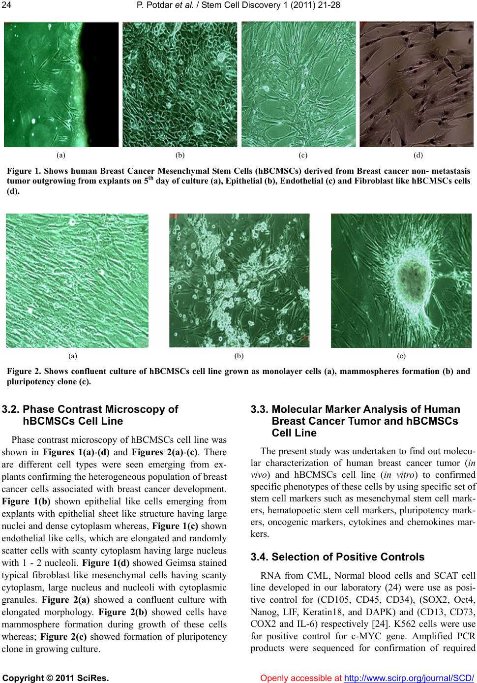 P. Potdar et al. / Stem Cell Discovery 1 (2011) 21-28 Copyright © 2011 SciRes. Openl y accessible at http://www.scirp.org/journal/SCD/ 24 (a) (b) (c) (d) Figure 1. Shows human Breast Cancer Mesenchymal Stem Cells (hBCMSCs) derived from Breast cancer non- metastasis tumor outgrowing from explants on 5th day of culture (a), Epithelial (b), Endothelial (c) and Fibroblast like hBCMSCs cells (d). (a) (b) (c) Figure 2. Shows confluent culture of hBCMSCs cell line grown as monolayer cells (a), mammospheres formation (b) and pluripotency clone (c). 3.2. Phase Contrast Microscop y of hBCMSCs Cell Line Phase contrast microscopy of hBCMSCs cell line was shown in Figures 1(a)-(d) and Figures 2(a)-(c). There are different cell types were seen emerging from ex- plants confirming the heterogeneous population of breast cancer cells associated with breast cancer development. Figure 1(b) shown epithelial like cells emerging from explants with epithelial sheet like structure having large nuclei and dense cytoplasm whereas, Figure 1(c) shown endothelial like cells, which are elongated and randomly scatter cells with scanty cytoplasm having large nucleus with 1 - 2 nucleoli. Figure 1(d) showed Geimsa stained typical fibroblast like mesenchymal cells having scanty cytoplasm, large nucleus and nucleoli with cytoplasmic granules. Figure 2(a) showed a confluent culture with elongated morphology. Figure 2(b) showed cells have mammosphere formation during growth of these cells whereas; Figure 2(c) showed formation of pluripotency clone in growing culture. 3.3. Molecular Marker Analysis of Human Breast Cancer Tumor and hBCMSCs Cell Line The present study was undertaken to find out molecu- lar characterization of human breast cancer tumor (in vivo) and hBCMSCs cell line (in vitro) to confirmed specific phenotypes of these cells by using specific set of stem cell markers such as mesenchymal stem cell mark- ers, hematopoetic stem cell markers, pluripotency mark- ers, oncogenic markers, cytokines and chemokines mar- kers. 3.4. Selection of Positive Controls RNA from CML, Normal blood cells and SCAT cell line developed in our laboratory (24) were use as posi- tive control for (CD105, CD45, CD34), (SOX2, Oct4, Nanog, LIF, Keratin18, and DAPK) and (CD13, CD73, COX2 and IL-6) respectively [24]. K562 cells were use for positive control for c-MYC gene. Amplified PCR products were sequenced for confirmation of required  P. Potdar et al. / Stem Cell Discovery 3 (2011) 21-28 Copyright © 2011 SciRes. Openl y accessible at http://www.scirp.org/journal/SCD/ 2525 gene sequences using specific primers. The sequences obtained for each gene were confirmed from NCBI da- tabase using BLAST software [data not shown] and shown 100% match for these specific genes reported here. On the basis of this, positive controls were selected for this study. 3.4.1. Mesenchymal and Hema topoietic Markers in Human Breast Tumor Cells and hBCMSCs Cell Line Mesenchymal and hematopoietic markers expression in human breast tumor cells and hBCMSC cells is shown in Figure 3. It was showed that tumor cells did not ex- press CD105, CD13 and CD73 genes whereas, hBC- MSCs cell line showed higher expression of all three genes, thus confirming their mesenchymal stem cell phenotypes. These cells also mildly expressed CD34 but showed negative expression of CD45 gene indicating that hBCMSCs cells may have hematopoetic phenotypes. Both these genes were not expressed in tumor tissue. 3.4.2. Pluripotency Markers in Human Breast Tumor Cells and hBCMSCs Cell Line We have studied expression of Oct4, Nanog, SOX2, LIF and Keratin18 genes as markers for pluripotency and differentiation in tumor cells and in hBCMSCs cell line respectively as shown in Figure 4. We have observed that Oct 4, Nanog and SOX2 were expressed at in vivo cells whereas, in hBCMSCs cell line Oct 4, Nanog were mildly expressed and SOX2 was shown to be down reg- ulated (Figure 4) indicate that SOX2 plays some role in pathogenesis of Breast cancer and needs further investi- gation. Differentiation markers LIF and keratin18 were expressed equally in vivo and in vitro cells (Figure 4). Figure 3. Shows expression of mes- enchymal and hematopoietic stem cell markers in Breast cancer tumor tissue (in vivo) and in hBCMSCs cell line (in vitro). Figure 4. Shows expression of pluri- potency stem cell markers in Breast cancer tumor tissue (in vivo) and in hBCMSCs cell line (in vitro). 3.4.3. Oncogenic Markers in Human Breast Tumor Cells and hBCMSCs Cell Line As hBCMSCs cell line was derived from non-metas- tasis tumor cells, we thought of studying oncogenic markers expression in these cells. We studied c-MYC, COX2, BCL2, CD44, EGFR, and DAPK genes in tumor as well as hBCMSCs cells as shown in Figure 5. It was observed that c-MYC, EGFR, CD44, DAPK and BCL2 genes were expressed in hBCMSCs cell line whereas only BCL2 and COX2 genes were expressed by tumor tissue (Figure 5). Within these expressions, CD44 was shown to be highly expressed in hBCMSCs cell line confirming, the presence of initiating breast cancer tu- mor cells. Expression of DAPK in tumor cells and hBCMSCs cell line also confirmed their premalignant status, as DAPK, a tumor suppressor gene expressed in normal and initiated cells (Figure 5). These results were further confirmed by studying chemokine receptor CXCR4 expression in tumor and hBCMSCs cells. It was also observed that CXCR4 gene was prominently ex- pressed in tumor but mildly expressed in hBCMSCs cell line indicating that the hBCMSCs cells isolated from tumor were not metastatic but they are breast cancer initiated cells. We also observed expression of SDF1 in tumor and hBCMSCs cell line (Figure 6(b)). As cyto- kines play very important role in mammary carcino- genesis, we have studied expression of IL6 and TNF in tumor and hBCMSCs cell line (Figure 6(a)). We ob- served that TNF was expressed prominently in tumor but shown mild expression in hBCMSCs cell line. Simi- larly tumor tissue cells expressed mild expression of IL6 whereas, hBCMSCs cells showed significantly higher expression of IL6 indicating specific role of IL6 in breast carcinogenesis as reported by various investigator [25].  P. Potdar et al. / Stem Cell Discovery 1 (2011) 21-28 Copyright © 2011 SciRes. Openl y accessible at http://www.scirp.org/journal/SCD/ 26 3.4.4. Expression of BRCA2 Gene Mutation in Human Breast Tumor Cells and hBCMSCs Cell Line As our laboratory has established a founder mutation in BRCA2 gene in Indian breast cancer patients [23], we thought of evaluated this BRCA2 mutation in tumor tissue as well as in hBCMSCs cell line by DNA se- quencing (Figure 6(c)). We did find the mutation at exon 11E of BRCA 2 gene in tumor as well as hBCMSCs cells indicating the cells isolated from tumor cells have similar characteristic, as it appeared in vivo. Thus, we can conclude that the cell line isolated from non-metas- tasis breast cancer tumor is none other than Initiated breast cancer stem cells causing tumor in humans. Figure 5. Shows expression of oncogenic cell markers in Breast cancer tumor tis- sue (in vivo) and in hBCMSCs cell line (in vitro). (a) (b) (c) Figure 6. (a) Shows expression of cytokine markers in Breast cancer tumor tissue (in vivo) and in hBCMSCs cell line (in vitro); (b) shows expression of chemokine markers in Breast cancer tumor tissue (in vivo) and in hBCMSCs cell line (in vitro); (c) shows electropherograms of exon 11E of BRCA2 gene mutation (A3624G) observed in tumor tissue as well as in hBCMSCs cell line. 4. DISCUSSION Breast cancer is a complex disease, with multiple ge- netic and environmental factors involved in its aetiology. Recent advances in stem cell research have demon- strated that the cancers originally develop from normal cells, which gain the ability to proliferate aberrantly and eventually turn malignant [1]. These malignant cells have been defined as cancer stem cells. These cells have exclusive ability to self-renewal and to differentiate into the heterogeneous lineages of cancer cells that comprise the tumor [1]. Two of major challenges in breast cancer studies are 1) Isolation of Breast cancer stem cells from breast tumor tissue and 2) identify breast cancer stem cells using specific stem cell markers. The technology for isolating Breast cancer stem cells from breast tumor has been shown that CD24low/− CD44+ are a define popu- lation of cells of potential breast cancer stem cells [5]. In present study, we have examined non-metastasis breast tumor for the presence of Breast cancer stem cells and characterized them using molecular markers. We are the first to isolate human breast cancer mesenchymal stem cells from non-metastasis breast tumor. We have desig- nated this cell line as human Breast Cancer Mesenchy- mal Stem Cells (hBCMSCs), which is presently at pas- sage 18. Recent studies have suggested the existence of stem cells in the human mammary gland and shown non-adherent mammospheres secretary cancer stem cells with progenitor cell properties [12,13]. In our study, we have observed similar phenomenon of formation of mammospheres by hBCMSCs cells indicating clearly that the cells isolated from non- metastatic tumor are Breast cancer initiated stem cells. Mesenchymal characteristic of these cells were con- firmed by examination of CD105, CD13 and CD73 markers, which are putative markers for Mesenchymal Stem Cells [14]. These cells also expressed hematopoi-  P. Potdar et al. / Stem Cell Discovery 3 (2011) 21-28 Copyright © 2011 SciRes. Openl y accessible at http://www.scirp.org/journal/SCD/ 2727 etic marker represented partial phenotype of hematopoi- etic stem cells [15]. Shi et al. (2006) have shown that transcription factors Oct-3/4, Nanog and SOX2 maintain the expression of Pluripotent factors at a steady state [26] whereas; Wilson et al. (1988) have shown that the LIF maintains the developmental potential of embryonic stem cells, which can be regarded as a pluripotency marker [20]. In present study Breast cancer tumor cells and hBCMSCs cells expressed Oct4, Nanog and LIF showing their pluripotency. Expression of keratin 18 in tumor cells and hBCMCSs cell line confirmed the epithelial origin of these cells [27]. After establishment of breast cancer stem cell line, it was very essential to know whether, the cell line pos- sesses normal or malignant phenotypes so we have util- ized oncogenic markers such as CD44, EGFR, c-MYC, DAPK, COX2, BCL2, and SOX2 to study the malig- nancy nature. Oncogenes overexpression is a common phenomenon in the development and progression of many human cancers hence it provide potential target for cancer gene therapy [28]. Breast cancer cells were het- erogeneous with respect to expression of a variety of cell surface markers including CD44 and CD24. CD44 is a complex transmembrane glycoprotein whose expression is associated with drug resistance and metastatic pro- gression in malignancy [21]. In present study, we have shown that significant expression of CD44 in hBCMSCs cell line and mild expression in tumor cells indicating that the cells isolated from heterogeneous population of tumor cells are breast cancer initiated cells. Studies have shown that oncogenes c-MYC, COX2, EGFR and DAPK plays important role in development of breast cancer whereas, their overexpression or loss of expres- sion is useful to study correlation with invasive potential and metastasis in aggressive tumors and neoplastic tis- sues[18,19,29,30]. Similarly expression of BCL2 and SOX2 may be associated with metastasis, proliferation and tumorigenesis of breast cancer cells [31,32]. Here in the present study, there was significant expression of c-MYC and EGFR in hBCMSCs cell line whereas, in vivo study of tumor cells showed mild expression of COX2 and down regulation of DAP Kinase gene sug- gesting that the hBCMSCs cell lines contains initiated breast cancer stem cells which may became transformed to metastatic cancer cells when triggered by endogenous factors. Cytokines, such as IL-6 and Tumor Necrosis Factor (TNFα), have an important role in regulating estrogen synthesis and shown in vitro expression by aggressive mesenchymal/basal-like breast cancer cell lines, tissues and xenograft which promote malignant features in breast cancer cells [25,33]. In present study, we found low expression of IL6 in tumor cells whereas, signifi- cantly high expression in hBCMSCs cell line which con- firmed with the finding of other investigators [25,33]. Chemokine receptor CXCR4 showed significant high expression in many solid tumors and also involved in cell migration and invasion, as well as in angiogenesis [34]. In present study, hBCMSCs cell line showed low expression of CXCR4 indicating non-metastatic nature of hBCMSCs cells isolated from tumor cells, which showed significant high expression of CXCR4 gene. We further thought of confirming cancer-initiated cells by studying gene mutation in BRCA2 gene. Our laboratory recently reported a specific founder mutation in Exon 11E of BRCA2 gene in Indian population as an early prognostic marker for Breast cancer [23]. In the present study, we have sequenced DNA samples of breast cancer tumor tissue and hBCMSCs cell line. Our study showed that this founder mutation was at in vivo and in vitro level indicating that the cells isolated from non-metastasis breast cancer tumor are noting but Breast cancer initiated cells which are responsible for develop- ment of breast cancer. Overall, this study establishes a technology for de- velopment of breast cancer stem cell lines from non-metastasis breast cancer tumor and represents an in vitro model to understand the mechanism of Breast car- cinogenesis. The identification of specific molecular markers in these cells allows us to plan targeted thera- pies for breast cancer. 5. ACKNOWLEDGEMENTS We acknowledged with thanks the management of Jaslok Hospital and Research Center, Mumbai, India for sanctioning and supporting financial assistance for this stem cell research project No. 491. We are also thankful to Dr. A. R. Khan, OT Manager for supplying us Breast cancer Tumor tissues and Mrs. Naina Rane for technical help for this study. REFERENCES [1] Smith, G.H. and Chepko, G. (2001) Mammary epithelial stem cells. Microscopy Research and Technique, 15, 190-203. [2] Pardal, R., Clarke, M.F. and Morrison, S.J. (2003) Ap- plying the principles of stem-cell biology to cancer. Na- ture Reviews Cancer, 3, 895-902. [3] Lapidot, T., Sirard, C., Vormoor, J., Murdoch, B., Hoang, T., Caceres-Cortes, J., Minden, M., Paterson, B., Caligi- uri, M.A. and Dick, J.E. (1994) A cell initiating human acute myeloid leukemia after transplantation into SCID mice. Nature, 367, 645-648. [4] Fidler, I.J. and Kripke, M.L. (1997) Metastasis results from preexisting variant cells within a malignant tumor. Science, 197, 893-895. [5] Al-Hajj, M., Wicha, M.S., Benito-Hernandez, A., Morri- son, S.J. and Clarke, M.F. (2003) Prospective identifica- tion of tumorigenic breast cancer cells. Proceedings of  P. Potdar et al. / Stem Cell Discovery 1 (2011) 21-28 Copyright © 2011 SciRes. http://www.scirp.org/journal/SCD/Openly accessible at 28 the National Academy of Sciences, 100, 3983-3988. [6] Bonnet, D. and Dick, J.E. (1997) Human acute myeloid leukemia is organized as a hierarchy that originates from a primitive hematopoietic cell. Nature Medicine, 3, 730-737. [7] Collins, A.T., Berry, P.A., Hyde, C., Stower, M.J. and Maitland, N.J. (2005) Prospective Identification of tu- morigenic prostate cancer stem cells. Cancer Research, 65, 10946-10951. [8] Hemmati, H.D., Nakano, I., Lazareff, J.A., Master- man-Smith, M., Geschwind, D.H., Bronner-Fraser M. and Komblurn, H.I. (2003) Cancerous stem cells can arise from pediatric brain tumors. Proceedings of the Na- tional Academy of Sciences, 100, 15178-15183. [9] Kim, C.F., Jackson, E.L., Woolfenden, A.E., Lawrence, S., Babar, I., Vogel, S., Crowley, D., Bronson, R.T. and Jacks, T. (2005) Identification of bronchioalveolar stem cells in normal lung and lung cancer. Cell, 121, 823-835. [10] O’Brien, C.A., Pollett, A., Gallinger, S. and Dick, J.E. (2007) A human colon cancer cell capable of initiating tumor growth in immunodeficient mice. Nature, 445, 106-110. [11] Potdar, P.D. and Subedi, R.P. (2011) Defining Molecular Phenotypes of Mesenchymal and hematopoietic Stem Cells derived from Peripheral blood of Acute Lympho- cytic Leukemia patients for regenerative stem cell ther- apy. Journal of Stem cells & Regenerative Medicine, 7, 29-40. [12] Grimshaw, M.J., Cooper, L., Papazisis, K., Coleman, J.A., Bohnenkamp, H.R., Chiapero-Stanke, L., Taylor-Papa- dimitriou, J. and Burchell, J.M. (2008) Mammosphere- culture of metastatic breast cancer cells enriches for tu- morigenic breast cancer cells. Breast Cancer Research, 10, 52. [13] Wicha, M., Liu, S. and Dontu, G. (2006) Cancer stem cells: an old idea – a paradigm shift. Cancer Research, 66, 1883-1890. [14] Barry, F.P., Boynton, R.E., Haynesworth, S., Murphy, J.M. and Zaia, J. (1999) The monoclonal antibody SH-2, raised against human mesenchymal stem cells, recog- nizes an epitope on endoglin (CD105). Biochemical and Biophysical Resear ch Comm un i c a t io n s, 265, 134-139. [15] Healy, L., May, G., Gale, K., Grosvel, F., Greaves, M. and Enver, T. (1995) The stem cell antigen CD34 func- tions as a regulator of hemopoietic cell adhesion. Pro- ceedings of the National Academy of Sciences, 92, 12240-12244. [16] Ogata, K., Satoh, C., Tachibana, M., Hyodo, H., Tamura, H., Dan, K., Kimura, T., Sonoda, Y. and Tsuji, T. (2005) Identification and hematopoietic potential of CD45- clonal cells with very immature phenotypes (CD45- Cd34-Cd38-Lin-) in patients with myelodysplastic syn- dromes. Stem Cell, 23, 619-630. [17] Olamura-Nakanishi, S., Saito, M., Niwa, H. and Isshil- cawa, F. (2005) Oct-3/4 and Sox2 regulate Oct-3/4 gene in embryonic stem cells. Journal of Biological Chemistry, 22, 5307-5317. [18] Prescott, S.M. and Fitzpatrick, F.A. (2000) Cyclooxy- genase-2 and carcinogenesis. Biochimica et Biophysica Acta, 1470, M69-M78. [19] Katzenellenbogen, R.A., Baylin, S.B. and Herman, J.G. (1999) Hypermethylation of the DAP-Kinase CpG island is a common alteration in B-cell malignancies. Blood, 93, 4347-4353. [20] William, R.L., Hilton, D.J., Pease, S., Willson, T.A., Stewart, C.L., Gearing, D.P., Wagner, E.F., Metcalf, D., Nicola, N.A. and Gough, N.M. (1988) Myeloid leukae- mia inhibitory factor maintains the developmental poten- tial of embryonic stem cells. Nature, 336, 684-687. [21] Draffin, J.E., McFarlane, S., Hill, A., Johnston, P.G. and Waugh, D.J. (2004) CD44 potentiates the adherence of metastatic prostate and breast cancer cells to bone mar- row endothelial cells. Cancer Research, 64, 5702-5711. [22] Schabeth, H., Runz, S., Joumaa, S. and Altevogt, P. (2006) CD24 affects CXCR4 function in pre-β lymphocytes and breast carcinoma cells. Journal of Cell Science, 119, 314-325. [23] Potdar, P.D. and Bisht, S. L. (2009) Identification and screening for novel mutations in exon 10 and exon 11 of BRCA 1 and BRCA 2 genes in hereditary and sporadic breast cancer patients in Indian population- concept for biomarkers for early detection. Annals of Oncology, 20, 51. [24] Potdar, P.D. and Sutar, J. P. (2010) Establishment and molecular characterization of mesenchymal stem cell lines derived from human visceral & subcutaneous adi- pose tissues. Journal of Stem cells & Regenerative Medi- cine, 6, 1-10. [25] Knupfer, H. and Preiss, R. (2007) Significance of inter- leukin-6 (IL-6) in breast cancer. Breast Cancer Res Treat, 102, 129-35. [26] Shi, W., Wang, H., PanYijie, G., Yunqian, G., Pei, G., (2006) Regulation of the Pluripotency Marker Rex-1 by NANOG and SOX2. Journal of Biological Chemistry, 281, 23319-23325. [27] Ku, N., et al. (1997) Mutation of human keratin 18 in association with cryptogenic cirrhosis. Journal of Clini- cal Investigation, 99, 19-23. [28] Malumbres, M. and Barbacid, M. (2001) To cycle or not to cycle: a critical decision in cancer. Nature Reviews Cancer, 1, 222-231. [29] Nesbit, C.E., Tersak, J.M. and Prochownik, E.V. (1999) MYC oncogenes and human neoplastic disease. Onco- gene, 18, 3004-3016. [30] Bhargava, R., Gerald, W.L., Li, A.R., Pan, Q., Lal, P., Ladanyi, M. and Chen, B. (2005) EGFR gene amplifica- tion in breast cancer: Correlation with epidermal growth factor receptor mRNA and protein expression and HER-2 status and absence of EGFR-activating mutations. Mod- ern Pathology, 18, 1027-1033. [31] Swellam, M., Ismail, M., Eissa, S., Hamdy, M. and Mok- htar, N. (2004) Emerging role of P53, Bcl-2 and telom- erase activity in egyptian breast cancer patients. IUBMB Life, 56, 483-490. [32] Chen, Y., Shi, L., Zhang, L., Li, R., Liang, J., Yu, W., Sun, L., Yang, X., Wang, Y., Zhang, Y. and Shang, Y. (2008) The molecular mechanism governing the oncogenic po- tential of SOX2 in breast cancer. The Journal of Bio- logical Chemistry, 283, 17969-17978. [33] Charafe-Jauffret, E., et al., (2006) Gene expression pro- filing of breast cell lines identifies potential new basal markers. Oncogene, 25, 2273-2284. [34] Smith, M.C.P., Luker, K.E., Garbow, J.R., et al., (2004) CXCR4 regulates growth of both primary and metastatic breast cancer. Cancer Research, 64, 8604-8612. |