

Paper Menu >>
Journal Menu >>
 Open Journal of Marine Science, 2011, 1, 31-35 doi:10.4236/ojms.2011.12003 Published Online July 2011 (http://www.SciRP.org/journal/ojms) Copyright © 2011 SciRes. OJMS Mechanisms of the Plurality of Scor paena porcus L. Serum Albumin A. M. Andreeva I. D. Papanin Institute for Biology of Inland Waters RAS E-mail: [email protected] Received March 18 2011; revised May 10, 2011; accepted May 20, 2011 Abstract The proteins, which bind albuminspecific dye Evans blue, are revealed in the low-molecular protein fraction of the blood serum from Scorpaena porcus L. and identified as serum albumin. They were represented by three bands in 2D-SDS-PAAG. MALDI-TOF-analysis revealed the fundamental similarity of the mass spec- trum of the fragments of tryptic cleavage of proteins with molecular weight 73 and 76 kDa. The role of du- plications and intragenic reconstructions in the creation of the plurality of scorpaena albumins is discussed. Keywords: Scorpaena, Blood, Tissue Fluids, Low-Molecular Prote ins, A lbum in, Ma ss Spectr um , Dupl icat ions 1. Introduction Serum albumins accomplish important functions in the organism of vertebrates, participating in the filtration of tissue fluid, in the transport of biomolecules and in the plastic metabolism. Mammalian albumins are simple monomeric proteins with the molecular weight about 67 kDa; they are represented, as a rule, by one component on the electrophoregram [1]. Fish albumins differ from mammalian ones in the diversity of organization ways, physical and chemical properties: there are simple pro- teins and glycoproteins, monomers, oligomers and ag- gregates among them. Usually their electrophoretic mo- bility does not co incid e with that of mamma lian alb umin, and they are often represented by the plural forms in the electrophoresis [2-20]. The ability of fish albumins to bind albuminspecific dyes, palmitic acid, incapacity to bind nickel, molecular weight and other characteristics are used for the fish albumin identification [9,11,14, 17,21,22]. We identified serum albumin of scorpaena Scorpaena porcus by the molecular weight, the ability to bind albuminspecific dyes and by means of MALDI- TOF-analysis; the results obtained were used to reveal the mechanisms of albumin plurality. 2. Material and Methods 2.1. Objects of Study The objects of this study were Scorpaenas Scorpaena porcus L. from the Black Sea. For comparison we used Mullus barbatus L., Uranoscopus scaber L., Symphodus tinca L., Gaidropsarus mediterraneus L., Neogobius melanostomus P. and Mesogobius batrachocephalus P. from the Black Sea and also roach Rutilus rutilus L. and perch Perca fluviatilis L. from the Rybinsk Reservoir. For the work we used proteins from blood serum and plasma and tissue fluids from the peritoneum, brain and white muscles. 2.2. Methods of Analysis The biological fluids obtaining. The blood was ob- tained from the caudal artery, tissue fluids were taken by pipetting or by the impregnation of the strip (0.5 × 4.0 mm) of the chromatographic paper Watmann 3 MM [23]. Protein concentration measurement. We used micro- biuret method to estimate the concentration of total pro- tein [24]. Electrophoresis methods. We analysed the albumins by disk- and 2D-electrophoresis (in gradient of PAGE concentration 5% - 40%, in PAGE with 8 M urea [17] and SDS [25]. For calculation of the molecular weights (MM) of proteins we used: myoglobin and the polymeric forms of human serum albumin HSA and ovalbumin OVA; the markers Fermentas PageRulerTM Prestained Protein Ladder Plus (11, 17, 28, 36, 55, 72, 95, 130, 250 kDa). Results were processed statistically with program package OneDscan. The binding of proteins by albuminspecific dyes. We studied the binding of proteins by Evans blue and brom- 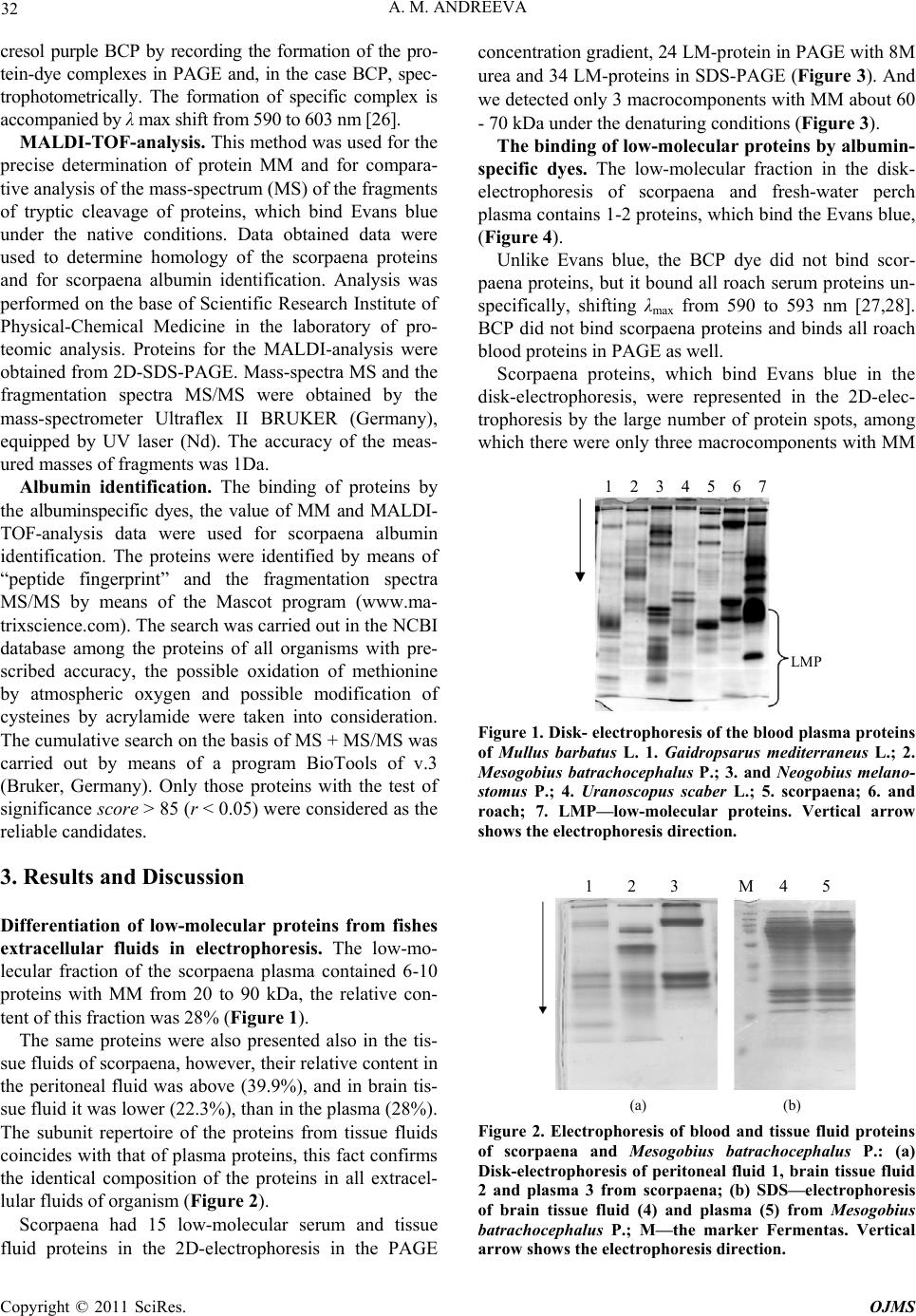 32 A. M. ANDREEVA cresol purple BCP by recording the formation of the pro- tein-dye complexes in PAGE and, in the case BCP, spec- trophotometrically. The formation of specific complex is accompanied by λ max shift from 590 t o 603 nm [26] . MALDI-TOF-analysis. This method was used for the precise determination of protein MM and for compara- tive analysis of the mass-spectrum (MS) of the fragments of tryptic cleavage of proteins, which bind Evans blue under the native conditions. Data obtained data were used to determine homology of the scorpaena proteins and for scorpaena albumin identification. Analysis was performed on the base of Scientific Research Institute of Physical-Chemical Medicine in the laboratory of pro- teomic analysis. Proteins for the MALDI-analysis were obtained from 2D-SDS-PAGE. Mass-spectra MS and the fragmentation spectra MS/MS were obtained by the mass-spectrometer Ultraflex II BRUKER (Germany), equipped by UV laser (Nd). The accuracy of the meas- ured masses of fragments was 1Da. Albumin identification. The binding of proteins by the albuminspecific dyes, the value of MM and MALDI- TOF-analysis data were used for scorpaena albumin identification. The proteins were identified by means of “peptide fingerprint” and the fragmentation spectra MS/MS by means of the Mascot program (www.ma- trixscience.com). The search was carried out in the NCBI database among the proteins of all organisms with pre- scribed accuracy, the possible oxidation of methionine by atmospheric oxygen and possible modification of cysteines by acrylamide were taken into consideration. The cumulative search on the basis of MS + MS/MS was carried out by means of a program BioTools of v.3 (Bruker, Germany). Only those proteins with the test of significance score > 85 (r < 0.05 ) were con sidered as the reliable candidates. 3. Results and Discussion Differentiation of low-molecular proteins from fishes extracellular fluids in electrophoresis. The low-mo- lecular fraction of the scorpaena plasma contained 6-10 proteins with MM from 20 to 90 kDa, the relative con- tent of this fraction was 28% (Figure 1). The same proteins were also presented also in the tis- sue fluids of scorpaena, however, their relative content in the peritoneal fluid was above (39.9%), and in brain tis- sue fluid it was lower (22.3%), than in the plasma (28%). The subunit repertoire of the proteins from tissue fluids coincides with that of plasma proteins, this fact confirms the identical composition of the proteins in all extracel- lular fluids of organism (Figure 2). Scorpaena had 15 low-molecular serum and tissue fluid proteins in the 2D-electrophoresis in the PAGE concentration grad ient, 24 LM-pro tein in PAGE with 8M urea and 34 LM-protein s in SDS-PAGE (Figure 3). And we detected only 3 macrocomponents with MM about 60 - 70 kDa under the denaturing conditions (Figure 3). The binding of low-molecular proteins by albumin- specific dyes. The low-molecular fraction in the disk- electrophoresis of scorpaena and fresh-water perch plasma contains 1-2 proteins, which bind the Evans blue, (Figure 4). Unlike Evans blue, the BCP dye did not bind scor- paena proteins, but it bound all roach serum proteins un- specifically, shifting λmax from 590 to 593 nm [27,28]. BCP did not bind scorpaena proteins and binds all roach blood proteins in PA GE as well. Scorpaena proteins, which bind Evans blue in the disk-electrophoresis, were represented in the 2D-elec- trophoresis by the large number of protein spots, among which there were only three macrocomponents with MM LMP 1 2 3 4 5 6 7 Figure 1. Disk- electrophoresis of the blood plasma proteins of Mullus barbatus L. 1. Gaidropsarus mediterraneus L.; 2. Mesogobius batrachocephalus P.; 3. and Neogobius melano- stomus P.; 4. Uranoscopus scaber L.; 5. scorpaena; 6. and roach; 7. LMP—low-molecular proteins. Vertical arrow shows the electrophoresis direction. 1 2 3 M 4 5 (a) (b) Figure 2. Electrophoresis of blood and tissue fluid proteins of scorpaena and Mesogobius batrachocephalus P.: (a) Disk-electrophoresis of peritoneal fluid 1, brain tissue fluid 2 and plasma 3 from scorpaena; (b) SDS—electrophoresis of brain tissue fluid (4) and plasma (5) from Mesogobius batrachocephalus P.; M—the marker Fermentas. Vertical arrow shows the electrophoresis direction. Copyright © 2011 SciRes. OJMS 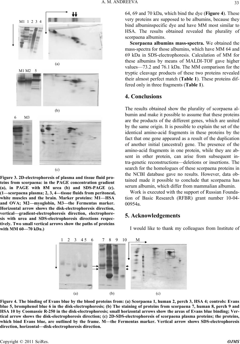 A. M. ANDREEVA Copyright © 2011 SciRes. OJMS 33 64, 69 and 70 kDa, which bind the dye (Figure 4). These very proteins are supposed to be albumins, because they bind albuminspecific dye and have MM most similar to HSA. The results obtained revealed the plurality of scorpaena albumins. M1 1 2 3 4 Scorpaena albumins mass-spectra. We obtained the mass-spectra for those albu mins, which have MM 64 and 69 kDa in SDS-electrophoresis. Calculation of MM for these albumins by means of MALDI-TOF gave higher values—73.2 and 76.1 kDa. The MM comparison for the tryptic cleavage products of these two proteins revealed their almost perfect match (Table 1). These proteins dif- fered only in three fragments (Table 1). (a) M1 M2 5 4. Conclusions The results obtained show the plurality of scorpaena al- bumin and make it possible to assume that these proteins are the products of the different genes, which are united by the same origin. It is possible to explain the set of the identical amino-acid fragments in these proteins by the fact that one gene appeared as a result of the duplication of another initial (ancestral) gene. The presence of the amino-acid fragments in one protein, while they are ab- sent in other protein, can arise from subsequent in- tra-genetic reconstructions—deletions or insertions. The search for the homologues of these scorpaena proteins in the NCBI database gave no results. However, data ob- tained made it possible to conclude that scorpaena has serum albumin, which differ from mammalian albumin. (b) 6 M3 (c) Figure 3. 2D-electrophoresis of plasma and tissue fluid pro- teins from scorpaena: in the PAGE concentration gradient (a), in PAGE with 8M urea (b) and SDS-PAGE (c). (1—scorpaena plasma; 2, 3, 4—tissue fluids from peritoneal, white muscles and the brain. Marker proteins: M1—HSA and OVA; M2—myoglobin, M3—the Fermentas marker. Horizontal arrow shows the disk-electrophoresis direction, vertical—gradient-electrophoresis direction, electrophore- sis with urea and SDS-electrophoresis directions respec- tively. Two small vertical arrows show the paths of proteins ith MM 60—70 kDa.) Work is executed with the suppor t of Russian Founda- tion of Basic Research (RFBR) grant number 10-04- 00954a. 5. Acknowledgements I would like to thank my colleagues from Institute of w 1 2 3 4 5 6 7 8 9 10 M (a) (b) (c) Figure 4. The binding of Evans blue by the blood proteins from: (a) Scorpaena 1, human 2, perch 3, HSA 4; controls: Evans blue 5, bromphenol blue 6 in the disk-electrophoresis; (b) The staining of proteins from scorpaena 7, human 8, perch 9 and HSA 10 by Coomassie R-250 in the disk-electrophoresis; small horizontal arrows show the areas of Evans blue binding; Ver- tical arrow shows the disk-electrophoresis direction; (c) 2D-SDS-electrophoresis of scorpaena plasma proteins; the proteins, which bind Evans blue, are outlined by the frame. M—the Fermentas marker. Vertical arrow shows SDS-electrophoresis direction, horizontal—disk-electrophoresis direction. 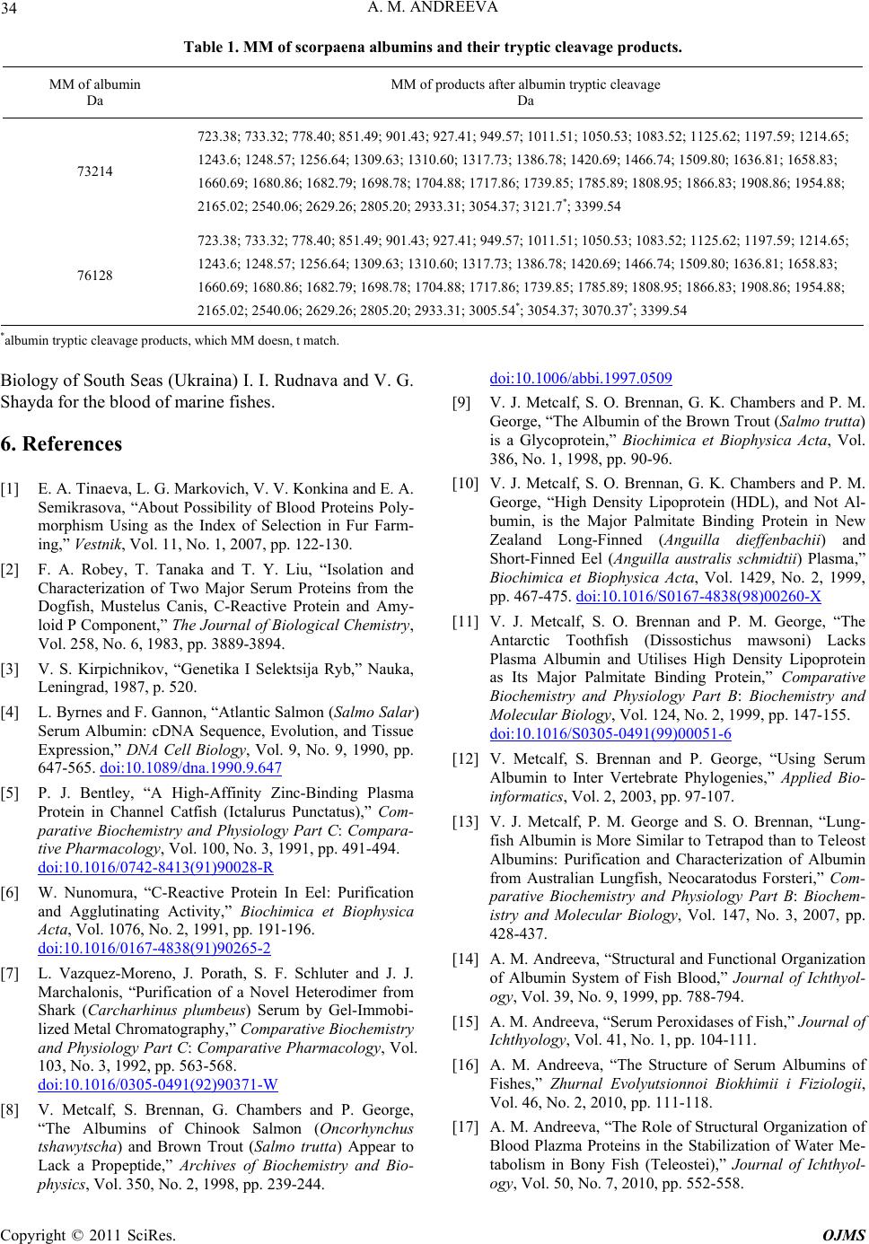 34 A. M. ANDREEVA Table 1. ММ of scorpaena albumins and their tryptic cleavage products. MM of albumin Da MM of products after albumin tryptic cleavage Da 73214 723.38; 733.32; 778.40; 851.49; 901.43; 927.41; 949.57; 1011.51; 1050.53; 1083.52; 1125.6 2; 1197.59; 1214.65; 1243.6; 1248.57; 1256.64; 130 9 .63; 1310.60; 1317.73; 1 38 6 .78; 1420.69; 1466.74; 1509.80; 1636.81; 1658.83; 1660.69; 1680. 8 6 ; 1682.79; 1698.78; 1704.88; 1717.86; 1739 .85; 1785.89; 1808.95; 186 6 .83; 1908.86; 1 954.88; 2165.02; 2540. 0 6; 2629.26; 2805.20; 2933.31; 3054.37; 3121.7*; 3399.54 76128 723.38; 733.32; 778.40; 851.49; 901.43; 927.41; 949.57; 1011.51; 1050.53; 1083.52; 1125.6 2; 1197.59; 1214.65; 1243.6; 1248.57; 1256.64; 130 9 .63; 1310.60; 1317.73; 1 38 6 .78; 1420.69; 1466.74; 1509.80; 1636.81; 1658.83; 1660.69; 1680. 8 6 ; 1682.79; 1698.78; 1704.88; 1717.86; 1739 .85; 1785.89; 1808.95; 186 6 .83; 1908.86; 1 954.88; 2165.02; 2540. 0 6; 2629.26; 2805.20; 2933.31; 3005.54*; 3054.37; 3070.37*; 3399.54 *albumin tryptic cleavage products, which MM doesn, t match. Biology of South Seas (U kraina) I. I. Rudnava and V. G. Shayda for the blood of marine fishes. 6. References [1] E. A. Tinaeva, L. G. Markovich, V. V. Konkina and E. A. Semikrasova, “About Possibility of Blood Proteins Poly- morphism Using as the Index of Selection in Fur Farm- ing,” Vestnik, Vol. 11, No. 1, 2007, pp. 122-130. [2] F. A. Robey, T. Tanaka and T. Y. Liu, “Isolation and Characterization of Two Major Serum Proteins from the Dogfish, Mustelus Canis, C-Reactive Protein and Amy- loid P Component,” The Journal of Biological Chemistry, Vol. 258, No. 6, 1983, pp. 3889-3894. [3] V. S. Kirpichnikov, “Genetika I Selektsija Ryb,” Nauka, Leningrad, 1987, p. 520. [4] L. Byrnes and F. Gannon, “Atlantic Salmon (Salmo Salar) Serum Albumin: cDNA Sequence, Evolution, and Tissue Expression,” DNA Cell Biology, Vol. 9, No. 9, 1990, pp. 647-565. doi:10.1089/dna.1990.9.647 [5] P. J. Bentley, “A High-Affinity Zinc-Binding Plasma Protein in Channel Catfish (Ictalurus Punctatus),” Com- parative Biochemistry and Physiology Part C: Compara- tive Pharmacology, Vol. 100, No. 3, 1991, pp. 491-494. doi:10.1016/0742-8413(91)90028-R [6] W. Nunomura, “C-Reactive Protein In Eel: Purification and Agglutinating Activity,” Biochimica et Biophysica Acta, Vol. 1076, No. 2, 1991, pp. 191-196. doi:10.1016/0167-4838(91)90265-2 [7] L. Vazquez-Moreno, J. Porath, S. F. Schluter and J. J. Marchalonis, “Purification of a Novel Heterodimer from Shark (Carcharhinus plumbeus) Serum by Gel-Immobi- lized Metal Chromatography,” Comparative Biochemistry and Physiology Part C: Comparative Pharmacology, Vol. 103, No. 3, 1992, pp. 563-568. doi:10.1016/0305-0491(92)90371-W [8] V. Metcalf, S. Brennan, G. Chambers and P. George, “The Albumins of Chinook Salmon (Oncorhynchus tshawytscha) and Brown Trout (Salmo trutta) Appear to Lack a Propeptide,” Archives of Biochemistry and Bio- physics, Vol. 350, No. 2, 1998, pp. 239-244. doi:10.1006/abbi.1997.0509 [9] V. J. Metcalf, S. O. Brennan, G. K. Chambers and P. M. George, “The Albumin of the Brown Trout (Salmo trutta) is a Glycoprotein,” Biochimica et Biophysica Acta, Vol. 386, No. 1, 1998, pp. 90-96. [10] V. J. Metcalf, S. O. Brennan, G. K. Chambers and P. M. George, “High Density Lipoprotein (HDL), and Not Al- bumin, is the Major Palmitate Binding Protein in New Zealand Long-Finned (Anguilla dieffenbachii) and Short-Finned Eel (Anguilla australis schmidtii) Plasma,” Biochimica et Biophysica Acta, Vol. 1429, No. 2, 1999, pp. 467-475. doi:10.1016/S0167-4838(98)00260-X [11] V. J. Metcalf, S. O. Brennan and P. M. George, “The Antarctic Toothfish (Dissostichus mawsoni) Lacks Plasma Albumin and Utilises High Density Lipoprotein as Its Major Palmitate Binding Protein,” Comparative Biochemistry and Physiology Part B: Biochemistry and Molecular Biology, Vol. 124, No. 2, 1999, pp. 147-155. doi:10.1016/S0305-0491(99)00051-6 [12] V. Metcalf, S. Brennan and P. George, “Using Serum Albumin to Inter Vertebrate Phylogenies,” Applied Bio- informatics, Vol. 2, 2003, pp. 97-107. [13] V. J. Metcalf, P. M. George and S. O. Brennan, “Lung- fish Albumin is More Similar to Tetrapod than to Teleost Albumins: Purification and Characterization of Albumin from Australian Lungfish, Neocaratodus Forsteri,” Com- parative Biochemistry and Physiology Part B: Biochem- istry and Molecular Biology, Vol. 147, No. 3, 2007, pp. 428-437. [14] A. M. Andreeva, “Structural and Functional Organization of Albumin System of Fish Blood,” Journal of Ichthyol- ogy, Vol. 39, No. 9, 1999, pp. 788-794. [15] A. M. Andreeva, “Serum Peroxidases of Fish,” Journal of Ichthyology, Vol. 41, No. 1, pp. 104-111. [16] A. M. Andreeva, “The Structure of Serum Albumins of Fishes,” Zhurnal Evolyutsionnoi Biokhimii i Fiziologii, Vol. 46, No. 2, 2010, pp. 111-118. [17] A. M. Andreeva, “The Role of Structural Organization of Blood Plazma Proteins in the Stabilization of Water Me- tabolism in Bony Fish (Teleostei),” Journal of Ichthyol- ogy, Vol. 50, No. 7, 2010, pp. 552-558. Copyright © 2011 SciRes. OJMS  A. M. ANDREEVA 35 [18] C. Szebedinszky and K. M. Gilmour, “The Buffering Power of Plasma in Brown Bullhead (Ameiurus nebulo- sus),” Comparative Biochemistry and Physiology Part B: Biochemistry and Molecular Biology, Vol. 131, No. 2, 2002, pp. 171-183. doi:10.1016/S1096-4959(01)00492-4 [19] Y. Xu and Z. Ding, “N-Terminal Sequence and Main Characteristics of Atlantic Salmon (Salmo salar) Albu- min,” Preparative Biochemistry and Biotechnology, Vol. 35, No. 4, 2005, pp. 283-290. doi:10.1080/10826060500218081 [20] A. M. Andreeva and R. A. Federov, “Features of the Or- ganization of Low-Molecular Weight Proteins from the Blood and Tissue Fluid of the Common Stingray Dasyatis Pastinaca L. (Chondroichthyes: Trigonidae),” Russian Journal of Marine Biology, Vol. 36, No. 6, 2010, pp. 469-472. [21] L. L.Sulya, B. E. Box and G. Gordon, “Plasma Proteins in the Blood of Fishes from the Gulf of Mexico,” Ameri- can Journal of Physiology, Vol. 200, No. 1, 1961, pp. 152-154. [22] H. De Smet, R. Blust and L. Moens, “Absence of Albu- min in the Plasma of the Common Carp Cyprinus Carpio Binding of Fatty Acids to High Density Lipoprotein,” Fish Physiology and Biochemistry, Vol. 19, No. 1, 1998, pp. 71-81. doi:10.1023/A:1007734127146 [23] A. M. Andreeva, I. P. Ryabtseva and V. V. Bolshakov, “Analysis of Permeability of Capillaries of Different De- partments of Microcirculatory System for Plasma Pro- teins in Some Representatives of Bony Fishes,” Zhurnal Evolyutsionnoi Biokhimii i Fiziologii, Vol. 44, No. 2, 2008, pp. 212-214. [24] R. F. Itzhaki and D. M. Gill, “A Micro-Biuret Method for Estimating Protein,” Analytical Biochemistry, Vol. 9, No. 4, 1964, pp. 401-410. doi:10.1016/0003-2697(64)90200-3 [25] U. K. Laemmli, “Cleavage of Structural Proteins during the Assembly of the Head of Bacteriophage,” Nature, Vol. 227, No. 5259, 1970, pp. 680-685. [26] A. E. Pinnel and B. E. Northam, “New Automated Dye-Binding Method for Serum Albumin Determination with Bromcresol Purple,” Clinical Chemistry, Vol. 24, No. 1, 1978, pp. 80-86. [27] A. M. Andreeva, “Identification of Serum Albumin and the Study Some of Its Physical Chemistry Properties in the Representatives of the Families Acipenseridae and Cyprinidae,” Inf Bull IBII AS USSR, Vol. 69, 1986, pp. 36-39. [28] A. M. Andreeva, “Physical Chemical Properties of Serum Albumin of the Blood from Acipenseridae and Cyprini- dae on the Example to Sterljad and Bream. Physiology and the Biochemistry of the Hydrobionts,” Yaroslavl, 1987, pp. 108-114. Copyright © 2011 SciRes. OJMS |