

Paper Menu >>
Journal Menu >>
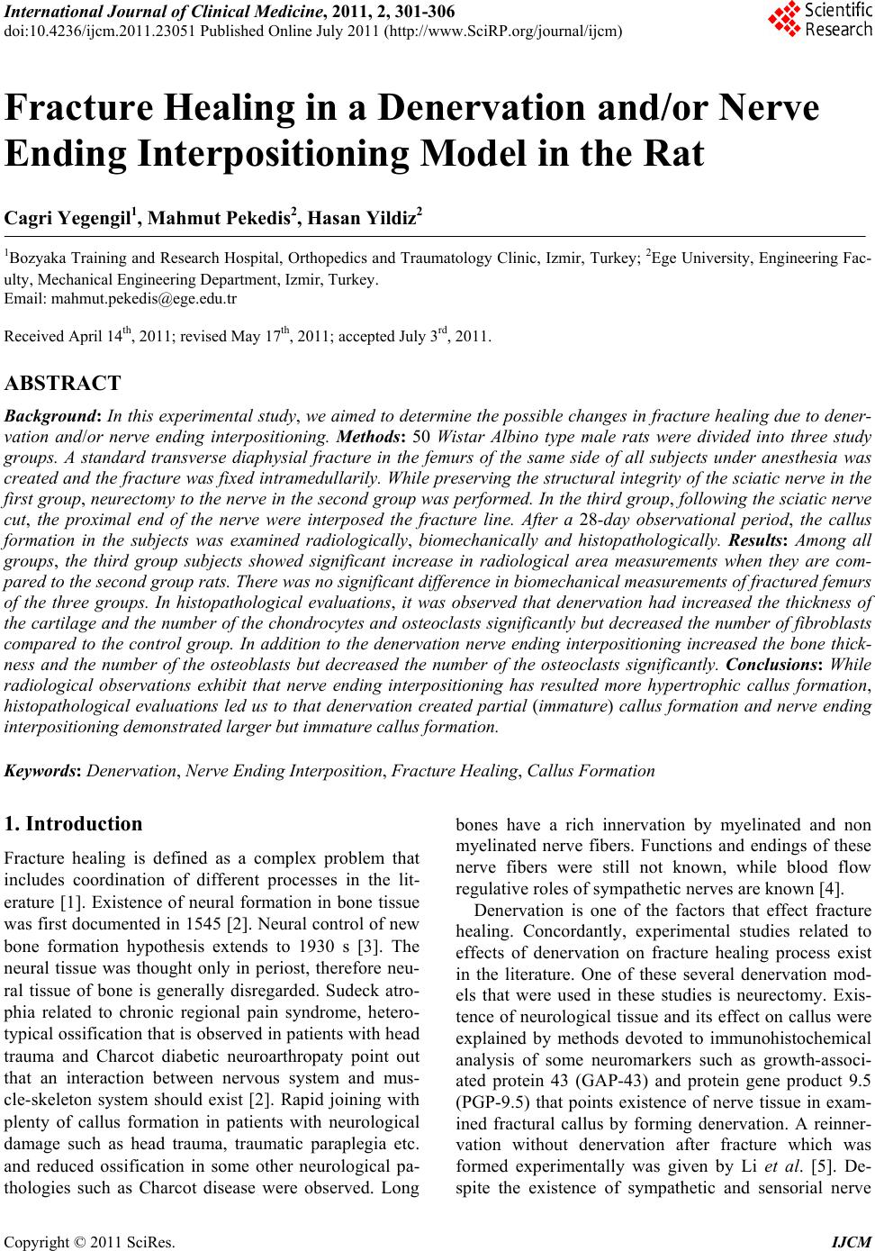 International Journal of Clinical Medicine, 2011, 2, 301-306 doi:10.4236/ijcm.2011.23051 Published Online July 2011 (http://www.SciRP.org/journal/ijcm) Copyright © 2011 SciRes. IJCM 301 Fracture Healing in a Denervation and/or Nerve Ending Interpositioning Model in the Rat Cagri Yegengil1, Mahmut Pekedis2, Hasan Yildiz2 1Bozyaka Training and Research Hospital, Orthopedics and Traumatology Clinic, Izmir, Turkey; 2Ege University, Engineering Fac- ulty, Mechanical Engineering Department, Izmir, Turkey. Email: [email protected] Received April 14th, 2011; revised May 17th, 2011; accepted July 3rd, 2011. ABSTRACT Background: In this experimental study, we aimed to determine the possib le changes in fracture healing du e to dener- vation and/or nerve ending interpositioning. Methods: 50 Wistar Albino type male rats were divided into three study groups. A standard transverse diaphysial fracture in the femurs of the same side of all subjects under anesthesia was created and the fracture was fixed intramedullarily. Wh ile preserving the structural integ rity of the sciatic nerve in the first group, neurectomy to the nerve in the second group was performed. In the third group, following the sciatic nerve cut, the proximal end of the nerve were interposed the fracture line. After a 28-day observational period, the callus formation in the subjects was examined radiologically, biomechanically and histopathologically. Results: Among all groups, the third group subjects showed significant increase in radiological area measurements when they are com- pared to the second g roup rats. Th ere was no sign ificant differen ce in biomechanical measuremen ts of fractu red femurs of the three groups. In histopathological evaluations, it was observed that denervation had increased the thickness of the cartilage an d the number of the chondrocytes and os teocla sts significantly but decreased the numb er of fibroblasts compared to the control group. In addition to the denervation nerve ending interpositioning increased the bone thick- ness and the number of the osteoblasts but decreased the number of the osteoclasts significantly. Conclusions: While radiological observations exhibit that nerve ending interpositioning has resulted more hypertrophic callus formation, histopathological evaluations led us to that denervation created partial (immature) callus formation and nerve ending interpositioning demon strated larger but immature callus formation. Keywords: Denervation, Nerve Ending Interposition, Fracture Healing, Callus Format ion 1. Introduction Fracture healing is defined as a complex problem that includes coordination of different processes in the lit- erature [1]. Existence of neural formation in bone tissue was first documented in 1545 [2]. Neural control of new bone formation hypothesis extends to 1930 s [3]. The neural tissue was thought only in periost, therefore neu- ral tissue of bone is generally disregarded. Sudeck atro- phia related to chronic regional pain syndrome, hetero- typical ossification that is observed in patients with head trauma and Charcot diabetic neuroarthropaty point out that an interaction between nervous system and mus- cle-skeleton system should exist [2]. Rapid joining with plenty of callus formation in patients with neurological damage such as head trauma, traumatic paraplegia etc. and reduced ossification in some other neurological pa- thologies such as Charcot disease were observed. Long bones have a rich innervation by myelinated and non myelinated nerve fibers. Functions and endings of these nerve fibers were still not known, while blood flow regulative roles of sympathetic nerves are known [4]. Denervation is one of the factors that effect fracture healing. Concordantly, experimental studies related to effects of denervation on fracture healing process exist in the literature. One of these several denervation mod- els that were used in these studies is neurectomy. Exis- tence of neurological tissue and its effect on callus were explained by methods devoted to immunohistochemical analysis of some neuromarkers such as growth-associ- ated protein 43 (GAP-43) and protein gene product 9.5 (PGP-9.5) that points existence of nerve tissue in exam- ined fractural callus by forming denervation. A reinner- vation without denervation after fracture which was formed experimentally was given by Li et al. [5]. De- spite the existence of sympathetic and sensorial nerve 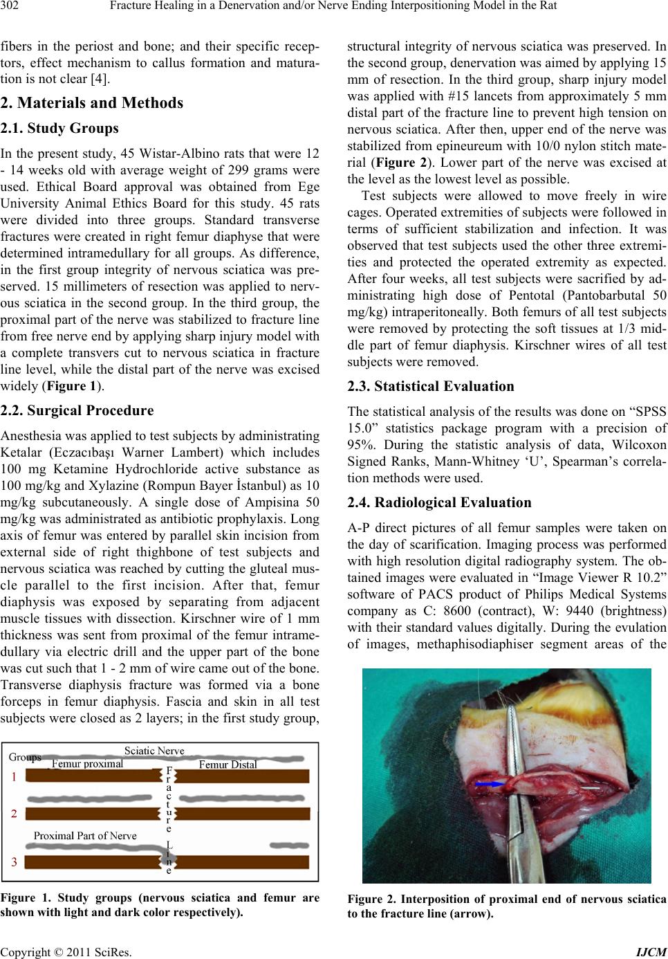 Fracture Healing in a Denervation and/or Nerve Ending Interpositioning Model in the Rat 302 fibers in the periost and bone; and their specific recep- tors, effect mechanism to callus formation and matura- tion is not clear [4]. 2. Materials and Methods 2.1. Study Groups In the present study, 45 Wistar-Albino rats that were 12 - 14 weeks old with average weight of 299 grams were used. Ethical Board approval was obtained from Ege University Animal Ethics Board for this study. 45 rats were divided into three groups. Standard transverse fractures were created in right femur diaphyse that were determined intramedullary for all groups. As difference, in the first group integrity of nervous sciatica was pre- served. 15 millimeters of resection was applied to nerv- ous sciatica in the second group. In the third group, the proximal part of the nerve was stabilized to fracture line from free nerve end by applying sharp injury model with a complete transvers cut to nervous sciatica in fracture line level, while the distal part of the nerve was excised widely (Figure 1). 2.2. Surgical Procedure Anesthesia was applied to test subjects by administrating Ketalar (Eczacıbaşı Warner Lambert) which includes 100 mg Ketamine Hydrochloride active substance as 100 mg/kg and Xylazine (Rompun Bayer İstanbul) as 10 mg/kg subcutaneously. A single dose of Ampisina 50 mg/kg was administrated as antibiotic prophylaxis. Long axis of femur was entered by parallel skin incision from external side of right thighbone of test subjects and nervous sciatica was reached by cutting the gluteal mus- cle parallel to the first incision. After that, femur diaphysis was exposed by separating from adjacent muscle tissues with dissection. Kirschner wire of 1 mm thickness was sent from proximal of the femur intrame- dullary via electric drill and the upper part of the bone was cut such that 1 - 2 mm of wire came out of the bone. Transverse diaphysis fracture was formed via a bone forceps in femur diaphysis. Fascia and skin in all test subjects were closed as 2 layers; in the first study group, Figure 1. Study groups (nervous sciatica and femur are shown with light and dark color respectively). structural integrity of nervous sciatica was preserved. In the second group, denervation was aimed by applying 15 mm of resection. In the third group, sharp injury model was applied with #15 lancets from approximately 5 mm distal part of the fracture line to prevent high tension on nervous sciatica. After then, upper end of the nerve was stabilized from epineureum with 10/0 nylon stitch mate- rial (Figure 2). Lower part of the nerve was excised at the level as the lowest level as possible. Test subjects were allowed to move freely in wire cages. Operated extremities of subjects were followed in terms of sufficient stabilization and infection. It was observed that test subjects used the other three extremi- ties and protected the operated extremity as expected. After four weeks, all test subjects were sacrified by ad- ministrating high dose of Pentotal (Pantobarbutal 50 mg/kg) intraperitoneally. Both femurs of all test subjects were removed by protecting the soft tissues at 1/3 mid- dle part of femur diaphysis. Kirschner wires of all test subjects were removed. 2.3. Statistical Evaluation The statistical analysis of the results was done on “SPSS 15.0” statistics package program with a precision of 95%. During the statistic analysis of data, Wilcoxon Signed Ranks, Mann-Whitney ‘U’, Spearman’s correla- tion methods were used. 2.4. Radiological Evaluation A-P direct pictures of all femur samples were taken on the day of scarification. Imaging process was performed with high resolution digital radiography system. The ob- tained images were evaluated in “Image Viewer R 10.2” software of PACS product of Philips Medical Systems company as C: 8600 (contract), W: 9440 (brightness) with their standard values digitally. During the evulation of images, methaphisodiaphiser segment areas of the Figure 2. Interposition of proximal end of nervous sciatica to the fracture line (arrow). Copyright © 2011 SciRes. IJCM 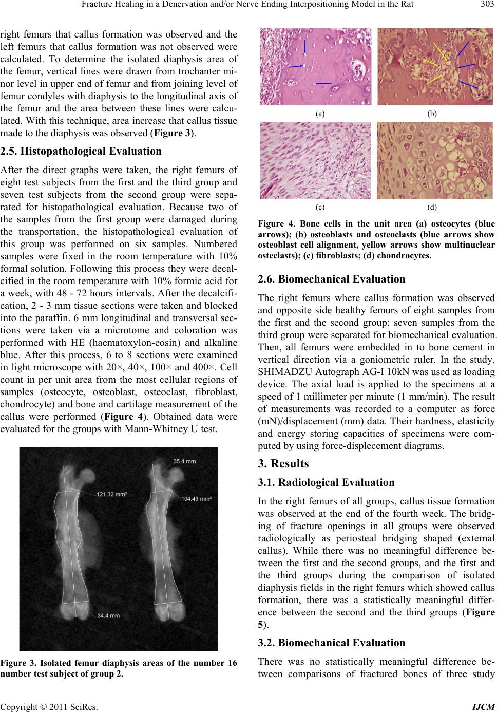 Fracture Healing in a Denervation and/or Nerve Ending Interpositioning Model in the Rat 303 right femurs that callus formation was observed and the left femurs that callus formation was not observed were calculated. To determine the isolated diaphysis area of the femur, vertical lines were drawn from trochanter mi- nor level in upper end of femur and from joining level of femur condyles with diaphysis to the longitudinal axis of the femur and the area between these lines were calcu- lated. With this technique, area increase that callus tissue made to the diaphysis was observed (Figure 3). 2.5. Histopathological Evaluation After the direct graphs were taken, the right femurs of eight test subjects from the first and the third group and seven test subjects from the second group were sepa- rated for histopathological evaluation. Because two of the samples from the first group were damaged during the transportation, the histopathological evaluation of this group was performed on six samples. Numbered samples were fixed in the room temperature with 10% formal solution. Following this process they were decal- cified in the room temperature with 10% formic acid for a week, with 48 - 72 hours intervals. After the decalcifi- cation, 2 - 3 mm tissue sections were taken and blocked into the paraffin. 6 mm longitudinal and transversal sec- tions were taken via a microtome and coloration was performed with HE (haematoxylon-eosin) and alkaline blue. After this process, 6 to 8 sections were examined in light microscope with 20×, 40×, 100× and 400×. Cell count in per unit area from the most cellular regions of samples (osteocyte, osteoblast, osteoclast, fibroblast, chondrocyte) and bone and cartilage measurement of the callus were performed (Figure 4). Obtained data were evaluated for the groups with Mann-Whitney U test. Figure 3. Isolated femur diaphysis areas of the number 16 number test subject of group 2. (a) (b) (c) (d) Figure 4. Bone cells in the unit area (a) osteocytes (blue arrows); (b) osteoblasts and osteoclasts (blue arrows show osteoblast cell alignment, yellow arrows show multinuclear osteclasts); (c) fibroblasts; (d) chondrocytes. 2.6. Biomechanical Evaluation The right femurs where callus formation was observed and opposite side healthy femurs of eight samples from the first and the second group; seven samples from the third group were separated for biomechanical evaluation. Then, all femurs were embedded in to bone cement in vertical direction via a goniometric ruler. In the study, SHIMADZU Autograph AG-I 10kN was used as loading device. The axial load is applied to the specimens at a speed of 1 millimeter per minute (1 mm/min). The result of measurements was recorded to a computer as force (mN)/displacement (mm) data. Their hardness, elasticity and energy storing capacities of specimens were com- puted by using force-displecement diagrams. 3. Results 3.1. Radiological Evaluation In the right femurs of all groups, callus tissue formation was observed at the end of the fourth week. The bridg- ing of fracture openings in all groups were observed radiologically as periosteal bridging shaped (external callus). While there was no meaningful difference be- tween the first and the second groups, and the first and the third groups during the comparison of isolated diaphysis fields in the right femurs which showed callus formation, there was a statistically meaningful differ- ence between the second and the third groups (Figure 5). 3.2. Biomechanical Evaluation There was no statistically meaningful difference be- tween comparisons of fractured bones of three study Copyright © 2011 SciRes. IJCM 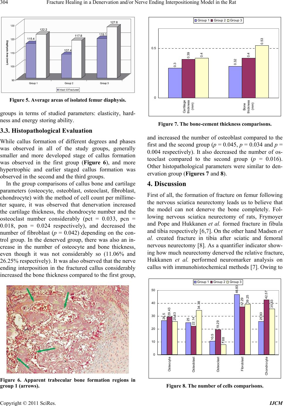 Fracture Healing in a Denervation and/or Nerve Ending Interpositioning Model in the Rat 304 Figure 5. Average areas of isolated femur diaphysis. groups in terms of studied parameters: elasticity, hard- ness and energy storing ability. 3.3. Histopathological Evaluation While callus formation of different degrees and phases was observed in all of the study groups, generally smaller and more developed stage of callus formation was observed in the first group (Figure 6), and more hypertrophic and earlier staged callus formation was observed in the second and the third groups. In the group comparisons of callus bone and cartilage parameters (osteocyte, osteoblast, osteoclast, fibroblast, chondrocyte) with the method of cell count per millime- ter square, it was observed that denervation increased the cartilage thickness, the chondrocyte number and the osteoclast number considerably (pct = 0.033, pcn = 0.018, pon = 0.024 respectively), and decreased the number of fibroblast (p = 0.042) depending on the con- trol group. In the denerved group, there was also an in- crease in the number of osteocyte and bone thickness, even though it was not considerably so (11.06% and 26.25% respectively). It was also observed that the nerve ending interposition in the fractured callus considerably increased the bone thickness compared to the first group, Figure 6. Apparent trabecular bone formation regions in group 1 (arrows). 0.3 0.32 0.39 0.4 0.4 0.53 0 0.5 Cartilage thickness (mm) Bone thickness (mm) Group 1Group 2Group 3 Figure 7. The bone-cement thickness comparisons. and increased the number of osteoblast compared to the first and the second group (p = 0.045, p = 0.034 and p = 0.004 respectively). It also decreased the number of os- teoclast compared to the second group (p = 0.016). Other histopathological parameters were similar to den- ervation group (Figures 7 and 8). 4. Discussion First of all, the formation of fracture on femur following the nervous sciatica neurectomy leads us to believe that the model can not denerve the bone completely. Fol- lowing nervous sciatica neurectomy of rats, Frymoyer and Pope and Hukkanen et al. formed fracture in fibula and tibia respectively [6,7]. On the other hand Madsen et al. created fracture in tibia after sciatic and femoral nervous neurectomy [8]. As a quantifier indicator show- ing how much neurectomy denerved the relative fracture, Hukkanen et al. performed neuromarker analysis on callus with immunohistochemical methods [7]. Owing to 26.5 25 10.5 46.83 25.83 29.43 22.14 19.29 37.29 42.57 25.63 34.38 7.88 39.25 35.63 0 10 20 30 40 50 Osteocyte Osteoblast Osteoclast Fibroblast Chondrocyte Group 1Group 2Group 3 Figure 8. The number of cells comparisons. Copyright © 2011 SciRes. IJCM  Fracture Healing in a Denervation and/or Nerve Ending Interpositioning Model in the Rat 305 their findings, tibial fracture callus is not solely den- erved by nervous sciatica, but it can also be innervated by other sources. For this reason, in the following study, Madsen et al. performed the neurectomy in both the nervous sciatica and femoral nerve [8]. The analysis of the callus with immunohistochemical methods in terms of neural tissue may clarify (as in more or less) the den- ervation amount in this study. How do the denervation models in rats affect the healing of fracture? When the fracture healing is evaluated in radiography and physical examination findings in clinics, quantifier evaluation may be difficult [9]. It is observed that this evaluation is done with radiographic, histopathologic, densitometric and biomechanical measurements on rat femurs [4,6-8,10,11]. Even though nervous sciatica den- ervation was bigger, flat and calcific looking, Aro et al. observed that in rat tibia fractures which are created as intramedullary lead to a formation of callus which ex- hibits more irregular density [4]. In the control groups, callus was oval shaped and showed more regular density distribution. Yüce et al. discovered radiologically that the fibula fractured fragments of rats which had sciatic denervation were combined on the 28th day and while a minimal opening had found in the control group 10. In their evaluation which was made with radiological scor- ing system, Hukkanen et al. and Madsen et al. discov- ered there was primer fracture healing in normal innerve fractures, and there was secondary healing in denerve cases which is characterized by large callus formation [7,8]. In the first group, considering the comparisons of fractured bones of the radiological findings at denerva- tion, no change was discovered which is similar to the literature. During the healing of fibula fractures of fibula frac- tures of rats whose nervous sciatica was denerved, Fry- moyer snd Pope found an increase in biomechanical properties such an elasticity, energy storing and hard- ness on the 15th and 20th days [6]. During the earlier stages of fracture healing (first 15 days), Aro et al. found that the maximum fracture force increased in denerve group 4. Hukkanen et al. and Madsen et al. found that maximum bending moment, energy storing capacity and hardness values in denerve group were somehow lessened on the 35th day when compared to control group [7,8]. In this study, the fact that no mean- ingful difference was observed between fractured and intact bone biomechanically resulting denervation and nerve end interposition model are compatible with the literature. This may be due to the fact that biomechanical evaluation in this study is done with axial lading test as opposed to fracturing from three points test in previous studies. The histopathological evaluation results of the dener- vation group, considering the stages of the fracture healing, point to the fact that denervation causes a delay in callus formation process. In the control group, a callus formation was observed and it is smaller than that of denervation and nerve ending interposition groups, but it was found to be is in more devoloped stage. In the heal- ing of fibula fractures of rats whose nervous sciatica was denerved, Frymoyer and Pope observed histological increase [6] on the 15th and the 20th days (using a scoring system which consists of hematom, callus, joining and compact bone formation measurements). Yüce et al. discovered lamellar bone formation and osteoclasts on the 28th day histologically, and on control side, an earlier stage of callus formation where the fibrosis and cartilage tissues were dominant [10]. In their study concerning the development of sensory innervations in rat tibias, Gajda et al. observed an increase in osteoclastic activity in denerved bones [12]. How does the performing of nervous sciatic denerva- tion with nerve ending interposition in rats affect the fracture healing in femur? No experimental models concerning the interposition of nerve end to fracture line were encountered in the literature. The fact that a more hypertrophic callus for- mation was observed in the nerve end interposition of this study radiologically when compared to denervation may be secondary to various physical and/or chemical stimulus in the fracture line of the nerve. Since neither immunohistochemical nor molecular analysis was done about this subject, no further comments can be made. In the histopathological evaluation, it can be observed that nerve end interposition leads to a bigger but immature callus formation when compared to denervation. REFERENCES [1] A. Schindeler, M. M. McDonald, P. Bokko and D. G. Little, “Bone Remodeling during Fracture Repair: The Cellular Picture,” Seminars in Cell and Development Bi- ology, Vol. 19, No. 5, 2008, pp. 459-466. doi:10.1016/j.semcdb.2008.07.004 [2] K. B. Jones, A. V. Mollano, J. A. Morcuende, R. R. Coo- per and C. L. Saltzman, “Bone and Brain: A Review of Neural, Hormonal, and Musculoskeletal Connections,” The Iowa Orthopaedic Journal, Vol. 24, 2004, pp. 123- 132. [3] D. J. Hurrell, “The Nerve Supply of Bone,” Journal of Anatomy, Vol. 72, No. Part 1, 1937, pp. 54-61. [4] H. Aro, E. Eerola, A. J. Aho and R. Penttinen, “Healing of Experimental Fractures in the Denervated Limbs of the Rat,” Clinical Orthopaedics and Related Research, Vol. 155, 1981, pp. 211-217. [5] J. Li, T. Ahmad, M. Spetea, M. Ahmed and A. Kreicbergs, Copyright © 2011 SciRes. IJCM  Fracture Healing in a Denervation and/or Nerve Ending Interpositioning Model in the Rat Copyright © 2011 SciRes. IJCM 306 “Bone Reinnervation after Fracture: A Study in the Rat,” Journal of Bone and Mineral Research, Vol. 16, No. 8, 2001, pp. 1505-1510. doi:10.1359/jbmr.2001.16.8.1505 [6] J. W. Frymoyer and M. H. Pope, “Fracture Healing in the Sciatically Denervated Rat,” The Journal of Trauma, Vol. 17, No. 5, 1977, pp. 355-361. doi:10.1097/00005373-197705000-00004 [7] M. Hukkanen, Y. T. Konttinen, S. Santavirta, L. Nordsletten, J. E. Madsen, R. Almaas, A. B. Oestreicher, T. Rootwelt and J. M. Polak, “Effect of Sciatic Nerve Section on Neural Ingrowth into the Rat Tibial Fracture Callus,” Clinical of Orthopaedics and Related Research. Vol. 311, 1995, pp. 247-257. [8] J. E. Madsen, M. Hukkanen, A. K. Aune, I. Basran, J. F. Møller, J. M. Polak and L. Nordsletten, “Fracture Healing and Callus Innervation after Peripheral Nerve Resection in Rats,” Clinical of Orthopaedics and Related Research, Vol. 351, 1998, pp. 230-240. doi:10.1097/00003086-199806000-00028 [9] R. A. Hayda, C. T. Brighton and J. L. Esterhai, “Patho- physiology of Delayed Healing,” Clinical of Orthopae- dics and Related Research, Vol. 355, 1998, pp. 31-40. doi:10.1097/00003086-199810001-00005 [10] N. Yüce, B. Gülman and F. Karagöz, “Periferik Sinir Lezyon-Larının Kırık Iyileşmesi Üzerine Etkisi (in Turk- ish),” Turkish Journal of Medical Research, Vol. 6, No. 5, 1988, pp. 391-396. [11] H. Aro, “Effect of Nerve Injury on Fracture Healing: Callus Formation Studied in the Rat,” Acta Orthopaedica Scandinavica, Vol. 56, No. 3, 1985, pp. 233-237. doi:10.3109/17453678508993002 [12] M. Gajda, J. A. Litwin, T. Cichocki, J. P. Timmermans and D. Adriaensen, “Development of Sensory Innervation in Rat Tibia: Co-Localization of CGRP and Substance P with Growth-Associated Protein 43 (GAP-43),” Journal of Anatomy, Vol. 207, No. 2, 2005, pp. 135-144. doi:10.1111/j.1469-7580.2005.00434.x |