

Paper Menu >>
Journal Menu >>
 New Journal of Glass and Ceramics, 2011, 1, 63-68 doi:10.4236/njgc.2011.12011 Published Online July 2011 (http://www.SciRP.org/journal/njgc) Copyright © 2011 SciRes. NGJC 1 Photoacoustic Studies of Colloidal Silica Particles after MeV Ion-Induced Shape Deformation* Ulises Morales1, Rosalba Castañeda-Guzmán2, Santiago Jesús Pérez-Ruiz2, Juan-Carlos Cheang-Wong1 1Instituto de Física, Universidad Nacional Autónoma de México, México, D.F., Mexico; 2Centro de Ciencias Aplicadas y Desarrollo Tecnológico, Universidad Nacional Autónoma de México, México, D.F., Mexico. Email: [email protected] Received May 17th, 2011; revised June 14th, 2011; accepted June 21st, 2011. ABSTRACT Ordered arrays of colloidal submicrometer-sized silica particles deposited onto silicon wafers were irradiated with MeV Si ions. The spherical silica particles tu rned into oblate particles as a result o f the increase of the particle dimen- sion perpendicular to the ion beam direction and the decrease in the parallel direction. Pulsed laser photoacoustic spectroscopy was used to study the structural changes of the silica particles after the ion-induced shape deformation. Our purpose is to correlate the mechanical vibrations generated by the pulsed laser as a function of the Si irradiation parameters: ion energy and fluence. Fast Fourier transform analysis of the photoacoustic signal was carried out in order to obtain the normal vibration modes of the system. The size, size distribution and shape of the silica particles were determined by scanning electron microscopy. Our results revealed significant structural differences between the spherical and the deformed silica particles. Keywords: P u l sed L a s e r Phot oacoustic Spectroscopy, Silica Particles, Ion Irradiation 1. Introduction Periodic arrays of colloidal silica particles are very at- tractive because of their potential applications in coating technology, optoelectronic/plasmonics devices, and na- nolithography [1,2]. The properties of these arrays of SiO2 particles depend on their shape, size and spatial distribution, which in turn determine the different roles they can play as electronic substrates, thin film substrates, electrical and thermal insulators, photonic band gap crystals, masks for lithographic patterning, etc. Changes in the mechanical properties of these colloidal silica par- ticles, as an integral part of the particle- substrate system, are important for the fundamental understanding of fric- tion, wear, and contact mechanics at the nanoscale, as well as for applications. Elastic parameters are sensi- tively related to the microstructure in these arrays. Ion irradiation is a widely used technique to modify the structure and composition of materials. It is well known that MeV ion-irradiated silica particles can un- dergo extreme deformations [3,4]. The ion-induced shape deformation has been described in terms of the ion hammering theory, in which an amorphous material undergoes a shape modification when bombarded with fast heavy ions [5]. In the case of spherical colloidal silica particles, ion irradiation turns them into oblate particles, as a result of the increase of the particle di- mension perpendicular to the ion beam and the de- crease in the parallel direction. Our previous works on this kind of samples showed that this anisotropic shape deformation increases with the ion fluence and with the silica particle size [4], and it depends on the impinging ion, the ion energy and the irradiation temperature in a way that merits further detailed studies [6,7]. Moreover, the mechanical or elastic properties of these structures are poorly studied. It seems that the mechanical char- acteristics of these deformed silica particles are not only size-dependent, but also shape dependent. Then, one of the aims of the present work is to study the structural changes induced by the ion irradiation on these silica particles as a function of the irradiation parameters (ion energy and fluence). We characterize the elastic properties of our arrays of silica particles by means of the Pulsed Laser Photo- acoustic (PLPA) spectroscopy. PLPA relies on the ab- sorption of a short laser pulse (nanosecond range) by a sample and the subsequent measurement of a nonradia- *This work was supported by DGAPA-UNAM projects IN-101210, IN-117208 and CONACYT grants No. 128274, 123143 and 82919. 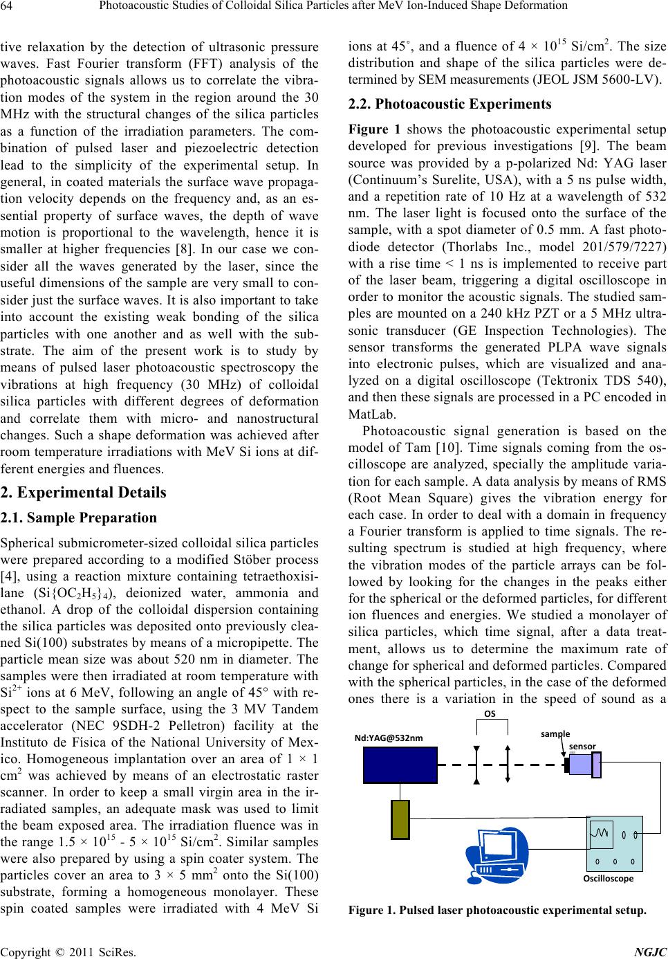 Photoacoustic Studies of Colloidal Silica Particles after MeV Ion-Induced Shape Deformation 64 tive relaxation by the detection of ultrasonic pressure waves. Fast Fourier transform (FFT) analysis of the photoacoustic signals allows us to correlate the vibra- tion modes of the system in the region around the 30 MHz with the structural changes of the silica particles as a function of the irradiation parameters. The com- bination of pulsed laser and piezoelectric detection lead to the simplicity of the experimental setup. In general, in coated materials the surface wave propaga- tion velocity depends on the frequency and, as an es- sential property of surface waves, the depth of wave motion is proportional to the wavelength, hence it is smaller at higher frequencies [8]. In our case we con- sider all the waves generated by the laser, since the useful dimensions of the sample are very small to con- sider just the surface waves. It is also important to take into account the existing weak bonding of the silica particles with one another and as well with the sub- strate. The aim of the present work is to study by means of pulsed laser photoacoustic spectroscopy the vibrations at high frequency (30 MHz) of colloidal silica particles with different degrees of deformation and correlate them with micro- and nanostructural changes. Such a shape deformation was achieved after room temperature irradiations with MeV Si ions at dif- ferent energies and fluences. 2. Experimental Details 2.1. Sample Preparation Spherical submicrometer-sized colloidal silica particles were prepared according to a modified Stöber process [4], using a reaction mixture containing tetraethoxisi- lane (Si{OC2H5}4), deionized water, ammonia and ethanol. A drop of the colloidal dispersion containing the silica particles was deposited onto previously clea- ned Si(100) substrates by means of a micropipette. The particle mean size was about 520 nm in diameter. The samples were then irradiated at room temperature with Si2+ ions at 6 MeV, following an angle of 45° with re- spect to the sample surface, using the 3 MV Tandem accelerator (NEC 9SDH-2 Pelletron) facility at the Instituto de Física of the National University of Mex- ico. Homogeneous implantation over an area of 1 × 1 cm2 was achieved by means of an electrostatic raster scanner. In order to keep a small virgin area in the ir- radiated samples, an adequate mask was used to limit the beam exposed area. The irradiation fluence was in the range 1.5 × 10¹⁵ - 5 × 10¹⁵ Si/cm2. Similar samples were also prepared by using a spin coater system. The particles cover an area to 3 × 5 mm2 onto the Si(100) substrate, forming a homogeneous monolayer. These spin coated samples were irradiated with 4 MeV Si ions at 45˚, and a fluence of 4 × 10¹⁵ Si/cm2. The size distribution and shape of the silica particles were de- termined by SEM measurements (JEOL JSM 5600-LV). 2.2. Photoacoustic Experiments Figure 1 shows the photoacoustic experimental setup developed for previous investigations [9]. The beam source was provided by a p-polarized Nd: YAG laser (Continuum’s Surelite, USA), with a 5 ns pulse width, and a repetition rate of 10 Hz at a wavelength of 532 nm. The laser light is focused onto the surface of the sample, with a spot diameter of 0.5 mm. A fast photo- diode detector (Thorlabs Inc., model 201/579/7227) with a rise time < 1 ns is implemented to receive part of the laser beam, triggering a digital oscilloscope in order to monitor the acoustic signals. The studied sam- ples are mounted on a 240 kHz PZT or a 5 MHz ultra- sonic transducer (GE Inspection Technologies). The sensor transforms the generated PLPA wave signals into electronic pulses, which are visualized and ana- lyzed on a digital oscilloscope (Tektronix TDS 540), and then these signals are processed in a PC encoded in MatLab. Photoacoustic signal generation is based on the model of Tam [10]. Time signals coming from the os- cilloscope are analyzed, specially the amplitude varia- tion for each sample. A data analysis by means of RMS (Root Mean Square) gives the vibration energy for each case. In order to deal with a domain in frequency a Fourier transform is applied to time signals. The re- sulting spectrum is studied at high frequency, where the vibration modes of the particle arrays can be fol- lowed by looking for the changes in the peaks either for the spherical or the deformed particles, for different ion fluences and energies. We studied a monolayer of silica particles, which time signal, after a data treat- ment, allows us to determine the maximum rate of change for spherical and deformed particles. Compared with the spherical particles, in the case of the deformed ones there is a variation in the speed of sound as a sensor sample Oscilloscope OS Nd:YAG@532nm Figure 1. Pulsed laser photoacoustic experimental setup. C opyright © 2011 SciRes. NGJC 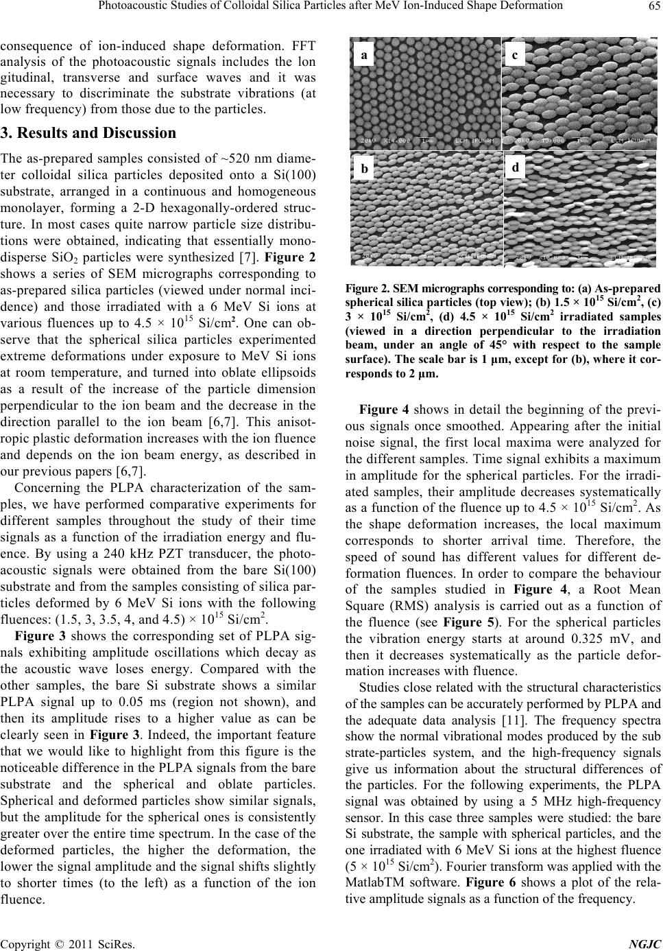 Photoacoustic Studies of Colloidal Silica Particles after MeV Ion-Induced Shape Deformation 65 consequence of ion-induced shape deformation. FFT analysis of the photoacoustic signals includes the lon gitudinal, transverse and surface waves and it was necessary to discriminate the substrate vibrations (at low frequency) from those due to the particles. 3. Results and Discussion The as-prepared samples consisted of ~520 nm diame- ter colloidal silica particles deposited onto a Si(100) substrate, arranged in a continuous and homogeneous monolayer, forming a 2-D hexagonally-ordered struc- ture. In most cases quite narrow particle size distribu- tions were obtained, indicating that essentially mono- disperse SiO2 particles were synthesized [7]. Figure 2 shows a series of SEM micrographs corresponding to as-prepared silica particles (viewed under normal inci- dence) and those irradiated with a 6 MeV Si ions at various fluences up to 4.5 × 10¹⁵ Si/cm2. One can ob- serve that the spherical silica particles experimented extreme deformations under exposure to MeV Si ions at room temperature, and turned into oblate ellipsoids as a result of the increase of the particle dimension perpendicular to the ion beam and the decrease in the direction parallel to the ion beam [6,7]. This anisot- ropic plastic deformation increases with the ion fluence and depends on the ion beam energy, as described in our previous papers [6,7]. Concerning the PLPA characterization of the sam- ples, we have performed comparative experiments for different samples throughout the study of their time signals as a function of the irradiation energy and flu- ence. By using a 240 kHz PZT transducer, the photo- acoustic signals were obtained from the bare Si(100) substrate and from the samples consisting of silica par- ticles deformed by 6 MeV Si ions with the following fluences: (1.5, 3, 3.5, 4, and 4.5) × 1015 Si/cm2. Figure 3 shows the corresponding set of PLPA sig- nals exhibiting amplitude oscillations which decay as the acoustic wave loses energy. Compared with the other samples, the bare Si substrate shows a similar PLPA signal up to 0.05 ms (region not shown), and then its amplitude rises to a higher value as can be clearly seen in Figure 3. Indeed, the important feature that we would like to highlight from this figure is the noticeable difference in the PLPA signals from the bare substrate and the spherical and oblate particles. Spherical and deformed particles show similar signals, but the amplitude for the spherical ones is consistently greater over the entire time spectrum. In the case of the deformed particles, the higher the deformation, the lower the signal amplitude and the signal shifts slightly to shorter times (to the left) as a function of the ion fluence. a b c d Figure 2. SEM micrographs corresponding to: (a) As-prepared spherical silica particles (top view); (b) 1.5 × 10¹⁵ Si/cm2, (c) 3 × 10¹⁵ Si/cm2, (d) 4.5 × 10¹⁵ Si/cm2 irradiated samples (viewed in a direction perpendicular to the irradiation beam, under an angle of 45° with respect to the sample surface). The scale bar is 1 μm, except for (b), where it cor- responds to 2 μm. Figure 4 shows in detail the beginning of the previ- ous signals once smoothed. Appearing after the initial noise signal, the first local maxima were analyzed for the different samples. Time signal exhibits a maximum in amplitude for the spherical particles. For the irradi- ated samples, their amplitude decreases systematically as a function of the fluence up to 4.5 × 10¹⁵ Si/cm2. As the shape deformation increases, the local maximum corresponds to shorter arrival time. Therefore, the speed of sound has different values for different de- formation fluences. In order to compare the behaviour of the samples studied in Figure 4, a Root Mean Square (RMS) analysis is carried out as a function of the fluence (see Figure 5). For the spherical particles the vibration energy starts at around 0.325 mV, and then it decreases systematically as the particle defor- mation increases with fluence. Studies close related with the structural characteristics of the samples can be accurately performed by PLPA and the adequate data analysis [11]. The frequency spectra show the normal vibrational modes produced by the sub strate-particles system, and the high-frequency signals give us information about the structural differences of the particles. For the following experiments, the PLPA signal was obtained by using a 5 MHz high-frequency sensor. In this case three samples were studied: the bare Si substrate, the sample with spherical particles, and the one irradiated with 6 MeV Si ions at the highest fluence (5 × 10¹⁵ Si/cm2). Fourier transform was applied with the MatlabTM software. Figure 6 shows a plot of the rela- tive amplitude signals as a function of the frequency. C opyright © 2011 SciRes. NGJC 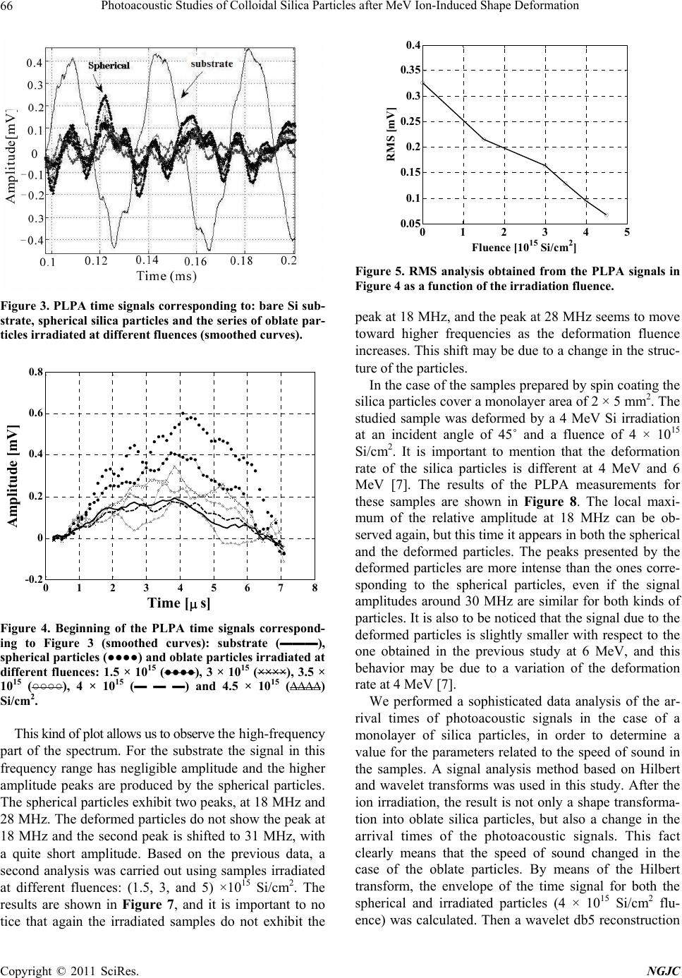 Photoacoustic Studies of Colloidal Silica Particles after MeV Ion-Induced Shape Deformation 66 Figure 3. PLPA time signals corresponding to: bare Si sub- strate, spherical silica particles and the series of oblate par- ticles irradiated at different fluences (smoothed curves). 0 1 2 34 5 6 78 -0.2 0 0.2 0.4 0.6 0.8 Time [ s] Amplitude [mV] Figure 4. Beginning of the PLPA time signals correspond- ing to Figure 3 (smoothed curves): substrate (▬▬▬), spherical particles (●●●●) and oblate particles irradiated at different fluences: 1.5 × 10¹⁵ (●●●●), 3 × 10¹⁵ (××××), 3.5 × 10¹⁵ (○○○○), 4 × 10¹⁵ (▬ ▬ ▬) and 4.5 × 10¹⁵ (∆∆∆∆) Si/cm2. This kind of plot allows us to observe the high-frequency part of the spectrum. For the substrate the signal in this frequency range has negligible amplitude and the higher amplitude peaks are produced by the spherical particles. The spherical particles exhibit two peaks, at 18 MHz and 28 MHz. The deformed particles do not show the peak at 18 MHz and the second peak is shifted to 31 MHz, with a quite short amplitude. Based on the previous data, a second analysis was carried out using samples irradiated at different fluences: (1.5, 3, and 5) ×1015 Si/cm2. The results are shown in Figure 7, and it is important to no tice that again the irradiated samples do not exhibit the 01 2 3 4 5 0.05 0.1 0.15 0.2 0.25 0.3 0.35 0.4 Fluence [10 15 Si/cm 2 ] RMS [mV] Figure 5. RMS analysis obtained from the PLPA signals in Figure 4 as a function of the irradiation fluence. peak at 18 MHz, and the peak at 28 MHz seems to move toward higher frequencies as the deformation fluence increases. This shift may be due to a change in the struc- ture of the particles. In the case of the samples prepared by spin coating the silica particles cover a monolayer area of 2 × 5 mm2. The studied sample was deformed by a 4 MeV Si irradiation at an incident angle of 45˚ and a fluence of 4 × 10¹⁵ Si/cm2. It is important to mention that the deformation rate of the silica particles is different at 4 MeV and 6 MeV [7]. The results of the PLPA measurements for these samples are shown in Figure 8. The local maxi- mum of the relative amplitude at 18 MHz can be ob- served again, but this time it appears in both the spherical and the deformed particles. The peaks presented by the deformed particles are more intense than the ones corre- sponding to the spherical particles, even if the signal amplitudes around 30 MHz are similar for both kinds of particles. It is also to be noticed that the signal due to the deformed particles is slightly smaller with respect to the one obtained in the previous study at 6 MeV, and this behavior may be due to a variation of the deformation rate at 4 MeV [7]. We performed a sophisticated data analysis of the ar- rival times of photoacoustic signals in the case of a monolayer of silica particles, in order to determine a value for the parameters related to the speed of sound in the samples. A signal analysis method based on Hilbert and wavelet transforms was used in this study. After the ion irradiation, the result is not only a shape transforma- tion into oblate silica particles, but also a change in the arrival times of the photoacoustic signals. This fact clearly means that the speed of sound changed in the case of the oblate particles. By means of the Hilbert transform, the envelope of the time signal for both the spherical and irradiated particles (4 × 10¹⁵ Si/cm2 flu- ence) was calculated. Then a wavelet db5 reconstruction C opyright © 2011 SciRes. NGJC 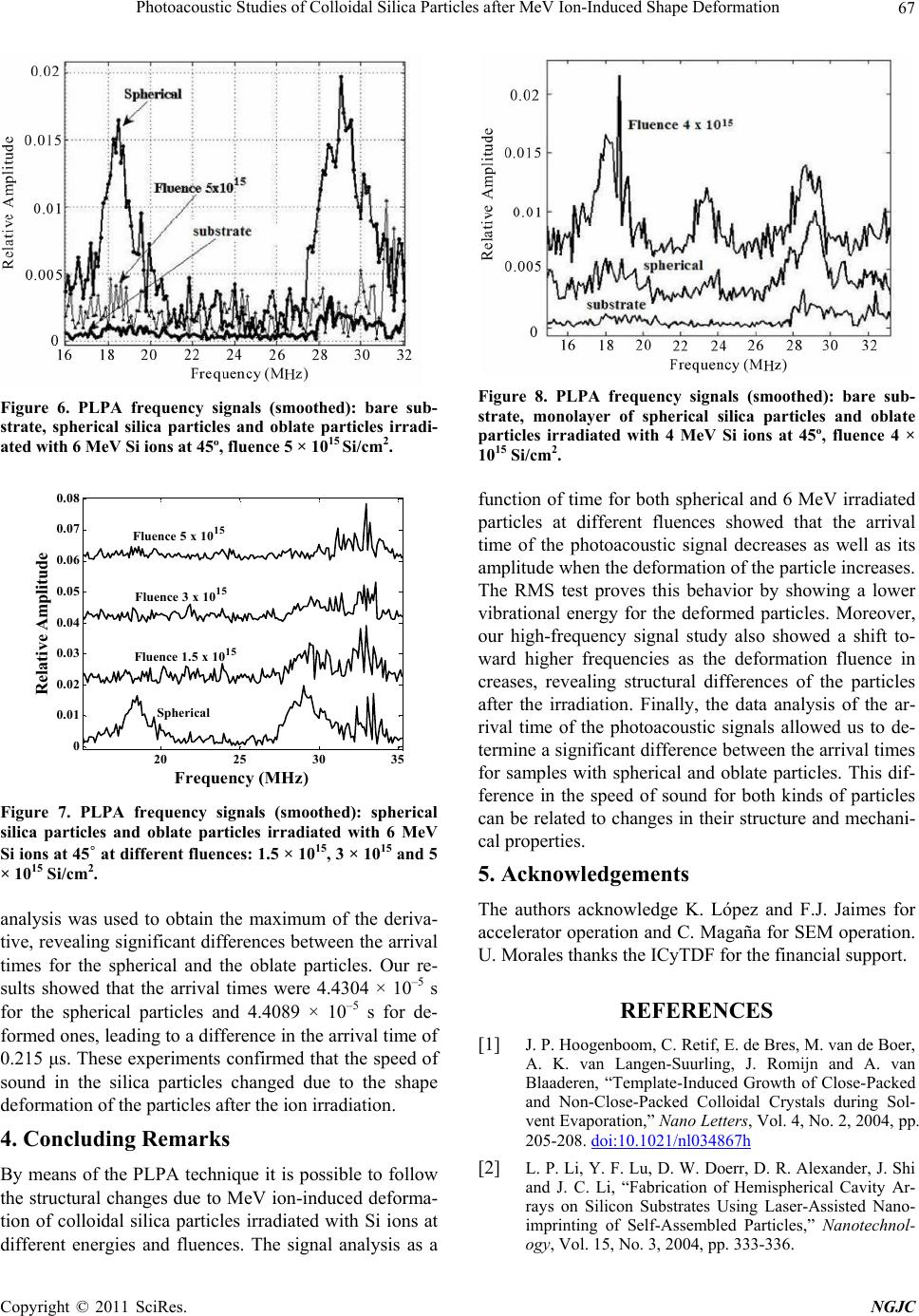 Photoacoustic Studies of Colloidal Silica Particles after MeV Ion-Induced Shape Deformation 67 Figure 6. PLPA frequency signals (smoothed): bare sub- strate, spherical silica particles and oblate particles irradi- ated with 6 MeV Si ions at 45º, fluence 5 × 10¹⁵ Si/cm2. 20 25 3035 0 0.01 0.02 0.03 0.04 0.05 0.06 0.07 0.08 Frequency (MHz) Relative Amplitude Fluence 5 x 10¹⁵ Fluence 3 x 10¹⁵ Fluence 1.5 x 10¹⁵ Spherical Figure 7. PLPA frequency signals (smoothed): spherical silica particles and oblate particles irradiated with 6 MeV Si ions at 45˚ at different fluences: 1.5 × 10¹⁵, 3 × 10¹⁵ and 5 × 10¹⁵ Si/cm2. analysis was used to obtain the maximum of the deriva- tive, revealing significant differences between the arrival times for the spherical and the oblate particles. Our re- sults showed that the arrival times were 4.4304 × 10⁻⁵ s for the spherical particles and 4.4089 × 10⁻⁵ s for de- formed ones, leading to a difference in the arrival time of 0.215 μs. These experiments confirmed that the speed of sound in the silica particles changed due to the shape deformation of the particles after the ion irradiation. 4. Concluding Remarks By means of the PLPA technique it is possible to follow the structural changes due to MeV ion-induced deforma- tion of colloidal silica particles irradiated with Si ions at different energies and fluences. The signal analysis as a Figure 8. PLPA frequency signals (smoothed): bare sub- strate, monolayer of spherical silica particles and oblate particles irradiated with 4 MeV Si ions at 45º, fluence 4 × 10¹⁵ Si/cm2. function of time for both spherical and 6 MeV irradiated particles at different fluences showed that the arrival time of the photoacoustic signal decreases as well as its amplitude when the deformation of the particle increases. The RMS test proves this behavior by showing a lower vibrational energy for the deformed particles. Moreover, our high-frequency signal study also showed a shift to- ward higher frequencies as the deformation fluence in creases, revealing structural differences of the particles after the irradiation. Finally, the data analysis of the ar- rival time of the photoacoustic signals allowed us to de- termine a significant difference between the arrival times for samples with spherical and oblate particles. This dif- ference in the speed of sound for both kinds of particles can be related to changes in their structure and mechani- cal properties. 5. Acknowledgements The authors acknowledge K. López and F.J. Jaimes for accelerator operation and C. Magaña for SEM operation. U. Morales thanks the ICyTDF for the financial support. REFERENCES [1] J. P. Hoogenboom, C. Retif, E. de Bres, M. van de Boer, A. K. van Langen-Suurling, J. Romijn and A. van Blaaderen, “Template-Induced Growth of Close-Packed and Non-Close-Packed Colloidal Crystals during Sol- vent Evaporation,” Nano Letters, Vol. 4, No. 2, 2004, pp. 205-208. doi:10.1021/nl034867h [2] L. P. Li, Y. F. Lu, D. W. Doerr, D. R. Alexander, J. Shi and J. C. Li, “Fabrication of Hemispherical Cavity Ar- rays on Silicon Substrates Using Laser-Assisted Nano- imprinting of Self-Assembled Particles,” Nanotechnol- ogy, Vol. 15, No. 3, 2004, pp. 333-336. C opyright © 2011 SciRes. NGJC  Photoacoustic Studies of Colloidal Silica Particles after MeV Ion-Induced Shape Deformation Copyright © 2011 SciRes. NGJC 68 doi:10.1088/0957-4484/15/3/017 [3] T. van Dillen, A. Polman, W. Fukarek and A. van Blaaderen, “Energy-Dependent Anisotropic Deforma- tion of Colloidal Silica Particles under MeV Au Irradia- tion,” Applied Physics Letters, Vol. 78, No. 7, 2001, pp. 910-912. doi:10.1063/1.1345827 [4] J. C. Cheang-Wong, U. Morales, E. Reséndiz, A. Oliver and L. Rodriguez-Fernández, “Deformation of Colloidal Silica Particles Using MeV Si Ion Irradiation,” Journal of Non-Crystalline Solids, Vol. 353, No. 18-21, 2007, pp. 1925-1929. doi:10.1016/j.jnoncrysol.2007.01.056 [5] S. Klaumünzer, “Ion Hammering of Silica Colloids,” Nuclear Instruments and Methods B, Vol. 215, No. 3-4, 2004, pp. 345-352. doi:10.1016/j.nimb.2003.08.031 [6] J. C. Cheang-Wong, U. Morales, A. Oliver, L. Rodríguez- Fernández and J. Rickards, “MeV Ion Beam Deforma- tion of Colloidal Silica Particles,” Nuclear Instruments and Methods B, Vol. 242, No. 1-2, 2006, pp. 452-454. doi:10.1016/j.nimb.2005.08.078 [7] J. C. Cheang-Wong, U. Morales, E. Reséndiz and A. López-Suárez, “Energy-Dependent Deformation of Col- loidal Silica Nanoparticles under Room Temperature Ir- radiation with MeV Si Ions,” Journal of Nano Research, Vol. 5, 2009, pp. 61-67. doi:10.4028/www.scientific.net/JNanoR.5.61 [8] D. Schneider and B. Schultrich, “Elastic Modulus: A Suitable Quantity for Characterization of Thin Films,” Surface and Coatings Technology, Vol. 98, No. 1-3, 1998, pp. 962-970. doi:10.1016/S0257-8972(97)00317-4 [9] R. Castañeda-Guzmán, A. Huanosta-Tera, L. Baños, M. Fernández-Zamora and S. J. Pérez-Ruiz, “Pulsed Photo- acoustic: A Reliable Technique to Investigate Diffuse phase Transitions and Associated Phenomena in Ferro- electrics,” Ferroelectrics, Vol. 386, No. 1, 2009, pp. 50-61. doi:10.1080/00150190902961298 [10] A. C. Tam, “Applications of Photoacoustic Sensing Techniques,” Reviews of Modern Physics, Vol. 58, No. 2, 1986, pp. 381-431. doi:10.1103/RevModPhys.58.381 [11] V. Torres-Zuñiga, R. Castañeda-Guzmán, S. J. Pérez- Ruiz, O. G. Morales-Saavedra and M. Zepahua- Camacho, “Optical Absorption Photoacoustic Measure- ments for Determination of Molecular Symmetries in a Dichroic Organic-Film,” Optics Express, Vol. 16, No. 25, 2008, pp. 20724-20733. doi:10.1364/OE.16.020724 |