

Paper Menu >>
Journal Menu >>
 International Journal of Clinical Medicine, 2011, 2, 269-271 doi:10.4236/ijcm.2011.23043 Published Online July 2011 (http://www.SciRP.org/journal/ijcm) Copyright © 2011 SciRes. IJCM 269 Anomalous Origin of the Right Coronary Artery from the Left Sinus Valsalva with Coronary Ectasia Nihat Söylemez1, Recep Demirbağ1, Tuncay Hazırolan2, Onur Akpınar3* 1Department of Cardiology, Harran University, Şanlıurfa, Turkey; 2Department of Radiology, Hacettepe University, Ankara, Turkey; 3Department of Cardiogy, Gaziantep Medical Park Hospital, Gaziantep, Turkey. Email: *[email protected] Received January 26th, 2011; revised April 6th, 2011; accepted June 27th, 2011. ABSTRACT The abnormal origin of the right coronary artery from the left aortic sinus coursing between the aorta and the pulmo- nary trunk is a rare congenital anomaly. It may remain asymptomatic or may result in cardiac morbidity or mortality. In the past, an anomalous origin of the right coronary artery from the left sinus of Valsalva was considered a benign finding; it is now evident that this anomaly can be associated with atypical chest pain, m y oca rdial ischemia, and sudden death. We thought that; the diagnosis of left sinus Valsalva aneurysm, described by transtora sic echocardiography and transeusophageal echocardiography, might need de m on st r at ed a dvanced scanning tech niques. Keywords: Congenital Anomaly, Coronary Ectasia, Echocardiography, Multi Detector Computerize Tomography 1. Introduction Coronary artery anomalies are a relatively rare condition. The clinical presentation of coronary artery anomalous is variable, ranging from being asymptomatic to symp- toms of syncope, unstable angina, myocardial ischemia and sudden death. The incidence of anomalous coronary arteries has been reported to be approximately 1% to 2% [1]. Variations in the origin and/or course of anomalous coronary arteries are well documented in the literature but in this case, “interestingly” there was an anomalous right coronary artery arising from the left coronary sinus with proximal ectasia. 2. Case Presentation A 56-year-old woman with a history of typical angina on effort and without history of smoking, hypertension and diabetes mellitus was admitted to our hospital. The pre- senting symptoms had begun approximately since one year however chest pain became more intensive dated from last one week. On admission, her heart rate were 68 beat/min, her blood pressure were 110/72 mmHg and her respiratory rate were 22 breath/min. Physical examina- tion was normal. Electrocardiography showed minimal ST depression and T wave inversion in DII-DIII and AVF leads. The results of the laboratory revealed crea- tine phosphokinase isoenzyme of 117 ng/mlt, troponin I of 0.02 ng/mlt, total cholesterol of 224 mg/dL, high den- sity lipoprotein cholesterol of 53 mg/dL, low density lipoprotein-cholesterol of 162 mg/dL, and triglycerides of 45 mg/d. Other biochemistry and blood test values were within normal limits. Exercise treadmill testing was posi- tive. Coronary angiography was recommended but the patient didn’t accept it. Normal systolic (Ejection fractions: 58% with Simp- son’s) and diastolic (E/A > 1) function, normal wall mo- tion and suspicious images for aneurysm of left sinus valsalva in transtorasic echocardiography (TTE). Tran- seusophageal echocardiography (TEE) findings were similar to TTE for the aneurysm of left sinus valsalva (Figure 1). Then the right coronary artery which arise from the left sinus valsalva and courses between the ascending aorta and pulmonary artery with proximal ectasia dem- onstrated by multi detector computerize tomography (MDCT) heart angiography (Figure 2 (a) and (b)). 3. Discussion In 0.03% - 0.17% of patients undergoing angiography, the right coronary arises from the left sinus of valsalva as a separate vessel or as a branch of a single coronary 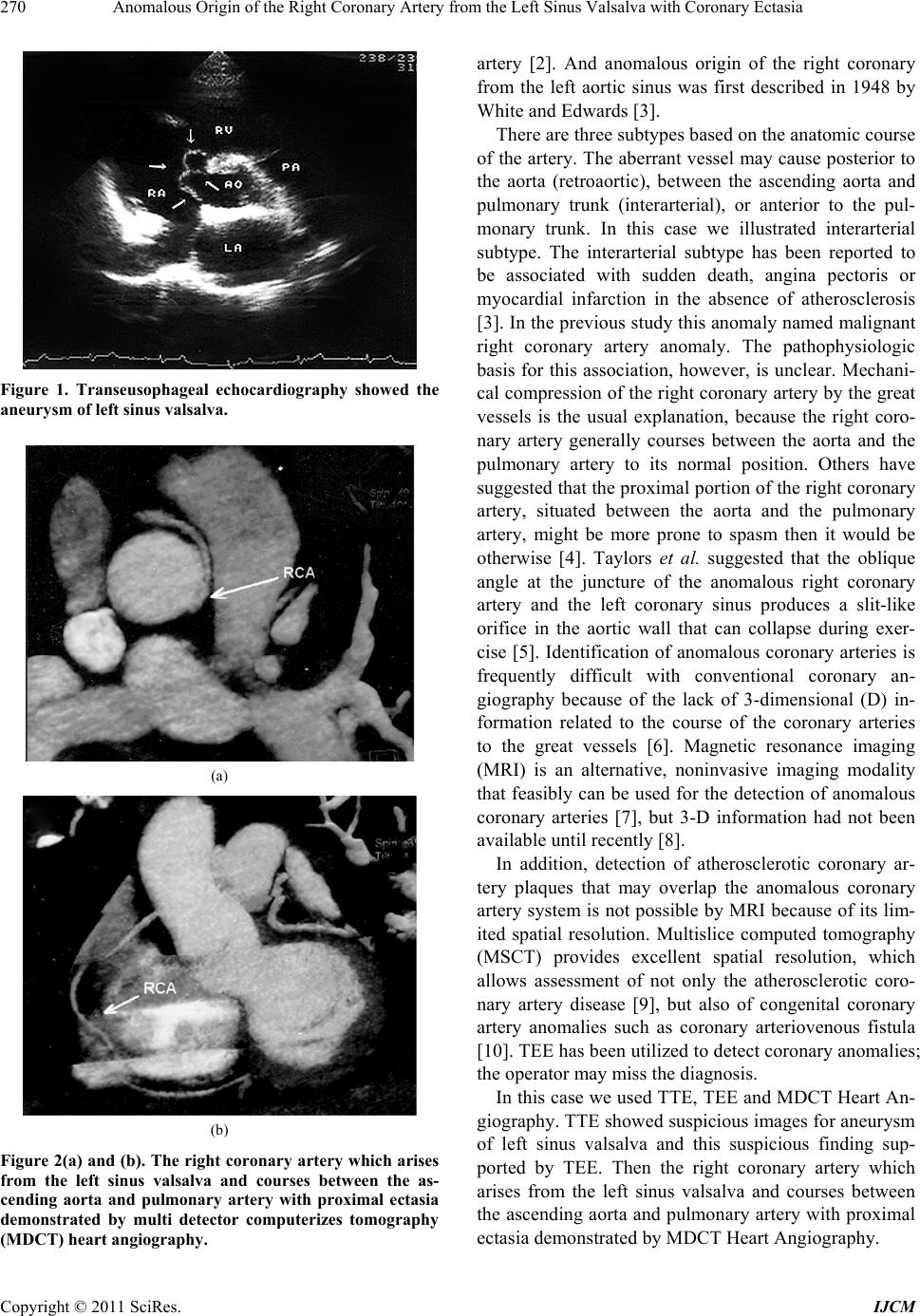 Anomalous Origin of the Right Coronary Artery from the Left Sinus Valsalva with Coronary Ectasia 270 Figure 1. Transeusophageal echocardiography showed the aneurysm of left sinus valsalva. (a) (b) Figure 2(a) and (b). The right coronary artery which arises from the left sinus valsalva and courses between the as- cending aorta and pulmonary artery with proximal ectasia demonstrated by multi detector computerizes tomography (MDCT) heart angiography. artery [2]. And anomalous origin of the right coronary from the left aortic sinus was first described in 1948 by White and Edwards [3]. There are three subtypes based on the anatomic course of the artery. The aberrant vessel may cause posterior to the aorta (retroaortic), between the ascending aorta and pulmonary trunk (interarterial), or anterior to the pul- monary trunk. In this case we illustrated interarterial subtype. The interarterial subtype has been reported to be associated with sudden death, angina pectoris or myocardial infarction in the absence of atherosclerosis [3]. In the previous study this anomaly named malignant right coronary artery anomaly. The pathophysiologic basis for this association, however, is unclear. Mechani- cal compression of the right coronary artery by the great vessels is the usual explanation, because the right coro- nary artery generally courses between the aorta and the pulmonary artery to its normal position. Others have suggested that the proximal portion of the right coronary artery, situated between the aorta and the pulmonary artery, might be more prone to spasm then it would be otherwise [4]. Taylors et al. suggested that the oblique angle at the juncture of the anomalous right coronary artery and the left coronary sinus produces a slit-like orifice in the aortic wall that can collapse during exer- cise [5]. Identification of anomalous coronary arteries is frequently difficult with conventional coronary an- giography because of the lack of 3-dimensional (D) in- formation related to the course of the coronary arteries to the great vessels [6]. Magnetic resonance imaging (MRI) is an alternative, noninvasive imaging modality that feasibly can be used for the detection of anomalous coronary arteries [7], but 3-D information had not been available until recently [8]. In addition, detection of atherosclerotic coronary ar- tery plaques that may overlap the anomalous coronary artery system is not possible by MRI because of its lim- ited spatial resolution. Multislice computed tomography (MSCT) provides excellent spatial resolution, which allows assessment of not only the atherosclerotic coro- nary artery disease [9], but also of congenital coronary artery anomalies such as coronary arteriovenous fistula [10]. TEE has been utilized to detect coronary anomalies; the operator may miss the diagnosis. In this case we used TTE, TEE and MDCT Heart An- giography. TTE showed suspicious images for aneurysm of left sinus valsalva and this suspicious finding sup- ported by TEE. Then the right coronary artery which arises from the left sinus valsalva and courses between the ascending aorta and pulmonary artery with proximal ectasia demonstrated by MDCT Heart Angiography. Copyright © 2011 SciRes. IJCM  Anomalous Origin of the Right Coronary Artery from the Left Sinus Valsalva with Coronary Ectasia Copyright © 2011 SciRes. IJCM 271 REFERENCES [1] O. Yamanaka and R. E. Hobbs, “Coronary Artery Anomalies in 126, 595 Patients Undergoing Coronary Angiography,” Catheterization and Cardiovascular Di- agnosis, Vol. 21, No. 1, 1990, pp. 28-40. [2] D. Kimbiris, A. S. Iskandrian, B. L. Segal and C. E. Bemis, “Anomalous Aortic Origin of Coronary Arteries,” Circulation, Vol. 58, No. 4, 1978, pp. 606-615. [3] N. K. White and J. E. Edwards, “Anomalies of the Coro- nary Arteries; Report of Four Cases,” Archives of Pa- thology, Vol. 45, No. 6, 1948, pp. 766-771. [4] A. J. Taylor, K. M. Rogan and R. Virmani, “Sudden Car- diac Death Associated with Isolated Congenital Coronary Artery Anomalies,” Journal of the American College of Cardiology, Vol. 20, No. 3, 1992, pp. 640-647. doi:10.1016/0735-1097(92)90019-J [5] B. Kaku, H. Kanaya, M. Ikeda, Y. Uno, S. Fujita and F. Kato, et al., “Acute Inferior Myocardial Infarction and Coronary Spasm in a Patient with an Anomalous Origin of the Right Coronary Artery from the Left Sinus of Val- salva,” Japanese Circulation Society, Vol. 64, No. 8, 2000, pp. 641-643. doi:10.1253/jcj.64.641 [6] H. Serota, C. Barth, C. A. Seuc, M. Vandormael, F. Aguirre and M. J. Kern, “Rapid Identification of the Course of Anomalous Coronary Arteries in Adults: The ‘Dot and Eye’ Method,” The American Journal of Cardi- ology, Vol. 65, No. 13, 1990, pp. 891-898. doi:10.1016/0002-9149(90)91432-6 [7] L. B. Haramati, J. S. Glickstein, H. Issenberg, N. Haramati and G. A. Crooke, “MR Imaging and CT of Vascular Anomalies and Connections in Patients with Congenital Heart Disease: Significance in Surgical Plan- ning,” Radio Graphics, Vol. 22, No. 2, 2002, pp. 337- 349. [8] N. H. Bunce, C. H. Lorenz, J. Keegan, J. Lesser, E. M. Reyes, D. N. Firmin, et al., “Coronary Artery Anomalies: Assessment with Free-Breathing Three-Dimensional Co- ronary MR Angiography,” Radiology, Vol. 227, No. 1, 2003, pp. 201-208. [9] S. Achenbach, T. Giesler, D. Ropers, S. Ulzheimer, H. Derlien, C. Schulte, et al., “Detection of Coronary Artery Stenoses by Contrast-Enhanced, Retrospectively Electro- cardiographically-Gated, Multislice Spiral Computed To- mography,” Circulation, Vol. 103, No. 21, 2001, pp. 2535-2538. [10] Y. Sato, M. Mitsui, H. Takahashi, T. Miyazawa, H. Okabe, F. Inoue, et al., “A Giant Left Circumflex Coro- nary Artery-Right Atrium Arteriovenous Fistula Detected by Multislice Spiral Computed Tomography,” Heart Vessels, Vol. 19, No. 1, 2004, pp. 55-56. doi:10.1007/s00380-003-0707-y |