

Paper Menu >>
Journal Menu >>
 International Journal of Clinical Medicine, 2011, 2, 264-268 doi:10.4236/ijcm.2011.23042 Published Online July 2011 (http://www.SciRP.org/journal/ijcm) Copyright © 2011 SciRes. IJCM Incidence of Incidental Thyroid Nodules on Computed Tomography (CT) Scan of the Chest Performed for Reasons Other than Thyroid Disease Kurt Scherer, Seth Means, Collins Chijioke, Paul Karmin, Mukta Panda Department of Radiology, Transitional Year Program and Department of Internal Medicine, College of Medicine, University of Tenne ssee, Chattanooga, USA. Email: [email protected] Received November 17th, 2010; revised April 5th, 2011; accepted April 20th, 2011. ABSTRACT Previous studies demonstrate an approximately 16% incidence of incidental thyroid nodules (ITNs) on Computed To- mography (CT) of the head/neck and thorax combined. Malignant disease is present in many cases. No study to date has quantified ITNs on CT of the thorax alone, an examination performed more frequently than head/neck CT. Our ob- jective was to determine the prevalence and significance of ITNs with further stratification based on size criteria of non-dominant (<10 mm) and dominant (>10 mm) on CT imaging of the chest performed for indications other than thy- roid disease and to assess if these were further evaluated. A retrospective analysisof 500 patients (257 men and 243 women; mean age, 58.3 +/– 16.7 years) with thoracic CT scans performed at Erlanger Health System from October 2007 to October 2008 was perform ed. ITNs were noted in 61 (12.2%) of patients. Nodules were so litary in 43 patients (70.5%) and multiple in 18 (29.5%). Thirty-three patients (6.6%) were found to have a dominant nodule > 10 mm. Six- teen of the 33 patients with dominant nodules received further workup showing benign pathology in 50%. More than 50% of th e 33 dominant nodules received no follow-up at all. Chest CT demonstrates many ITNs . The incidence in this study was 12.2% with 6.6% being potentially malignant dominant thyroid nodules. Partial thyroid glands were visual- ized in 58.2% exams, implying an incidence of 12.2% ITNs is an underestimate. It may be beneficial for routine chest CT to be extended 2 cm superiorly in order to ensure full visualization of the thyroid gland and related pathology. Keywords: Incidental Thyroid Nodules, Thyroid Malignancy 1. Introduction Thyroid abnormalities, specifically thyroid nodules are routinely identified on CT imaging. To date, studies have shown that incidental findings of the thyroid occur at the rate of 16% on CT imaging and 27% on sonogra- phy [1,2]. However, the incidence of these nodules has only been studied with imaging of the head and neck [3]. The frequency of chest imaging is increasing. Wittram, et al. report a significant increase over a decade (with P = 0.04 for inpatient and P = 0.01 for outpatients) [4]. With this increase, there actually may be an increase in the previously reported incidence of these abnormalities. Furthermore, the indications for chest CT are much wider than for head and neck CT scans, which fre- quently include chest CT solely for detection of metas- tases in malignant disease of the head and neck. The true incidence of thyroid disease in CT chest exams with no head and neck malignancy is unknown. In patients with a known malignancy other than thyroid, incidental thy- roid nodules were found to be malignant in 24%, which is above the expected rate of 5% in traditionally discov- ered nodules [5]. From the reported data, it was unclear whether these incidental findings were initially made on head and ne ck CT or ch e st CT alon e [5]. In general it has been reported that among incidentally detected lesions of the thyroid gland, there was at least a 3.9% rate of malignancy and 7.4% rate of malignant potential, with at least an 11.3% prevalence of malignant or potentially malignant lesions among incidental thy- roid abnormalities detected on CT [6]. The overall in- creased use of CT chest may demonstrate more thyroid nodules to be managed and subsequently require that all CT chest scans include the thyroid gland as a means of coincidental screening for thyroid disease, benign and 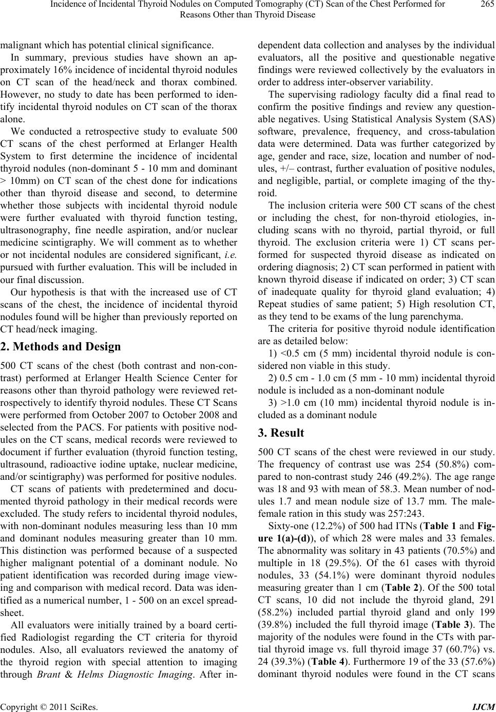 Incidence of Incidental Thyroid Nodules on Computed Tomography (CT) Scan of the Chest Performed for 265 Reasons Other than Thyroid Disease malignant which has potential clinical significance. In summary, previous studies have shown an ap- proximately 16% incidence of incidental thyroid nodules on CT scan of the head/neck and thorax combined. However, no study to date has been performed to iden- tify incidental thyroid nodules on CT scan of the thorax alone. We conducted a retrospective study to evaluate 500 CT scans of the chest performed at Erlanger Health System to first determine the incidence of incidental thyroid nodules (non-dominant 5 - 10 mm and dominant > 10mm) on CT scan of the chest done for indications other than thyroid disease and second, to determine whether those subjects with incidental thyroid nodule were further evaluated with thyroid function testing, ultrasonography, fine needle aspiration, and/or nuclear medicine scintigraphy. We will comment as to whether or not incidental nodules are considered significant, i.e. pursued with further evaluation. This will be included in our final discussion. Our hypothesis is that with the increased use of CT scans of the chest, the incidence of incidental thyroid nodules found will be higher than previously reported on CT head/neck imaging. 2. Methods and Design 500 CT scans of the chest (both contrast and non-con- trast) performed at Erlanger Health Science Center for reasons other than thyroid pathology were reviewed ret- rospectively to identify thyroid nodules. These CT Scans were performed from October 2007 to October 2008 and selected from the PACS. For patients with positive nod- ules on the CT scans, medical records were reviewed to document if further evaluation (thyroid function testing, ultrasound, radioactive iodine uptake, nuclear medicine, and/or scintigraphy) was performed for positive nodules. CT scans of patients with predetermined and docu- mented thyroid pathology in their medical records were excluded. The study refers to incidental thyroid nodules, with non-dominant nodules measuring less than 10 mm and dominant nodules measuring greater than 10 mm. This distinction was performed because of a suspected higher malignant potential of a dominant nodule. No patient identification was recorded during image view- ing and comparison with medical record. Data was iden- tified as a numerical number, 1 - 500 on an excel spread- sheet. All evaluators were initially trained by a board certi- fied Radiologist regarding the CT criteria for thyroid nodules. Also, all evaluators reviewed the anatomy of the thyroid region with special attention to imaging through Brant & Helms Diagnostic Imaging. After in- dependent data collection and analyses by the individual evaluators, all the positive and questionable negative findings were reviewed collectively by the evaluators in order to address inter-observer variability. The supervising radiology faculty did a final read to confirm the positive findings and review any question- able negatives. Using Statistical Analysis System (SAS) software, prevalence, frequency, and cross-tabulation data were determined. Data was further categorized by age, gender and race, size, location and number of nod- ules, +/– contrast, further evaluation of positive nodules, and negligible, partial, or complete imaging of the thy- roid. The inclusion criteria were 500 CT scans of the chest or including the chest, for non-thyroid etiologies, in- cluding scans with no thyroid, partial thyroid, or full thyroid. The exclusion criteria were 1) CT scans per- formed for suspected thyroid disease as indicated on ordering diagnosis; 2) CT scan performed in patient with known thyroid disease if indicated on order; 3) CT scan of inadequate quality for thyroid gland evaluation; 4) Repeat studies of same patient; 5) High resolution CT, as they tend to be exams of the lung parenchyma. The criteria for positive thyroid nodule identification are as detailed below: 1) <0.5 cm (5 mm) incidental thyroid nodule is con- sidered non viable in this study. 2) 0.5 cm - 1.0 cm (5 mm - 10 mm) incidental thyroid nodule is included as a non-dominant nodule 3) >1.0 cm (10 mm) incidental thyroid nodule is in- cluded as a dominant nodule 3. Result 500 CT scans of the chest were reviewed in our study. The frequency of contrast use was 254 (50.8%) com- pared to non-contrast study 246 (49.2%). The age range was 18 and 93 with mean of 58.3. Mean number of nod- ules 1.7 and mean nodule size of 13.7 mm. The male- female ration in this study was 257:243. Sixty-one (12.2 %) of 500 h ad ITNs (Table 1 and Fig- ure 1(a)-(d)), of which 28 were males and 33 females. The abnormality was solitary in 43 patients (70.5%) and multiple in 18 (29.5%). Of the 61 cases with thyroid nodules, 33 (54.1%) were dominant thyroid nodules measuring greater than 1 cm (Table 2). Of the 500 total CT scans, 10 did not include the thyroid gland, 291 (58.2%) included partial thyroid gland and only 199 (39.8%) included the full thyroid image (Table 3). The majority of the nodules were found in the CTs with par- tial thyroid image vs. full thyroid image 37 (60.7%) vs. 24 (39.3%) (Table 4). Furthermore 19 of the 33 (57.6%) dominant thyroid nodules were found in the CT scans Copyright © 2011 SciRes. IJCM 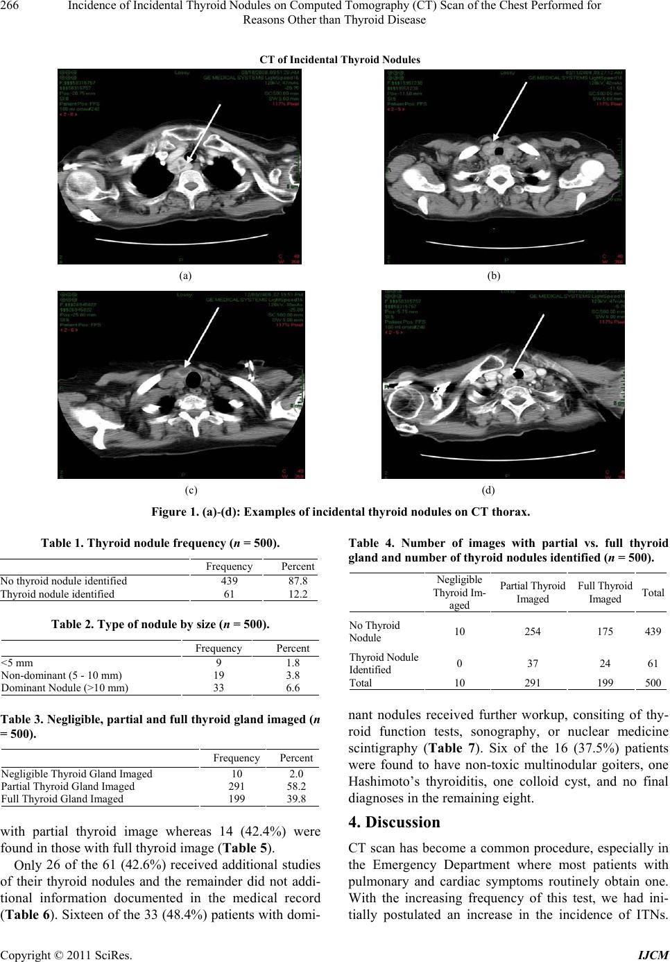 Incidence of Incidental Thyroid Nodules on Computed Tomography (CT) Scan of the Chest Performed for Reasons Other than Thyroid Disease Copyright © 2011 SciRes. IJCM 266 CT of Incidental Thyroid Nodules (a) (b) (c) (d) Figure 1. (a)-(d): Examples of incidental thyroid nodules on CT thorax. Table 1. Thyroid nodule frequency (n = 500). Frequency Percent No thyroid nodule identified 439 87.8 Thyroid nodule identified 61 12.2 Table 4. Number of images with partial vs. full thyroid gland and number of thyroid nodules identified (n = 500). Table 2. Type of nodule by size (n = 500). Frequency Percent <5 mm 9 1.8 Non-dominant (5 - 10 mm) 19 3.8 Dominant Nodule (>10 mm) 33 6.6 Negligible Thyr oi d Im - aged Partial T hyr oi d Imaged Full Thyroid Imaged Total No Thyroid Nodule 10 254 175 439 Thyr oi d No dul e Identified 0 37 24 61 Total 10 291 199 500 Table 3. Negligible, partial and full thyroid gland imaged (n = 500). Frequency Percent Negligible Thyroid Gland Imaged 10 2.0 Partial Thyroid Gland Imaged 291 58.2 Full Thyroid Gland Imaged 199 39.8 nant nodules received further workup, consiting of thy- roid function tests, sonography, or nuclear medicine scintigraphy (Table 7). Six of the 16 (37.5%) patients were found to have non-toxic multinodular goiters, one Hashimoto’s thyroiditis, one colloid cyst, and no final diagnoses in the remaining eight. 4. Discussion with partial thyroid image whereas 14 (42.4%) were found in those with full thyroid image (Table 5). CT scan has become a common procedure, especially in the Emergency Department where most patients with pulmonary and cardiac symptoms routinely obtain one. With the increasing frequency of this test, we had ini- tially postulated an increase in the incidence of ITNs. Only 26 of the 61 (42.6%) received additional studies of their thyroid nodules and the remainder did not addi- tional information documented in the medical record (Table 6). Sixteen of the 33 (48.4%) patients with domi-  Incidence of Incidental Thyroid Nodules on Computed Tomography (CT) Scan of the Chest Performed for 267 Reasons Other than Thyroid Disease Table 5. Negligible, partial and full thyroid gland imaged classified by thyroid nodule sizes (n = 500). Negli g i bl e T hyroid Gl a n d ImagedPart ia l T hy r o i d Gl and Imaged Full T hyroid G la n d Imaged Tota l <5 mm 0 5 4 9 Non-dominant (5 - 10 mm) 0 13 6 19 Dominant Nodule (>10 mm) 0 19 14 33 No Nodule Identified 10 254 175 439 Total 10 291 199 500 Table 6. Additional studies on cases with thyroid nodule identified (n = 61). FrequencyPercent No studies noted in medical record 35 7.0 Presence of additional FU (TFT, U/S, RAIU, NucMed, others) 26 5.2 FU: follow up; TFT: thyroid funtion test; U/S: Ultrasonography; RAIU: Radioactive Iodine Uptake Test; NucMed: Nuclear medicine. Table 7. Description of type s of additional studie s performe d (n = 26). No studies noted in medical record Presence of additional FU (TFT, U/S, RA IU, N ucMe d, others) <5 mm 8 1 Non-dominant (5 - 10 mm) 10 9 Dominant Nodule (>10 mm) 17 16 The current literature evaluating the incidence of ITNs found on routine CT scans is paucity. The previously reported incidence of ITNs is 16% [2, 3]. As has been previously reported, there is an 11.3% prevalence of malignant or potential malignant lesions among incidental thyroid abnormalities detected on CT scans [6]. In our study we found similar percentages. The incidence of ITNs in our study is 12.2% with more than 50% being dominant nodules. About fifty percent received further evaluation of the nodules. To our knowledge, there are no guidelines for further evaluation of incidental nodules found on routine CT scans. This study indicated that less than 50% of the 61 cases of ITNs had the full thyroid gland imaged. Given this fact, we suggest that it may be beneficial for CT scans of the chest to be extended superiorly 2 cm in order to visualize more of the thyroid gland and thus increase the frequency of detecting incidental thyroid pathology without exposing the patients to unnecessary amount of additional radiation. Though this study is a single center, retrospective, observational, dependent on accuracy of orders for CTs and convenience sample, it still, however, identified some relatively simple system changes that may assist in early identification and follow up of thyroid nodules especially in high risk patients. We recommend a future prospective study to further evaluate ITNs by obtaining history of previous malignancy. The overall increased use of CT chest may demon- strate more thyroid nodules to be managed and subse- quently require that all CT chest scans include the thy- roid gland as a means of coincidental screening for thy- roid disease, benign and malignant which has potential clinical significance The study also suggested that the use of contrast does not improve the detection of thyroid nodules and hence may not be use in chest CTs except when indicated for other reasons. This study serves as a pilot for future study to further evaluate the incidence and consequence of ITNs that may assist to provide standardized guidelines for clini- cians in facilitating further management of ITNs. The strengths of the study include detailed exclusion criteria, limited variability as all scans were obtained from the same hospital and CT scanners, limited vari- ability as all scans were evaluated on the same PACS system and by evaluators with similar thyroid nodule criteria training. The limitiations of the study include a single center study, retrospective study, observational study, depend- ency on accuracy of orders for CTs, and convenience sample. 5. Conclusions Given the separate findings including that the incidence of ITNs in our study is 12.2% with more than 50% being dominant nodules, the previously described 11.3% ma- lignant potential of these incidental thyroid nodules, and the fact that 50% of the 61 cases of ITNs did not receive complete visualization of the thyroid gland on cross- sectional imaging, we recommend extending superiorly by 2 cm the extent of the CT scans of the thorax in order to visualize more of the thyroid gland and thus increase the frequency of detecting incidental thyroid pathology without exposing the patients to unnecessary amount of additional radiation. REFERENCES [1] A. Brander, P. Viikinkoski, J. Nickels and L. Kivisaari, Copyright © 2011 SciRes. IJCM  Incidence of Incidental Thyroid Nodules on Computed Tomography (CT) Scan of the Chest Performed for 268 Reasons Other than Thyroid Disease “Thyroid Gland: US Screening in a Random Adult Popu- lation,” Radio logy, Vol. 181, No. 3, 1991, pp. 683-687. [2] D. M. Yousem, T. Huang, L. A. Loevner and C. P. Lan- glotz, “Clinical and Economic Impact of Incidental Thy- roid Lesions Found with CT and MR,” American Journal of Neuroradiology, Vol. 18, No. 8, 1997, pp. 1423-1428. [3] Y. S. Rho and H. Y. Ahn, “The Prevalence and Signifi- cance of Incidental Thyroid Nodules Identified on Com- puted Tomography,” Journal of Computer Assisted To- mograph, Vol. 32, No. 5, 2008, pp. 810-815. doi:10.1097/RCT.0b013e318157fd38 [4] Wittram, M. B. Conrad, J. Margaret, et al., “Trends in Thoracic Radiology Over Decade at a Large Academic Medical Center,” Journal of Thoracic Imaging, Vol. 19, No. 3, 2004, pp. 164-170. doi:10.1097/01.rti.0000117623.02841.e6 [5] S. M. Wilhelm, A. V. Robinson, S. S. Krishnamurthi and H. L. Reynolds, “Evaluations and Management of Inci- dental Thyroid Nodules in Patients with Another Primary Malignancy,” Surge ry, Vol. 142, No. 4, 2007, pp. 581-586, discussion pp. 586-587. doi:10.1016/j.surg.2007.06.033 [6] S. K. Shetty, M. M. Maher, P. F. Hahn, E. F. Halper n and S. L. Aquino, “Significance of Incidental Thyroid Lesions Detected on CT: Correlation among CT, Sonography, and Pathology,” American Journal of Roentgenology, Vol. 187, No. 5, 2006, pp. 1349-1356. doi:10.2214/AJR.05.0468 Copyright © 2011 SciRes. IJCM |