Paper Menu >>
Journal Menu >>
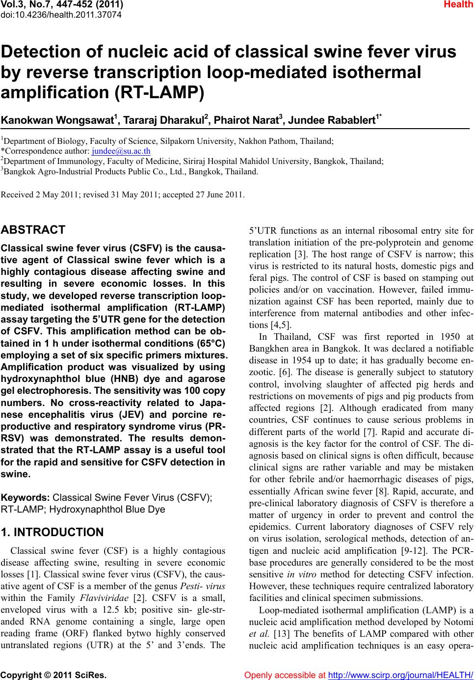
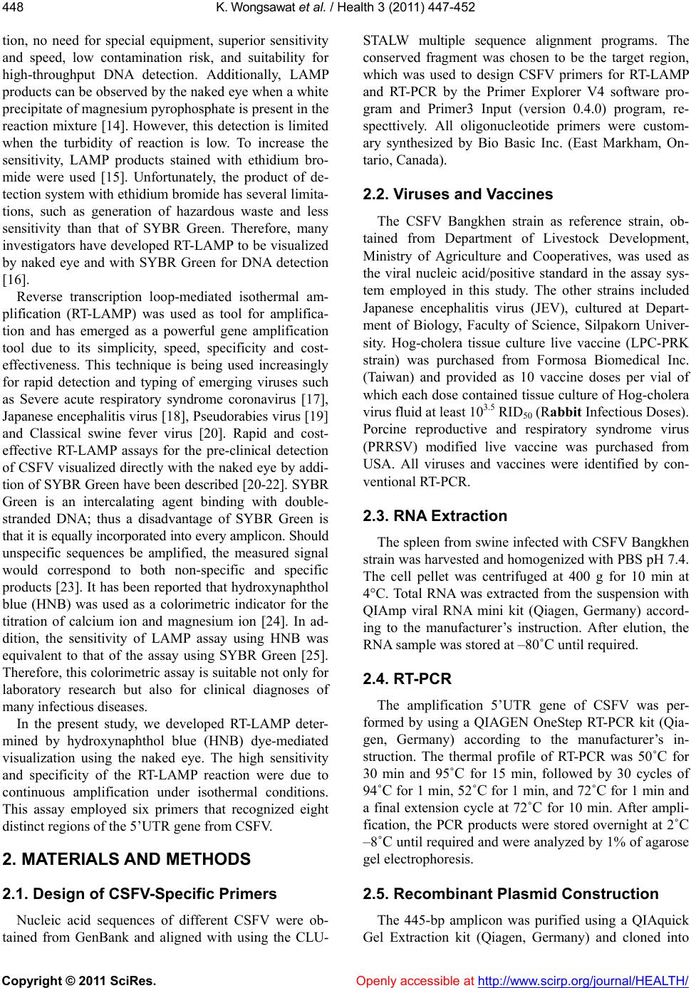
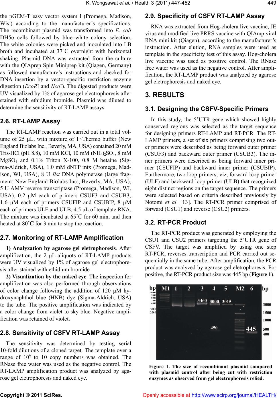
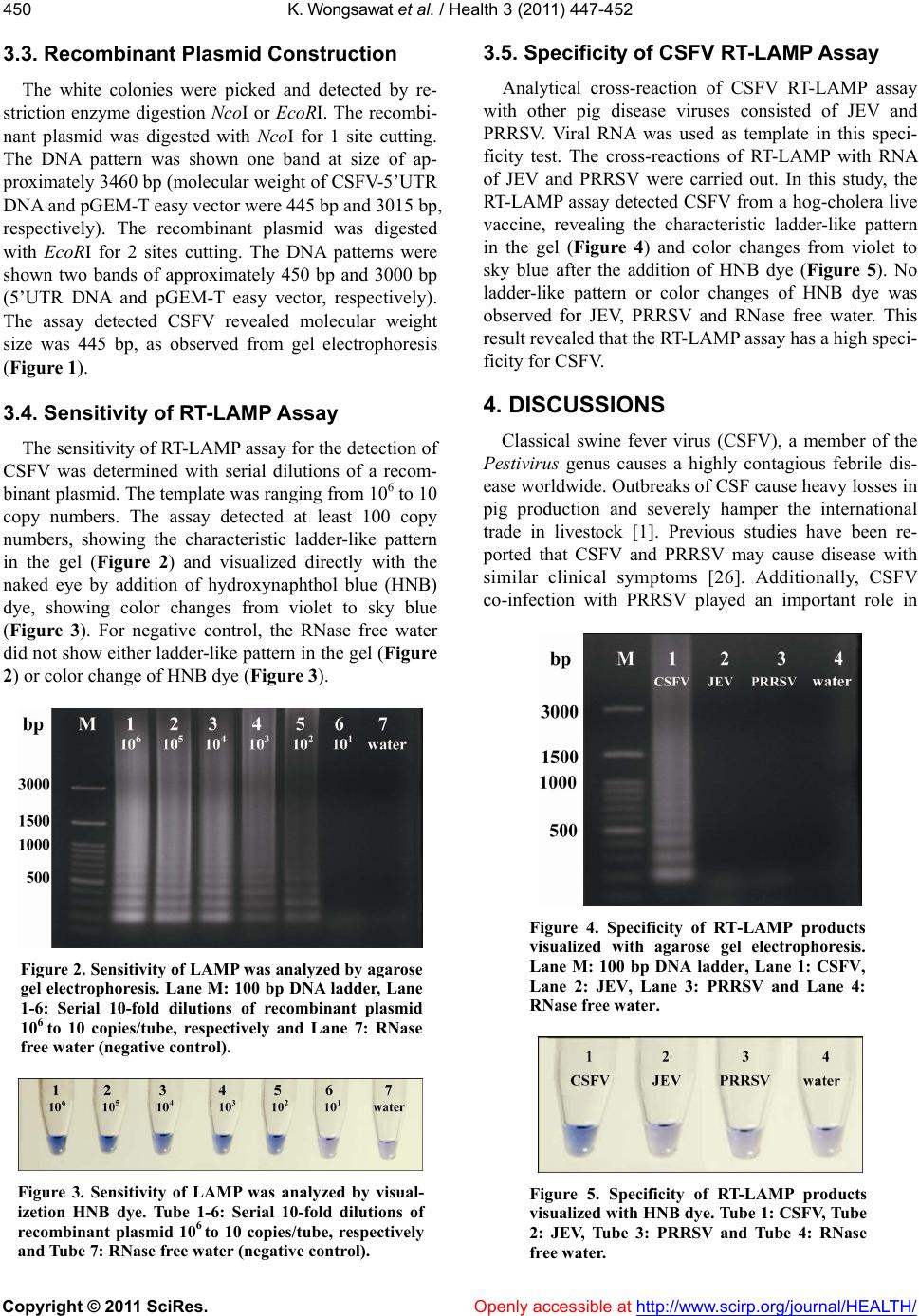
![]() Vol.3, No.7, 447-452 (2011) doi:10.4236/health.2011.37074 Copyright © 2011 SciRes. Openly accessible at http://www.scirp.org/journal/HEALTH/ Health Detection of nucleic acid of classical swine fever virus by reverse transcription loop-mediated isothermal amplification (RT-LAMP) Kanokwan W ongsawa t1, Tararaj Dharakul2, Phairot Narat3, Jundee Rabablert 1* 1Department of Biology, Faculty of Science, Silpakorn University, Nakhon Pathom, Thailand; *Correspondence author: [email protected] 2Department of Immunology, Faculty of Medicine, Siriraj Hospital Mahidol University, Bangkok, Thailand; 3Bangkok Agro-Industrial Products Public Co., Ltd., Bangkok, Thailand. Received 2 May 2011; revised 31 May 2011; accepted 27 June 2011. ABSTRACT Classical swine fever virus (CSFV) is the causa- tive agent of Classical swine fever which is a highly contagious disease affecting swine and resulting in severe economic losses. In this study, we developed reverse transcription loop- mediated isothermal amplification (RT-LAMP) assay targ eting t he 5’UTR gene for the detecti on of CSFV. This amplification method can be ob- tained in 1 h under isothermal conditions (65°C) employing a set of six specific primers mixtures. Amplification product was visualized by using hydroxynaphthol blue (HNB) dye and agarose gel electrophoresis. The sensitivity was 100 copy numbers. No cross-reactivity related to Japa- nese encephalitis virus (JEV) and porcine re- productive and respiratory syndrome virus (PR- RSV) was demonstrated. The results demon- strated that the RT-LAMP assay is a useful tool for the rapid and sensitive for CSFV detection in swine. Keywords: Classical Swine Fever Virus (CSFV); RT-LAMP; Hydroxynaphthol Blue Dye 1. INTRODUCTION Classical swine fever (CSF) is a highly contagious disease affecting swine, resulting in severe economic losses [1]. Classical swine fever virus (CSFV), the caus- ative agent of CSF is a member of the genus Pesti- virus within the Family Flav iviridae [2]. CSFV is a small, enveloped virus with a 12.5 kb; positive sin- gle-str- anded RNA genome containing a single, large open reading frame (ORF) flanked bytwo highly conserved untranslated regions (UTR) at the 5’ and 3’ends. The 5’UTR functions as an internal ribosomal entry site for translation initiation of the pre-polyprotein and genome replication [3]. The host range of CSFV is narrow; this virus is restricted to its natural hosts, domestic pigs and feral pigs. The control of CSF is based on stamping out policies and/or on vaccination. However, failed immu- nization against CSF has been reported, mainly due to interference from maternal antibodies and other infec- tions [4,5]. In Thailand, CSF was first reported in 1950 at Bangkhen area in Bangkok. It was declared a notifiable disease in 1954 up to date; it has gradually become en- zootic. [6]. The disease is generally subject to statutory control, involving slaughter of affected pig herds and restrictions on movements of pigs and pig products from affected regions [2]. Although eradicated from many countries, CSF continues to cause serious problems in different parts of the world [7]. Rapid and accurate di- agnosis is the key factor for the control of CSF. The di- agnosis based on clinical signs is often difficult, because clinical signs are rather variable and may be mistaken for other febrile and/or haemorrhagic diseases of pigs, essentially African swine fever [8]. Rapid, accurate, and pre-clinical laboratory diagnosis of CSFV is therefore a matter of urgency in order to prevent and control the epidemics. Current laboratory diagnoses of CSFV rely on virus isolation, serological methods, detection of an- tigen and nucleic acid amplification [9-12]. The PCR- base procedures are generally considered to be the most sensitive in vitro method for detecting CSFV infection. However, these techniques require centralized laboratory facilities and clinical specimen submissions. Loop-mediated isothermal amplification (LAMP) is a nucleic acid amplification method developed by Notomi et al. [13] The benefits of LAMP compared with other nucleic acid amplification techniques is an easy opera- ![]() K. Wongsaw at et al. / Health 3 (2011) 447-452 Copyright © 2011 SciRes. Openly accessible at http://www.scirp.org/journal/HEALTH/ 448 tion, no need for special equipment, superior sensitivity and speed, low contamination risk, and suitability for high-throughput DNA detection. Additionally, LAMP products can be observed by the naked eye when a white precipitate of magnesium pyrophosphate is present in the reaction mixture [14]. However, this detection is limited when the turbidity of reaction is low. To increase the sensitivity, LAMP products stained with ethidium bro- mide were used [15]. Unfortunately, the product of de- tection system with ethidium bromide has several limita- tions, such as generation of hazardous waste and less sensitivity than that of SYBR Green. Therefore, many investigators have developed RT-LAMP to be visualized by naked eye and with SYBR Green for DNA detection [16]. Reverse transcription loop-mediated isothermal am- plification (RT-LAMP) was used as tool for amplifica- tion and has emerged as a powerful gene amplification tool due to its simplicity, speed, specificity and cost- effectiveness. This technique is being used increasingly for rapid detection and typing of emerging viruses such as Severe acute respiratory syndrome coronavirus [17], Japanese encephalitis virus [18], Pseudorabies virus [19] and Classical swine fever virus [20]. Rapid and cost- effective RT-LAMP assays for the pre-clinical detection of CSFV visualized directly with the naked eye by addi- tion of SYBR Green have been described [20-22]. SYBR Green is an intercalating agent binding with double- stranded DNA; thus a disadvantage of SYBR Green is that it is equally incorporated into every amplicon. Should unspecific sequences be amplified, the measured signal would correspond to both non-specific and specific products [23]. It has been reported that hydroxynaphthol blue (HNB) was used as a colorimetric indicator for the titration of calcium ion and magnesium ion [24]. In ad- dition, the sensitivity of LAMP assay using HNB was equivalent to that of the assay using SYBR Green [25]. Therefore, this colorimetric assay is suitable not only for laboratory research but also for clinical diagnoses of many infectious diseases. In the present study, we developed RT-LAMP deter- mined by hydroxynaphthol blue (HNB) dye-mediated visualization using the naked eye. The high sensitivity and specificity of the RT-LAMP reaction were due to continuous amplification under isothermal conditions. This assay employed six primers that recognized eight distinct regions of the 5’UTR gene from CSFV. 2. MATERIALS AND METHODS 2.1. Design of CSFV-Specific Primers Nucleic acid sequences of different CSFV were ob- tained from GenBank and aligned with using the CLU- STALW multiple sequence alignment programs. The conserved fragment was chosen to be the target region, which was used to design CSFV primers for RT-LAMP and RT-PCR by the Primer Explorer V4 software pro- gram and Primer3 Input (version 0.4.0) program, re- specttively. All oligonucleotide primers were custom- ary synthesized by Bio Basic Inc. (East Markham, On- tario, Canada). 2.2. Viruses and Vaccines The CSFV Bangkhen strain as reference strain, ob- tained from Department of Livestock Development, Ministry of Agriculture and Cooperatives, was used as the viral nucleic acid/positive standard in the assay sys- tem employed in this study. The other strains included Japanese encephalitis virus (JEV), cultured at Depart- ment of Biology, Faculty of Science, Silpakorn Univer- sity. Hog-cholera tissue culture live vaccine (LPC-PRK strain) was purchased from Formosa Biomedical Inc. (Taiwan) and provided as 10 vaccine doses per vial of which each dose contained tissue culture of Hog-cholera virus fluid at least 103.5 RID50 (Rabbit Infectious Doses). Porcine reproductive and respiratory syndrome virus (PRRSV) modified live vaccine was purchased from USA. All viruses and vaccines were identified by con- ventional RT-PCR. 2.3. RNA Extraction The spleen from swine infected with CSFV Bangkhen strain was harvested and homogenized with PBS pH 7.4. The cell pellet was centrifuged at 400 g for 10 min at 4°C. Total RNA was extracted from the suspension with QIAmp viral RNA mini kit (Qiagen, Germany) accord- ing to the manufacturer’s instruction. After elution, the RNA sample was stored at –80˚C until required. 2.4. RT-PCR The amplification 5’UTR gene of CSFV was per- formed by using a QIAGEN OneStep RT-PCR kit (Qia- gen, Germany) according to the manufacturer’s in- struction. The thermal profile of RT-PCR was 50˚C for 30 min and 95˚C for 15 min, followed by 30 cycles of 94˚C for 1 min, 52˚C for 1 min, and 72˚C for 1 min and a final extension cycle at 72˚C for 10 min. After ampli- fication, the PCR products were stored overnight at 2˚C –8˚C until required and were analyzed by 1% of agarose gel electrophoresis. 2.5. Recombinant Plasmid Construction The 445-bp amplicon was purified using a QIAquick Gel Extraction kit (Qiagen, Germany) and cloned into ![]() K. Wongsaw at et al. / Health 3 (2011) 447-452 Copyright © 2011 SciRes. Openly accessible at http://www.scirp.org/journal/HEALTH/ 449 the pGEM-T easy vector system I (Promega, Madison, Wis.) according to the manufacturer’s specifications. The recombinant plasmid was transformed into E. coli DH5α cells followed by blue–white colony selection. The white colonies were picked and inoculated into LB broth and incubated at 37˚C overnight with horizontal shaking. Plasmid DNA was extracted from the culture with the QIAprep Spin Miniprep kit (Qiagen, Germany) as followed manufacture’s instructions and checked for DNA insertion by a vector-specific restriction enzyme digestion (EcoRI and NcoI). The digested products were UV visualized by 1% of agarose gel electrophoresis after stained with ethidium bromide. Plasmid was diluted to determine the sensitivity of RT-LAMP assays. 2.6. RT-LAMP Assay The RT-LAMP reaction was carried out in a total vol- ume of 25 μL, with mixture of 1×Thermo buffer (New England Biolabs Inc., Beverly, MA, USA) contained 20 mM Tris-HCl (pH 8.8), 10 mM KCl, 10 mM (NH4)2SO4, 8 mM MgSO4 and 0.1% Triton X-100, 0.8 M betaine (Sig- ma-Aldrich, USA), 1.0 mM dNTP mix (Promega, Mad- ison, WI, USA), 8 U Bst DNA polymerase (large frag- ment; New England Biolabs Inc., Beverly, MA, USA), 5 U AMV reverse transcriptase (Promega, Madison, WI, USA), 0.2 μM each of primers CSUF3 and CSUB3, 1.6 μM each of primers CSUFIP and CSUBIP, 8 μM each of primers ULF and ULB, 4.5 μL of template RNA. The mixture was incubated at 65˚C for 60 min, and then heated at 80˚C for 3 min to stop the reaction. 2.7. Monitoring of RT-LAMP Amplification 1) Analyzation by agarose gel eletrophoresis. After amplification, the 2 μL aliquots of RT-LAMP products were UV visualized by 1% of agarose gel electrophore- sis after stained with ethidium bromide 2) Visualiza ti on by th e n ak ed eye. The inspection for amplification was also performed through observations of color change following the addition of 120 μM hy- droxynaphthol blue (HNB) dye (Sigma-Aldrich, USA) to the tube. The positive amplification was indicated by a color change from violet to sky blue. Negative ampli- fication was retained of violet. 2.8. Sensitivity of CSF V RT-LAMP Assay The sensitivity was determined by testing serial 10-fold dilutions of a cloned target. The template over a range of 106 to 10 copy numbers was obtained. The RNase free water was used as the negative control. The RT-LAMP amplification product was analyzed by aga- rose gel eletrophoresis and naked eye. 2.9. Specificity of CSFV RT-LAMP Assay RNA was extracted from Hog-cholera live vaccine, JE virus and modified live PRRS vaccine with QIAmp viral RNA mini kit (Qiagen), according to the manufacturer’s instruction. After elution, RNA samples were used as template in the specificity test of this assay. Hog-cholera live vaccine was used as positive control. The RNase free water was used as the negative control. After ampli- fication, the RT-LAMP product was analyzed by agarose gel eletrophoresis and naked eye. 3. RESULTS 3.1. Designing the CSFV-Specific Primers In this study, the 5’UTR gene which showed highly conserved regions was selected as the target sequence for designing primers RT-LAMP and RT-PCR. The RT- LAMP primers, a set of six primers comprising two out- er primers were described as being forward outer primer (CSUF3) and backward outer primer (CSUB3). The in- ner primers were described as being forward inner pri- mer (CSUFIP) and backward inner primer (CSUBIP). Furthermore, two loop primers, viz, forward loop primer (ULF) and backward loop primer (ULB) that recognized eight distinct regions on the target sequence. The primers were selected based on criteria described previously by Notomi et al. [13]. The RT-PCR primer comprised of forward (CSU1) and reverse (CSU2) primers. 3.2. RT-PCR Product The RT-PCR product was generated by employing the CSU1 and CSU2 primers targeting the 5’UTR gene of CSFV. The target was amplified by using one step RT-PCR, reverses transcription and PCR carried out se- quentially in the same tube. After amplification, the PCR product was analyzed by agarose gel eletrophoresis. For positive, the RT-PCR product size was 445 bp (Figure 1). Figure 1. The size of recombinant plasmid compared with plasmid control after being cut with restriction enzymes as observed from gel electrophoresis relied. ![]() K. Wongsaw at et al. / Health 3 (2011) 447-452 Copyright © 2011 SciRes. Openly accessible at http://www.scirp.org/journal/HEALTH/ 450 3.3. Recombinant Plasmid Construction The white colonies were picked and detected by re- striction enzyme digestion NcoI or EcoRI. The recombi- nant plasmid was digested with NcoI for 1 site cutting. The DNA pattern was shown one band at size of ap- proximately 3460 bp (molecular weight of CSFV-5’UTR DNA and pGEM-T easy vector were 445 bp and 3015 bp, respectively). The recombinant plasmid was digested with EcoRI for 2 sites cutting. The DNA patterns were shown two bands of approximately 450 bp and 3000 bp (5’UTR DNA and pGEM-T easy vector, respectively). The assay detected CSFV revealed molecular weight size was 445 bp, as observed from gel electrophoresis (Figure 1). 3.4. Sensitivity of RT-LAMP Assay The sensitivity of RT-LAMP assay for the detection of CSFV was determined with serial dilutions of a recom- binant plasmid. The template was ranging from 106 to 10 copy numbers. The assay detected at least 100 copy numbers, showing the characteristic ladder-like pattern in the gel (Figure 2) and visualized directly with the naked eye by addition of hydroxynaphthol blue (HNB) dye, showing color changes from violet to sky blue (Figure 3). For negative control, the RNase free water did not show either ladder-like pattern in the gel (Figure 2) or color change of HNB dye (Figure 3). Figure 2. Sensitivity of LAMP was analyzed by agarose gel electrophoresis. Lane M: 100 bp DNA ladder, Lane 1-6: Serial 10-fold dilutions of recombinant plasmid 106 to 10 copies/tube, respectively and Lane 7: RNase free wat er (negative control). Figure 3. Sensitivity of LAMP was analyzed by visual- izetion HNB dye. Tube 1-6: Serial 10-fold dilutions of recombinant plasmid 106 to 10 copies/tube, respectively and Tube 7: RNase free water (negative control). 3.5. Specificity of CSFV RT-LAMP Assay Analytical cross-reaction of CSFV RT-LAMP assay with other pig disease viruses consisted of JEV and PRRSV. Viral RNA was used as template in this speci- ficity test. The cross-reactions of RT-LAMP with RNA of JEV and PRRSV were carried out. In this study, the RT-LAMP assay detected CSFV from a hog-cholera live vaccine, revealing the characteristic ladder-like pattern in the gel (Figure 4) and color changes from violet to sky blue after the addition of HNB dye (Figure 5). No ladder-like pattern or color changes of HNB dye was observed for JEV, PRRSV and RNase free water. This result revealed that the RT-LAMP assay has a high speci- ficity for CSFV. 4. DISCUSSIONS Classical swine fever virus (CSFV), a member of the Pestivirus genus causes a highly contagious febrile dis- ease worldwide. Outbreaks of CSF cause heavy losses in pig production and severely hamper the international trade in livestock [1]. Previous studies have been re- ported that CSFV and PRRSV may cause disease with similar clinical symptoms [26]. Additionally, CSFV co-infection with PRRSV played an important role in Figure 4. Specificity of RT-LAMP products visualized with agarose gel electrophoresis. Lane M: 100 bp DNA ladder, Lane 1: CSFV, Lane 2: JEV, Lane 3: PRRSV and Lane 4: RNase free water. Figure 5. Specificity of RT-LAMP products visualized with HNB dye. Tube 1: CSFV, Tube 2: JEV, Tube 3: PRRSV and Tube 4: RNase free water. ![]() K. Wongsaw at et al. / Health 3 (2011) 447-452 Copyright © 2011 SciRes. Openly accessible at http://www.scirp.org/journal/HEALTH/ 451 reproducing typical post weaning multisystemic wasting syndrome [27]. Taxonomically, JEV belongs to the same Flaviviridae family as CSFV [20]. Therefore, the devel- opment of a simple and rapid diagnostic tool that can detect CSFV and differentiae it from PRRSV in the same samples would be of significant importance in the epi- demiologic surveillance and the prediction of severity of economically important viral diseases in swine herds. It is very important to find a conserved nucleic acid fragment to design specific RT-LAMP primers. In this study, the nucleic acid sequence of 33 CSFV in Gen- Bank was analyzed with the CLUSTALW multiple se- quence alignment programs. The most conserved of 445 bp was found in the 5’ untranslated gene which appeared highly conserved. The CSFV primers for RT-LAMP and RT-PCR targeting the conserved sequence of 5’ untrans- lated genes were designed successfully with the Primer Explorer V4 software program and Primer3 Input (ver- sion 0.4.0) program, respectively. The RT-LAMP assay is a simple diagnostic tool in which the reaction is car- ried out in a single tube by a mixing of the buffer, prim- ers, reverse transcriptase, and DNA polymerase, and incubating the mixture at 65˚C for 60 min. Besides, the higher amplification efficiency of the RT-LAMP reaction yields a large amount of a by-product, pyrophosphate ion, leading to white precipitate of magnesium pyro- phosphate in the reaction mixture. Since the increase in the turbidity of the reaction mixture according to the production of precipitate correlates with the amount of the DNA synthesized, monitoring of the RT-LAMP reac- tion can be achieved with the naked eye or in a real-time with a turbidimeter [14]. Another useful feature was that RT-LAMP products could be directly observed by the addition of hydroxynaphthol blue (HNB) dye to the am- plified products. Therefore, this technique is effective due to the high specificity and amplification efficiency, and may facilitate the application of RT-LAMP, espe- cially in the field. The results revealed that RT-LAMP assay detected at least 100 copy numbers of recombinant plasmid. Fur- thermore, the specificity of CSFV RT-LAMP did not show cross-reactivity with JEV and PRRSV, suggesting that this method is highly specific among the viral strains used in this study. The higher sensitivity and spe- cificity of the RT-LAMP reaction were attributed to its continuous amplification under the isothermal condi- tions employing six primers that recognized eight dis- tinct regions of the target. 5. CONCLUSIONS Our studies presented the developed RT-LAMP assay is an extremely rapid, cost-effective, sensitive, and spe- cific method for the detection of CSFV RNA. The me- thod requires only simple conditions and less time to obtain a result using the HNB dye, compared with the traditional gel electrophoresis. Therefore, the assay is more suitable for use under field conditions for rapid diagnosis of CSFV, which would allow emergency con- trol measures to be implemented to prevent spread of infection. 6. ACKNOWLEDGEMENTS We are thank Department of Biology Faculty of Sci- ence Silpakorn University and Department of Immunol- ogy Faculty of Medicine Siriraj Hospital Mahidol Uni- versity, Omnoisophonchanupathom school, The Institute for the Promotion of Teaching Science and Technology, Thailand Research Fund (TRF no. DIG 5180004), Par- tial fund from MRG-WII525S092 and RGP 2552-06 for the financial supports, Department of Livestock Devel- opment, Ministry of Agriculture and Cooperatives for CSFV Bangkhen strain. We also thank to Dr. Pahol Ko- siyachinda for reviewing the manuscript, Miss Siri- porn Kaewklom for photographing. REFERENCES [1] Edwards, S., Fukusho, A., Lefèvre, P.C., Lipowski, A., Pejsak, Z., Roehe, P. and Westergaard, J. (2000) Classical swine fever: The global situation. Veterinary Microbiol- ogy, 73, 103-119. doi:10.1016/S0378-1135(00)00138-3 [2] Lowings, P., Ibata, G., Needham, J. and Paton, D. (1996) Classical swine fever virus diversity and evolution. Jour- nal of General Virology, 77, 1311-1321. doi:10.1099/0022-1317-77-6-1311 [3] Stadejek, T., Vilcek, S., Lowings, J.P., Pordany, A.B., Paton, D.J. and Belak, S. (1997) Genetic heterogeneity of classical swine fever virus in Central Europe. Virus Re- search, 52, 195-204. doi:10.1016/S0168-1702(97)00118-4 [4] Kaden, V., Lange, E., Müller, T., Teuffert, J., Teifke, J.P. and Riebe, R. (2006) Protection of gruntlings against classical swine fever virus-infection after oral vaccina- tion of sows with C-strain vaccine. Journal of Veterinary Medicine, Series B, 53, 455-460. doi:10.1111/j.1439-0450.2006.00993.x [5] Suradhat, S., Kesdangsakonwut, S., Sada, W., Bu- ranapraditkun, S., Wongsawang, S. and Thanawongnu- wech, R. (2006) Negative impact of porcine reproductive and respiratory syndrome virus infection on the efficacy of classical swine fever vaccine. Vaccine, 24, 2634-2642. doi:10.1016/j.vaccine.2005.12.010 [6] Damrongwatanapokin, S., Pinyochon, W., Parchariyanon, S. and Inui, K. (1999) Classical Swine Fever and Emer- ging Diseases in Southeast Asia. Proceedings of an In- ternational Workshop of the Australian Centre for Inter- national Agricultura l Resear ch, 94, 109-110. [7] Paton, D.J., McGoldrick, A., Greiser-Wilke, I., Parchari- yanon, S., Song, J.Y., Liou, P.P., Stadejek, T., Lowings, J.P., BjoÈrklund, H. and BelaÂk, S. (2000) Genetic typ- ![]() K. Wongsaw at et al. / Health 3 (2011) 447-452 Copyright © 2011 SciRes. http://www.scirp.org/journal/HEALTH/Openly accessible at 452 ing of classical swine fever virus. Veterinary Microbiol- ogy, 73, 137-157. doi:10.1016/S0378-1135(00)00141-3 [8] Moennig, V., Niesmann, G.F. and Wilke, I.G. (2003) Clinical signs and epidemiology of classical swine fever. The Veterinary Journal, 165, 11-20 doi:10.1016/S1090-0233(02)00112-0 [9] Clavijo, A., Zhou, E.M., Vydelingum, S. and Heckert, R. (1998) Development and evaluation of a novel antigen capture assay for the detection of classical swine fever virus antigens. Veterinary Microbiology, 60, 155-168. doi:10.1016/S0378-1135(98)00160-6 [10] Zupancić, Z., Jukić, B., Lojkić, M., Cac, Z., Jemersić, L. and Staresina, V. (2002) Prevalence of antibodies to clas- sical swine fever, Aujeszky’s disease, porcine reproduc- tive and respiratory syndrome, and bovine viral diarrhoea viruses in wild boars in Croatia. Journal of Veterinary Medicine, Series B, 49, 253-256. doi:10.1046/j.1439-0450.2002.00562.x [11] Risatti, G.R., Callahan, J.D., Nelson, W.M. and Borca, M.V. (2003) Rapid detection of classical swine fever vi- rus by a portable real-time reverse transcriptase PCR as- say. Journal of Clinical Microbiology, 41, 500-505. doi:10.1128/JCM.41.1.500-505.2003 [12] Handel, K., Kehler, H., Hills, K. and Pasick, J. (2004) Comparison of reverse transcriptase polymerase chain reaction, virus isolation, and immunoperoxidase assays for detecting pigs infected with low, moderate, and high virulent strains of classical swine fever virus. Journal of Veterinary Diagnostic Investigation, 16, 132-138. doi:10.1177/104063870401600207 [13] Notomi, T., Okayama, H., Masubuchi, H., Yonekawa, T., Watanabe, K., Amino, N. and Hase, T. (2000) Loop me- diated isothermal amplification of DNA. Nucleic Acids Research, 28, e63. doi:10.1093/nar/28.12.e63 [14] Mori, Y., Nagamine, K., Tomita, N. and Notomi, T. (2001) Detection of loop-mediated isothermal amplification re- action by turbidity derived from magnesium pyrophos- phate formation. Biochemical and Biophysical Research Communications, 289, 150-154. doi:10.1006/bbrc.2001.5921 [15] Pham, H.M., Nakajima, C., Ohashi, K. and Onuma, M. (2005) Loop-mediated isothermal amplification rapid detection of Newcastle disease virus. Journal of Clinical Microbiology, 43, 1646-1650. [16] Iwamoto, T., Sonobe, T. and Hayashi, K. (2003) Loop- mediated isothermal amplification for direct detection of Mycobacterium tuberculosis complex, M. avium, and M. intracellulare in sputum samples. Journal of Clinical Microbiology, 41, 2616-2622. doi:10.1128/JCM.41.6.2616-2622.2003 [17] Hong, T.C., Mai, Q.L., Cuong, D.V., Parida, M., Mine- kawa, H., Notomi, T., Hasebe, F. and Morita1 K. (2004) Development and evaluation of a novel loop-mediated isothermal amplification method for rapid detection of severe acute respiratory syndrome coronavirus. Journal of Clinical Microbiology, 42, 1956-1961. [18] Parida, M.M., Santhosh, S.R., Dash, P.K., Tripathi, N.K., Saxena, P., Ambuj, S., Sahni, A.K., Lakshmana Rao, P.V. and Morita, K. (2006) Development and evaluation of reverse transcription-loop-mediated isothermal amplifi- cation assay for rapid and real-time detection of Japanese encephalitis virus. Journal of Clinical Microbiology, 44, 4172-4178. doi:10.1128/JCM.01487-06 [19] En, F.X., Wei, X., Jian, L. and Qin, C. (2008) Loop-me- diated isothermal amplification establishment for detec- tion of pseudorabies virus. Journal of Virological Meth- ods, 151, 35-39. doi:10.1016/j.jviromet.2008.03.028 [20] Chen, H.T., Zhang, J., Ma, L.N., Ma, Y.P., Ding, Y.Z., Liu, X.T., Chen, L., Ma, L.Q., Zhang, Y.Q. and Liu, Y.S. (2009) Rapid pre-clinical detection of classical swine fe- ver by reverse transcription loop-mediated isothermal amplification. Molecular and Celularl Probes, 23, 71-74. doi:10.1016/j.mcp.2008.12.001 [21] Yin, S., Shang, Y., Zhou, G., Tian, H., Liu, Y., Cai, X. and Liu, X. (2010) Development and evaluation of rapid de- tection of classical swine fever virus by reverse tran- scription loop-mediated isothermal amplification (RT- LAMP). Journal of Biotechnology, 146, 147-150. doi:10.1016/j.jbiotec.2009.11.006 [22] Zhang, X.J., Sun, Y., Liu, L., Belák, S. and Qiu, H.J. (2010) Validation of a loop-mediated isothermal amplifi- cation assay for visualised detection of wild-type classi- cal swine fever virus. Journal of Virological Methods, 167, 74-78. doi:10.1016/j.jviromet.2010.03.013 [23] Gachon, C., Mingam, A. and Charrier, B. (2004) Real- -time PCR: What relevance to plant studies. Journal of Experimental Botany, 55, 1445-1454. doi:10.1093/jxb/erh181 [24] Ito, A. and Ueno, K. (1970) Successive chelatometric titration of calcium and magnesium using hydroxy naph- thol blue (HNB) indicator. Japan Analys t, 19, 393-397. [25] Goto, M., Honda, E., Ogura, A., Nomoto, A. and Hanaki, K. (2009) Colorimetric detection of loop mediated iso- thermal amplification reaction by using hydroxy naph- thol blue. Bio Te chniqu es, 46, 167-172. doi:10.2144/000113072 [26] Krumbholz, A., Wurm, R., Scheck, O., Birch-Hirschfeld, E., Egerer, R., Henke, A., Wutzler, P. and Zell, R. (2003) Detection of porcine teschoviruses and enteroviruses by LightCycler real-time PCR. Journal of Virological Me- thods, 11 3, 51-63. doi:10.1016/S0166-0934(03)00227-1 [27] Rovira, A., Balasch, M., Segales, J., Garcia, L., Pla- na-Duran, J., Rosell, C., Ellerbrok, H., Mankertz, A. and Domingo, M. (2002) Experimental inoculation of con- ventional pigs with porcine reproductive and respiratory syndrome virus and porcine circovirus 2. Journal of Vi- rology, 76, 3232-3239. |