

Paper Menu >>
Journal Menu >>
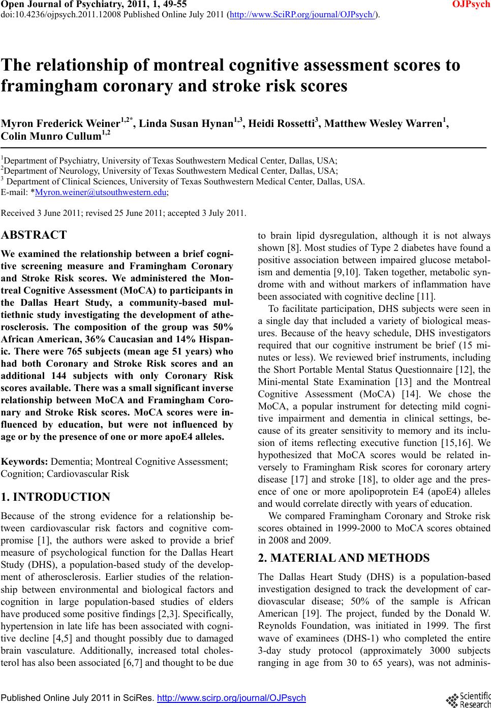 Open Journal of Psychiatry, 2011, 1, 49-55 OJPsych doi:10.4236/ojpsych.2011.12008 Published Online July 2011 (http://www.SciRP.org/journal/OJPsych/). Published Onl ine July 2011 in SciRes. http://www.scirp.org/journal/OJPsych The relationship of montreal cognitive assessment scores to framingham coronary and stroke risk scores Myron Frederick Weiner 1,2*, Linda Susan Hynan1,3, Heidi Rossetti3, Matthew Wesley Warren1, Colin Munro Cullum1,2 1Department of Psychiatry, Universit y of Texas Southwestern Medical Center, Dallas, USA; 2Department of Neurology, University of Texa s Southwestern Medical Center, Dallas, USA; 3 Department of Clinical Sciences, University of Texas Southwestern Medical Center, Dallas, USA. E-mail: *[email protected]; Received 3 June 2011; revised 25 June 2011; accepted 3 July 2011. ABSTRACT We examined the relationship between a brief cogni- tive screening measure and Framingham Coronary and Stroke Risk scores. We administered the Mon- treal Cognitive Assessment (MoCA) to pa r ticipants in the Dallas Heart Study, a community-based mul- tiethnic study investigating the development of athe- rosclerosis. The composition of the group was 50% Africa n American, 36% Caucasian and 14% Hispan- ic. There were 765 subjects (mean age 51 years) who had both Coronary and Stroke Risk scores and an additional 144 subjects with only Coronary Risk scores available. There was a small significant inverse relationship between MoCA and Framingham Coro- nary and Stroke Risk scores. MoCA scores were in- fluenced by education, but were not influenced by age or by the presence of one or more apoE4 alleles. Keywords: Dementia; Montreal Cognitive Assessment; Cognition; Cardiovascular Risk 1. INTRODUCTION Because of the strong evidence for a relationship be- tween cardiovascular risk factors and cognitive com- promise [1], the authors were asked to provide a brief measure of psychological function for the Dallas Heart Study (DHS), a population-based study of the develop- ment of atherosclerosis. Earlier studies of the relation- ship between environmental and biological factors and cognition in large population-based studies of elders have produced some positive findings [2,3]. Specifically, hypertension in late life has been associated with cogni- tive decline [4,5] and thought possibly due to damaged brain vasculature. Additionally, increased total choles- terol has also been associated [6,7] and thoug h t to be due to brain lipid dysregulation, although it is not always shown [8 ]. Most stud ies of Type 2 diabete s have found a positive association between impaired glucose metabol- ism and dementia [9,10]. Ta k en together, metabolic syn- drome with and without markers of inflammation have been associated with cognitive declin e [11]. To facilitate participation, DHS subjects were seen in a single day that included a variety of biological meas- ures. Because of the heavy schedule, DHS investigators required that our cognitive instrument be brief (15 mi- nutes or less). We reviewed brief instruments, including the Short Portable Mental Status Questionnaire [12], the Mini-mental State Examination [13] and the Montreal Cognitive Assessment (MoCA) [14]. We chose the MoCA, a popular instrument for detecting mild cogni- tive impairment and dementia in clinical settings, be- cause of its greater sensitivity to memory and its inclu- sion of items reflecting executive function [15,16]. We hypothesized that MoCA scores would be related in- versely to Framingham Risk scores for coronary artery disease [17] and stroke [18], to older age and the pres- ence of one or more apolipoprotein E4 (apoE4) alleles and would correlate directly with years of edu c ation . We compared Framingham Coronary and Stroke risk scores obtained in 1999-2000 to MoCA scores obtained in 2008 and 2009. 2. MATERIAL AND METHODS The Dallas Heart Study (DHS) is a population-based investigation designed to track the development of car- diovascular disease; 50% of the sample is African American [19]. The project, funded by the Donald W. Reynolds Foundation, was initiated in 1999. The first wave of examinees (DHS-1) who completed the entire 3-day study protocol (approximately 3000 subjects ranging in age from 30 to 65 years), was not adminis- 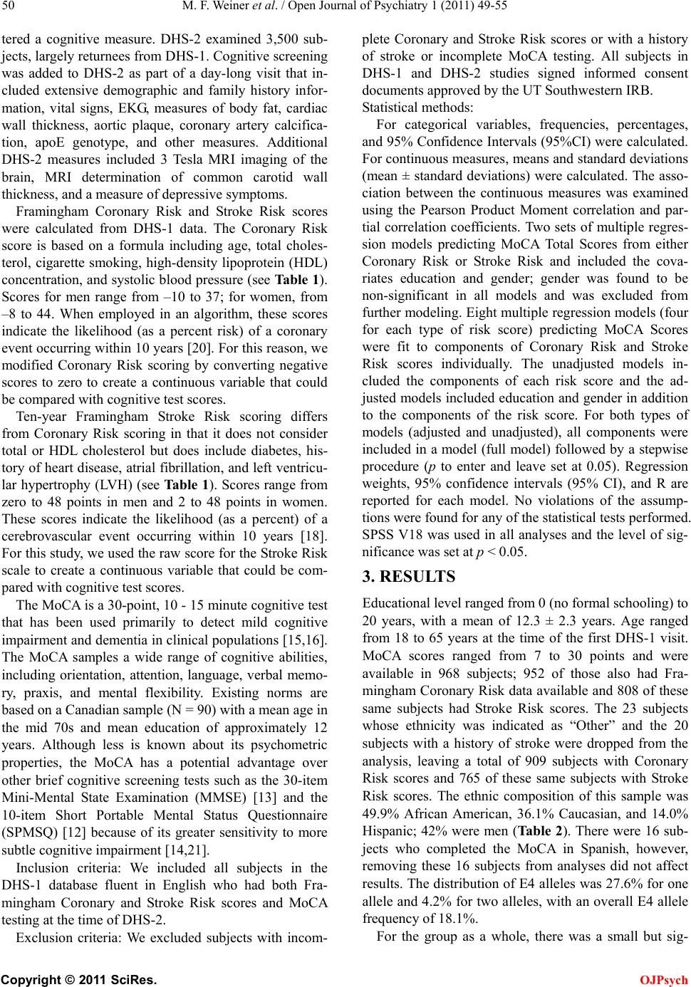 M. F. Weiner et al. / Open Journal of Psychiatry 1 (2011) 49-55 Copyright © 2011 SciRes. OJPsych 50 tered a cognitive measure. DHS-2 examined 3,500 sub- jects, largely returnees from DHS-1. Cognitive screening was added to DHS-2 as part of a day-long visit that in- cluded extensive demographic and family history infor- mation, vital signs, EKG, measures of body fat, cardiac wall thickness, aortic plaque, coronary artery calcifica- tion, apoE genotype, and other measures. Additional DHS-2 measures included 3 Tesla MRI imaging of the brain, MRI determination of common carotid wall thickness, and a measure of depressive symptoms. Framingham Coronary Risk and Stroke Risk scores were calculated from DHS-1 data. The Coronary Risk score is based on a formula including age, total choles- terol, cigarette smoking, high-density lipoprotein (HDL) concentration, and systolic blood pressure (see Table 1). Scores for men range from –10 to 37; for women, from –8 to 44. When employed in an algorithm, these scores indicate the likelihood (as a percent risk) of a coronary event occurring within 10 years [20]. For this reason, we modified Coronary Risk scoring by converting negative scores to zero to create a continuous variable that could be compared with cognitive test scores. Ten-year Framingham Stroke Risk scoring differs from Coronary Risk scoring in that it does not consider total or HDL cholesterol but does include diabetes, his- tory of heart disease, atrial fibrillation , and left ventricu- lar hypertrophy (LVH) (see Ta ble 1). Scores range from zero to 48 points in men and 2 to 48 points in women. These scores indicate the likelihood (as a percent) of a cerebrovascular event occurring within 10 years [18]. For this study, we used the raw score for the Stroke Risk scale to create a continuous variable that could be com- pared with cognitive test scores. The MoCA is a 30-point, 10 - 15 minute cognitive test that has been used primarily to detect mild cognitive impairment and dementia in clinical populations [15,16]. The MoCA samples a wide range of cognitive abilities, including orientation, attention, language, verbal memo- ry, praxis, and mental flexibility. Existing norms are based on a Canadian sample (N = 90) with a mean age in the mid 70s and mean education of approximately 12 years. Although less is known about its psychometric properties, the MoCA has a potential advantage over other brief cognitive screening tests such as the 30-item Mini-Mental State Examination (MMSE) [13] and the 10-item Short Portable Mental Status Questionnaire (SPMSQ) [12] because of its greater sensitivity to more subtle cognitive impairment [14,21]. Inclusion criteria: We included all subjects in the DHS-1 database fluent in English who had both Fra- mingham Coronary and Stroke Risk scores and MoCA testing at the time of DHS-2. Exclusion criteria: We excluded subjects with incom- plete Coronary and Stroke Risk scores or with a history of stroke or incomplete MoCA testing. All subjects in DHS-1 and DHS-2 studies signed informed consent docum e nt s ap proved by the UT Southwestern IRB. Statistical methods: For categorical variables, frequencies, percentages, and 95% Confidence Intervals (95%CI) w ere calculated. For continuous measures, means and standard deviations (mean ± standard deviations) were calculated. The asso- ciation between the continuous measures was examined using the Pearson Product Moment correlation and par- tial correlation coefficients. Two sets of multiple regres- sion models predicting MoCA Total Scores from either Coronary Risk or Stroke Risk and included the cova- riates education and gender; gender was found to be non-significant in all models and was excluded from further modeling. Eight multiple regression models (four for each type of risk score) predicting MoCA Scores were fit to components of Coronary Risk and Stroke Risk scores individually. The unadjusted models in- cluded the components of each risk score and the ad- justed models included education and gender in addition to the components of the risk score. For both types of models (adjusted and unadjusted), all components were included in a model (full model) followed by a stepwise procedure (p to enter and leave set at 0.05). Regression weights, 95% confidence intervals (95% CI), and R are reported for each model. No violations of the assump- tions were found for any of the statistical tests performed. SPSS V18 was used in all analyses and the level of sig- nificance was set at p < 0.0 5. 3. RESULTS Educational level ranged from 0 (no formal schooling) to 20 years, with a mean of 12.3 ± 2.3 years. Age ranged from 18 to 65 years at the time of the first DHS-1 visit. MoCA scores ranged from 7 to 30 points and were available in 968 subjects; 952 of those also had Fra- mingham Coronary Risk data available and 808 of these same subjects had Stroke Risk scores. The 23 subjects whose ethnicity was indicated as “Other” and the 20 subjects with a history of stroke were dropped from the analysis, leaving a total of 909 subjects with Coronary Risk scores and 765 of these same subjects with Stroke Risk scores. The ethnic composition of this sample was 49.9% African American, 36.1% Caucasian, and 14.0% Hispanic; 42% were men (Ta ble 2). There were 16 sub- jects who completed the MoCA in Spanish, however, removing these 16 subjects from analyses did not affect results. The distribution of E4 alleles was 27 .6% for one allele and 4.2% for two alleles, with an overall E4 allele frequenc y of 1 8.1%. For the group as a whole, there was a small but sig- 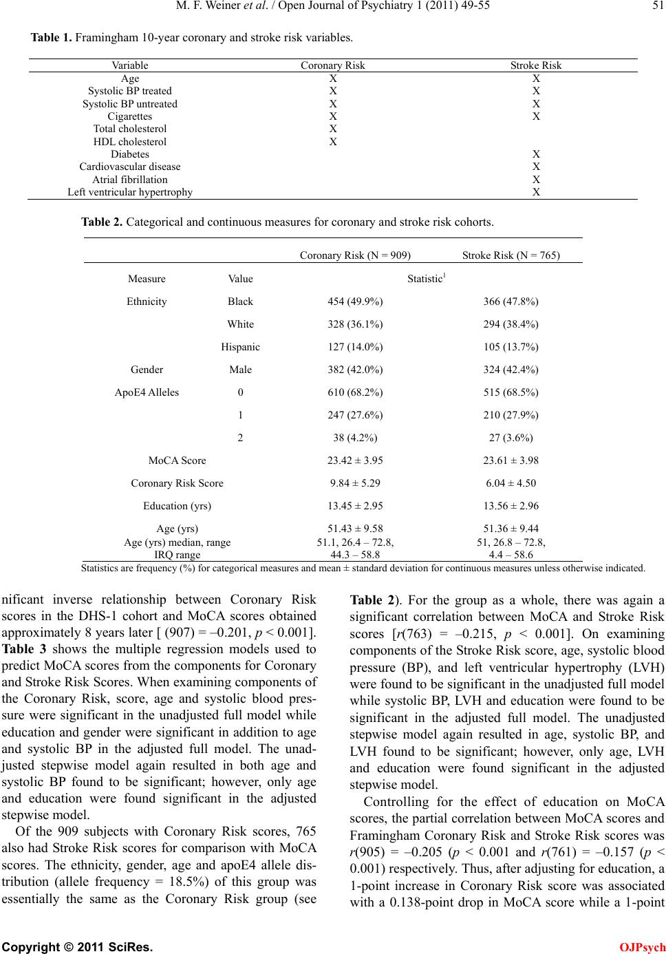 M. F. Weiner et al. / Open Journal of Psychiatry 1 (2011) 49-55 Copyright © 2011 SciRes. OJPsych 51 Table 1. Framingham 10-year coronary and stroke risk variables. Variable Coronary Risk Stroke Risk Age X X Systolic BP treated X X Systolic BP untreated X X Cigarettes X X To tal cholesterol X HDL cholesterol X Diabetes X Cardiovascular disease X Atrial fibrillation X Left ventricular hypertrophy X Table 2. Categorical and continuous measures for coronary and stroke risk cohorts. Coronary Ri s k (N = 909) Stroke Risk (N = 765) Measure Value Statistic1 Ethnicity Black 454 (49.9%) 366 (47.8%) White 328 (36.1%) 294 (38.4%) Hispanic 127 (14.0%) 105 (13.7%) Gender Male 382 (42.0%) 324 (42.4%) ApoE4 Alleles 0 610 (68.2%) 515 (68.5%) 1 247 (27.6%) 210 (27.9%) 2 38 (4.2%) 27 (3.6%) MoCA Score 23.42 ± 3.95 23.61 ± 3.98 Coronary Risk Score 9.84 ± 5.29 6.04 ± 4.50 Education (yrs) 13.45 ± 2.95 13.56 ± 2.96 Age (yrs) 51.43 ± 9.58 51.36 ± 9.44 Age (yrs) median, range IRQ range 51.1, 26.4 – 72.8, 44.3 – 58.8 51, 26.8 – 72.8, 4.4 – 58.6 Statistics are frequency (%) for categorical measures and mean ± standard deviation for continuous measures unless otherwise indicated. nificant inverse relationship between Coronary Risk scores in the DHS-1 cohort and MoCA scores obtained approximately 8 years later [ (907) = –0.201, p < 0.001]. Table 3 shows the multiple regression models used to predict MoCA scores from the components for Coronary and Stroke Risk Scores. When examining components of the Coronary Risk, score, age and systolic blood pres- sure were significant in the unadjusted full model while education and gender were significant in addition to age and systolic BP in the adjusted full model. The unad- justed stepwise model again resulted in both age and systolic BP found to be significant; however, only age and education were found significant in the adjusted stepwise model. Of the 909 subjects with Coronary Risk scores, 765 also had Stroke Risk scores for comparison with MoCA scores. The ethnicity, gender, age and apoE4 allele dis- tribution (allele frequency = 18.5%) of this group was essentially the same as the Coronary Risk group (see Table 2). For the group as a whole, there was again a significant correlation between MoCA and Stroke Risk scores [r(763) = –0.215, p < 0.001]. On examining components of the Stroke Risk score, age, systolic blood pressure (BP), and left ventricular hypertrophy (LVH) were found to be significant in the unadjusted full model while systolic BP, LVH and education were found to be significant in the adjusted full model. The unadjusted stepwise model again resulted in age, systolic BP, and LVH found to be significant; however, only age, LVH and education were found significant in the adjusted stepwise model. Controlling for the effect of education on MoCA scores, the partial correlation between MoCA scores and Framingham Coronary Risk and Stroke Risk scores was r(905) = –0.205 (p < 0.001 and r(761) = –0.157 (p < 0.001) respectively. Thus, after adjusting for educatio n, a 1-point increase in Coronary Risk score was associated with a 0.138-point drop in MoCA score while a 1-point 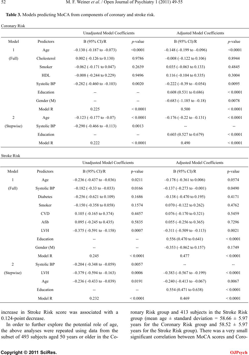 M. F. Weiner et al. / Open Journal of Psychiatry 1 (2011) 49-55 Copyright © 2011 SciRes. OJPsych 52 Table 3. Models predicting MoCA from components of coronary and stroke risk. Coronary Risk Unadjusted Model Coefficients Adjusted Model Coefficients Model Predictors B (95% CI)/R p-value B (95% CI)/R p-value 1 Age –0.130 (–0.187 to –0.073) <0.0001 –0.148 (–0.199 to –0.096) <0.0001 (Full) Cholesterol 0.002 (–0.126 to 0.130) 0.9786 –0.008 (–0.122 to 0. 106) 0.8944 Smoker –0.062 (–0.171 to 0. 047) 0.2639 0.035 (–0.063 to 0.133) 0.4845 HDL –0.008 (–0.244 to 0. 229) 0.9496 0.116 (–0.104 to 0.335) 0.3004 Systolic BP –0.282 (–0.460 to –0.103) 0.0020 –0.222 (–0.39 to –0.054) 0.0095 Education -- -- 0.608 (0.531 to 0.6 86) < 0.0001 Gender (M) -- -- –0.683 (–1.185 to –0.18) 0.0078 Model R 0.225 < 0.0001 0.500 < 0.0001 2 Age –0.123 (–0.177 to –0.07) < 0.0001 –0.176 (–0.22 to –0.131) < 0.0001 (Stepwise) Systolic BP –0.290 (–0.466 to –0.113) 0.0013 -- -- Education -- -- 0.603 (0.527 to 0.6 79) < 0.0001 Model R 0.222 < 0.0001 0.490 < 0.0001 Stroke Risk Unadjusted Model Coefficients Adjusted Model Coefficients Model Predictors B (95% CI)/R p-value B (95% CI)/R p-value 1 Age –0.236 (–0.437 to –0.036) 0.0211 –0.178 (–0.361 to 0.006) 0.0574 (Full) Systolic BP –0.182 (–0.33 to –0.033) 0.0166 –0.137 (–0.273 to –0.001) 0.0490 Diabetes –0.256 (–0.621 to 0.109) 0.1686 –0.138 (–0.4 70 to 0.19 5) 0.4171 Smoker –0.150 (–0.358 to 0.058) 0.1574 0. 07 0 (–0.122 to 0.262) 0.4762 CVD 0.105 (–0.165 to 0.374) 0.4457 0.076 (–0 .170 to 0.321) 0.5459 Afib 0.095 (–0.245 to 0.435) 0.5835 0.055 (–0 .256 to 0.365) 0.7296 LVH –0.375 (–0.591 to –0.158) 0.0007 –0.311 (–0.5 09 to –0.113) 0.0021 Education -- -- 0. 5 56 (0.470 to 0.6 4 1) < 0.0001 Gender (M) -- -- –0.353 (–0.862 to 0.157) 0.1749 Model R 0.245 < 0.0001 0.477 < 0.0001 2 Systolic BP –0.204 (–0.348 to –0.059) 0.0057 -- -- (Stepwise) LVH –0.379 (–0.594 to –0.163) 0.0006 –0.383 (–0.567 to –0.199) < 0.0001 Age –0.236 (–0.433 to –0.039) 0.0191 –0.240 (–0.413 to –0.067) 0.0067 Education -- -- 0. 5 54 (0.471 to 0.6 3 8) < 0.0001 Model R 0.232 < 0.0001 0.469 < 0.0001 increase in Stroke Risk score was associated with a 0.124-p oi nt decrea se. In order to further explore the potential role of age, the above analyses were repeated using data from the subset of 493 subjects aged 50 years or older in the Co- ronary Risk group and 413 subjects in the Stroke Risk group (mean age ± standard deviation = 58.66 ± 5.97 years for the Coronary Risk group and 58.52 ± 5.97 years for the Stroke Risk group). The re was a v ery small significant correlation between MoCA scores and Coro-  M. F. Weiner et al. / Open Journal of Psychiatry 1 (2011) 49-55 Copyright © 2011 SciRes. OJPsych 53 nary Risk scores [r(491) = –0.139, p = 0.002] and Stroke Risk scores [r(411) = –0.214, p < 0.001] in these older subjects. Controlling for the effect of education on Mo- CA scores, the partial correlation between Framingham Coronary Risk and Stroke Risk scores in older subjects was r(4 89) = –0.059 (p = .191) and r(409) = –0.110 (p = 0.026) respectively. Thus, after adjusting for education in older subjects, a 1-point increase in Coronary Risk score was associated with a 0.067-point drop in MoCA score while a 1-point increase in Stroke Risk score was associated with a 0.082-point decrease. There were no significant differences in MoCA scores, Coronary Risk s cores, or Stroke Risk scor es for subjects with or without apoE4 alleles (p = 0.110, p = 0.312, and p = 0.874, respectively) in the entire cohort or among subjects age 50 + (p = 0.103, p = 0.623, and p = 0.526, respectively). Also , there were no significant d ifferences between men and women in the relationship between MoCA and Coronary or Stroke Risk scores when ex- amined for the entire sample or the older group (data not shown). 4. DISCUSSION The uniqueness of our study is that it examines vascular risk factors in a population-based sample that includes 50% African Americans. It is possible that the risk fac- tors we examined were not sufficiently sensitive to detect and quantify the effects of subclinical atheroscle- rosis. We have now begun exploring the relationship between more direct biological measures of atheroscle- rosis including the concentration of atherosclero- sis-related inflammatory substances such as CRP and direct measures of atherosclerosis such as coronary ar- tery calcium. Our cognitive measure, the MoCA, was designed to be used in clinical settings in which there is great vari a- tion in cognitive function [22]. It has been suggested that, as a screening tool, it may have limited v alue in popula- tions where prevalence of mild cognitive impairment is low [23]. However, the range of MoCA scores in this study was 7-30 (mean ± standard deviation = 23.38 ± 4). Other investigators have found more robust relationships between cardiovascular disease and cognitive function using more detailed neurocognitive measures [24,25] and also with very crude measures. For example, one study found that SPMSQ scores were lower in the pres- ence of apoE4 [26]. These subjects had lower initial SPMSQ scores, and there was increased disparity be- tween E4 carriers and non-E4 carriers over a period of 4 years. We found that the influence of Coronary or Stroke Risk scores on MoCA scores did not increase with age. Because the mean age of this study population was rela- tively young, it may be that the impact of coronary and stroke risk factors are limited at th is age, indicating such patients either need continued fo llow up at a later time or more sensitive tests early on. We also did not find the negative effect of the apoE4 allele on cognition found in the another study [27] or in a meta-analysis of 77 studies in which apoE4 carriers performed more poorly on tests of global cognitive function, and the disparity between E4 and non-E4 carriers increased with age [28]. One study found significant interactions between the presence of E4 and verbal memory, verbal organization, nonverbal memory, set shifting and complex attention in a community-based group of subjects with an average age of 61 years, but systolic blood pr essure was the only individual risk factor significantly related to these cogni- tive measures [25]. Because of the disparity of our find- ings from those of others in the literature, we reviewed data from non-demented older adults persons followed yearly at the UT Southwestern Alzheimer’s Disease Center (ADC). We examined MMSE data from all 219 subjects who had both MMSE scores and apoE4 allele determination, of whom 81 (40%) had one or more apoE4 alleles. We found no significant difference in MMSE scores in no n-demented subjects w ith or without an apoE4 allele. Our findings concerning the impact of coronary and stroke risk factors and E4 on cognition may be related to differences in the populations studied and in the psy- chological measure employed. Both our “young” (mean age = 51 years) and our “old” (mean age = 58 years) cohorts were relatively young in relation to the sample examined by Haan et al. [29] and the more recent me- ta-analytic study [28]. The relationship of apoE4 to cog- nition in other studies may be partially explainable by the possible inclusion in older populations of persons with incipient Alzheimer disease [30], which is less likely in our DHS sample. Another study, which ex- amined the relationship of the apoE4 allele to MMSE scores in persons 659 persons followed over 22 years in a large community-based study, found no relationship between apoE4 status and MMSE scores, but there was a significant difference in delayed recall in persons < 65 years of age [31]. They suggested survival bias as an explanation of the difference in apoE 4 influence on cognition. Other studies have suggested that vascular disease in- fluences performance on cognitive tasks associated with frontal lobe function more than those associated with other cortical areas [24]. The MoCA contains few items relating to this cognitive domain, and score ranges for those items are limited. Supporting this explanation is the finding that a delayed recall test was more sensitive than the MMSE in detecting cognitive decline in elders  M. F. Weiner et al. / Open Journal of Psychiatry 1 (2011) 49-55 Copyright © 2011 SciRes. OJPsych 54 [31]. We plan to re-test a subgroup of DHS-2 subjects with additional brief measures that may increase sensi- tivity to executive and memory function and we will also follow them prospectively to examine the rate of cogni- tive change. Although the MoCA samples many of the same cog- nitive areas than less sensitive instruments, several less sensitive instruments have shown stronger correlations of global cognitive function with cardiovascular risk factors, including age and apoE status. Our failure to find correlations of similar strength may be attributable to some limitation of the MoCA, the relative youth of the population we studied or differences between per- sons with presymptomatic and symptomatic cardiovas- cular disease. REFERENCES [1] Beeri, M.S., Ravona-Springer, R., Silverman, J.M. and Haroutunian, V. (2009) The effects of cardiovascular risk factors on cognitive compromise. Dialogues in Clinical Neuroscience, 11, 201-212. [2] Middleton, L.E., Barnes, D.E., Lui, L.Y. and Yaffe, K. (2010) Physical activity over the life course and its asso- ciation with cognitive performance and impairment in old age. Journal of the American Geriatrics Society, 58, 1322-1326. doi:10.1111/j.1532-5415.2010.02903.x [3] Yaffe, K., Vittinghoff, E., Lindquist, K., et al. (2010) Posttraumatic stress disorder and risk of dementia among us veterans. Archives of General Psychiatry, 67, 608-613. doi:10.1001/archgenpsychiatry.2010.61 [4] Tzourio, C. , D uf oui l, C ., D uc im etier e, P. and Alperovitch, A. (1999) Cognitive decline in individuals with high blood pressure: A longitudinal study in the elderly. Eva study group. Epidemiology of vascular aging. Neurology, 53, 1948-1952. [5] Mielke, M. M., Rosenbe r g, P. B., Tschanz, J., et al. (2007) Vascular factors predict rate of progression in alzheimer disease. Neurology, 69, 1850-1858. doi:10.1212/01.wnl.0000279520.59792.fe [6] Solomon, A., Kareholt, I., Ngandu, T., et al. (2007) Serum cholesterol changes after midlife and late-life cognition: Twenty-one-year follow-up study. Neurology, 68, 751-756. doi:10.1212/01.wnl.0000256368.57375.b7 [7] Yaffe, K., Barr ett-Connor , E., Lin, F. and Grady , D . (2002) Serum lipoprotein levels, statin use, and cognitive func- tion in older women. Archives of Neurology, 59, 378-384. doi:10.1001/archneur.59.3.378 [8] Reitz, C., Luchsinger, J., Tang, M.X., Manly, J. and Mayeux, R. (2005) Impact of plasma lipids and time on memory performance in healthy elderly withou t dementia. Neurology, 64, 1378-1383. doi:10.1212/01.WNL.0000158274.31318.3C [9] Whitmer, R. A., Sidney, S., Selby, J., Johnston, S. C. and Yaffe, K. (2005) Midlife cardiovascular risk factors and risk of dementia in late life. Neurology, 64, 277-281. doi:10.1212/01.WNL.0000149519.47454.F2 [10] Schnaider Beeri, M., Goldbour t, U., Silverman, J. M., et al. (2004) Diabetes mellitus in midlife and the risk of de- mentia three decades later. Neurology, 63, 1902-1907. [11] Yaffe, K., Kanaya, A., Lindquist, K., et al. (2004) The metabolic syndrome, inflammation, and risk of cognitive decline. Journal of the American Medical Association, 292, 2237-2242. doi:10.1001/jama.292.18.2237 [12] Pfeiffer, E. (1975) A short portable mental status ques- tionnaire for the assessment of organic brain deficit in elderly patients. Journal of the American Geriatrics So- ciety, 23, 433-441. [13] Folstein, M.F., Folstein, S.E. and McHugh, P.R. (1975) “Mini-mental state”. A practical method for grading the cognitive state of patients for the clinician. Journal of Psychiatric R es earch, 12, 189-198. doi:10.1016/0022-3956(75)90026-6 [14] Nasreddine, Z.S., Phillips, N. A ., Bedirian, V., et al. (2005) The montreal cognitive assessment, moca: A brief screening tool for mild cognitive impairment. Journal of the American Geriatrics Soci ety, 53, 695-699. doi:10.1111/j.1532-5415.2005.53221.x [15] Smith, T., Gildeh, N. and Holmes , C. (2007) The montreal cognitive assessment: Validity and utility in a memory clinic setting. Canadian Journal of Psychiatry, 52, 329-332. [16] Hoops, S., Nazem, S., Siderowf, A. D., et al. (2009) Va- lidity of the MoCA and mmse in the detection of mci and dementia in parkinson disease. Neurology, 73, 1738-1745. doi:10.1212/WNL.0b013e3181c34b47 [17] D'Agostino, R. B., Sr., Grundy, S., Sullivan, L. M. and Wilson, P. (2001) Validation of the framingham coronary heart disease prediction scores: Results of a multiple eth- nic groups investigation. Journal of the American Medical Association, 286, 180-187. doi:10.1001/jama.286.2.180 [18] D’Agostino, R.B., Wolf, P.A., Belanger, A.J. and Kannel, W.B. (1994) Stroke risk profile: Adjustment for antihy- pertensive medication. The framingham study. Stroke, 25, 40-43. doi:10.1161/01.STR.25.1.40 [19] Victor , R. G., Hal ey, R. W., W illett, D . L., et al. (2004) The dallas heart study: A population-based probability s ampl e for the multidisciplinary study of ethnic differences in cardiovascular health. American Journal of Cardiology, 93, 1473-1480. [20] Wilson, P.W., D’Agostino, R.B., Levy, D., et al. (1998) Prediction of coronary heart disease using risk factor categories. Circulation, 97, 1837-1847. [21] Aggarwal, A. and Kean, E. (2010) Comparison of the folstein mini mental state examination (mmse) to the montreal cognitive assessment (MoCA) as a cognitive screening tool in an inpatient rehabilitation setting. Neu- roscience and Medicine, 1, 39-42. doi:10.4236/nm.2010.12006 [22] Bernstein, I.H., Lacritz, L., Barlow, C.E., Weiner, M.F. and Defina, L. F. (2011) Psychometric evaluation of the montreal cognitive assessment (MoCA) in three diverse samples. Clinical Neuropsychology, 25, 119-126. doi:10.1080/13854046.2010.533196 [23] McLennan, S.N., Mathias, J.L., Brennan, L.C. and Ste- wart, S. (2010) Validity of the montreal cognitive as- sessment (MoCA) as a screening test for mild cognitive impairment (MCI) in a cardiovascular population. Journal of Geriatric Psychiatry and Neurology, 24, 33-38. [24] Kuczynski, B., Jagust, W., Chui, H.C. and Re ed, B. (2009) An inverse association of cardiovascular risk and frontal  M. F. Weiner et al. / Open Journal of Psychiatry 1 (2011) 49-55 Copyright © 2011 SciRes. OJPsych 55 lobe glucose metabolism. Neurology, 72, 738-743. doi:10.1212/01.wnl.0000343005.35498.e5 [25] Zade, D., Beiser, A., McGlinchey, R., et al. (2010) Inter- active effects of apolipoprotein E type 4 genotype and cerebrovascular risk on neuropsychological performance and structural brain changes. Journal of Stroke and Ce- rebrovascular Diseases, 19, 261-268. doi:10.1016/j.jstrokecerebrovasdis.2009.05.001 [26] Fillenbaum, G.G., Landerman, L.R., Blazer, D.G., et al. (2001) The relationship of APOE genotype to cognitive functioning in older African-American and caucasian community residents. Journal of the American Geriatrics Society, 49, 1148-1155. doi:10.1212/01.WNL.0000149643.91367.8A [27] Blair, C.K., Folsom, A.R., Knopman, D.S., et al. (2005) APOE genotype and cognitive decline in a middle-aged cohort. Neurology, 64, 268-276. doi:10.1212/01.WNL.0000149643.91367.8A [28] Wisdom, N.M., Callahan, J.L. and Hawkins, K.A. (2011) The effects of apolipoprotein e on non-im paired cognitive functioning: A meta-analysis. Neurobiology of Aging, 32, 63-74. doi:10.1001/jama.282.1.40 [29] Haan, M.N., Shemanski, L., Jagust, W.J., Manolio, T.A. and Kuller, L. (1999) The role of APOE epsilon4 in modulating effects of other risk factors for cognitive de- cline in elderly persons. Journal of the American Medical Association, 282, 40-46. doi:10.1001/jama.282.1.40 [30] Qiu, C. and Fratiglioni, L. (2010) Apolipoprotein E epsi- lon4 status and cognitive decline with and without de- mentia. Archives of Neurology, 67, 1036. doi:10.1001/archneurol.2010.163 [31] Kozauer, N.A., Mielke, M.M., Chan, G.K., Rebok, G.W. and Lyketsos, C.G. (2008) Apolipoprotein e genotype and lifetime cogni tive decline. International Psychogeriatrics, 20, 109-123. doi:10.1017/S104161020700587X |