

Paper Menu >>
Journal Menu >>
 American Journal of Plant Sciences, 2011, 2, 148-155 doi:10.4236/ajps.2011.22016 Published Online June 2011 (http://www.SciRP.org/journal/ajps) Copyright © 2011 SciRes. AJPS Identification of EDTA-Soluble Polysaccharides from Pea Epicotyl Cell Walls and Their Interaction with Xyloglucan Elias A.-H. Baydoun1, Oula C. Mansour1, Sandra E. Rizk2, Christopher T. Brett3 1Department of Biology, American University of Beirut, Beirut, Lebanon; 2Department of Natural Sciences, Lebanese American University, Beirut, Lebanon; 3Institute of Biomedical Life Sciences, University of Glasgow, Glasgow, United Kingdom. Email: [email protected], oula[email protected], sandra.rizk@lau.edu.lb, [email protected] Received December 9th, 2010; revised March 23rd, 2011; accepted March 31st, 2011. ABSTRACT Nascent pectin and glucuronoxylan were prepared from membrane-bound enzymes obtained from pea epicotyls. They had previously been shown to exhibit a protein- and pH-dependent pattern of binding to cell wall ghosts and to xy- loglucan extracted from cell walls prepared from pea epicotyls; maximum binding required a pH of 3-4, and the pres- ence of cell wall proteins, namely assemblins. To determine whether wall polysaccharides deposited in cell walls be- have in the same manner as nascent polymers, radioactively labeled EDTA-soluble polymers were prepared from newly-deposited pea epicotyl cell walls. Different enzyme treatments followed by column chromatography, in addition to complete acid hydrolysis followed by paper and thin layer chromatography, indicated the presence of pectin, to- gether with smaller amounts of glucuronoxylan, in this EDTA-soluble extract. These radioactively labeled polysaccha- rides were found to bind to cell wall ghosts and to xyloglucan extracted from the second and third internodes of pea epicotyls cell walls in a pH-dependent manner, similar to the binding pattern obtained with nascent polymers. Maxi- mum bindi ng occurre d at pH 3-4, and also r e qu ired the pr esence of p ro tein. Keywords: Assemblin, Cell Wall Assembly, Glucuronoxylan, Pectin, Pisum Sativum, Xyloglucan 1. Introduction The plant cell wall contains cellulose microfibrils em- bedded in a non-crystalline matrix. Matrix polysaccha- rides are synthesized in the Golgi apparatus and trans- ported in vesicles to the wall [1]. There, the newly syn- thesized matrix polysaccharides associate with each other and with newly synthesized cellulose to form the new innermost layer of the cell wall. Xyloglucan, the principal hemicellulose of the primary cell walls of dicotyledonous plants, plays an essential role in cell wall elongation due to its strong association to cellulose microfibrils [2,3]. Nascent EDTA-soluble 14C-polysaccharides prepared from pea seedlings (Pisum sativum) consist of radioactively labeled pectins and glu- curonarabinoxylans (GAX), with a significantly greater abundance of pectins [4]. These polysaccharides bound to xyloglucan in a pH-dependant manner, with the high- est binding occurring at pH 3 - 4, which corresponds to the pH of a growing wall. The binding decreased to al- most zero at pH 6 [5]. This binding pattern indicates a significant role in cell-wall assembly during deposition and cell wall extension during growth. Since pre-treatment of nascent GAX with protease greatly de- creased the binding at acidic pH and abolished the pH-dependant binding pattern, the protein attached to nascent GAX, named assemblin, appeared to have an essential role in the deposition of GAX into the cell wall [5]. However, cell wall polymers are synthesized in the Golgi apparatus and undergo several modifications prior to their excretion into the cell wall space [6]. The present study aims to determine whether newly-deposited poly- saccharides extracted from preformed cell walls behave in the same manner as nascent polymers, with respect to their interaction with cell-wall ghosts and with xyloglu- can. 2. Materials and Methods 2.1. Plant Material Peas (Pisum sativum L.cv Meteor, obtained form Sharpes International, Sleaford, UK) were soaked overnight at  Identification of EDTA-Soluble Polysaccharides from Pea Epicotyl Cell Walls and 149 Their Interaction with Xyloglucan room temperature and grown on damp vermiculite at 25˚C in continuous darkness. For xyloglucan extraction, the peas were grown for 8 - 9 days. For the preparation of cell wall ghosts, nascent cell wall polymers and newly-deposited cell wall polymers, the growth period was 6 days. 2.2. Preparation of Cell Wall Ghosts and Extraction of Xyloglucan A modification of the methods of Hayashi and Maclach- lan (1984) and Ogawa et al. (1990) was used [7,8]. Third internodes (50 g) were harvested from 8-9-day-old peas and extracted three times with 70% (v/v) ethanol (150 ml, 30 min, 70˚C). The tissue was chopped with a razor blade, homogenized using a pestle and mortar in Tris-HCl buffer (0.1 M, pH 7.0, 75 ml), and centrifuged (8000 g, 10 min). The cell wall pellets were extracted three times with EDTA buffer (0.1 M, pH 7.0, 75 ml, 30 min, 85˚C), and three times with 0.71 M KOH/26 mM NaBH4 (75 ml, 1 h, 25˚C) in a shaking incubator. Insolu- ble material (cell wall ghosts) was obtained by centrifu- gation (8000 g, 10 min). To prepare xyloglucan, the cell wall ghosts were extracted twice with 4.3 M KOH/26 mM NaBH4 (25 ml, 4 h, 25˚C) in the same incubator. The combined 4.3 M KOH-soluble fractions were neu- tralized with acetic acid. Ethanol was added 70% (v/v), and xyloglucan allowed to precipitate overnight. The precipitated material was treated with salivary amylase (24 h, 40˚C) and protease (Type XIV, Sigma, 24 h, 37˚C). Xyloglucan content was determined by iodine-sodium sulphate method [9,10]. 2.3. Preparation of Nascent EDTA-Soluble 14C-Polysaccharides The procedure for particulate enzyme preparation was similar to that of Waldron and Brett (1987) and Rizk et al. (2000) [5,11]. Epicotyls (6 - 9 cm long) were cut off, the hooks discarded and the remaining tissue cooled on ice. Subsequent operations were carried out at 0˚C - 4˚C. Epicotyls were homogenized using a pestle and mortar in Tris-Mes buffer (10 mM, pH 6.0). The homogenate was strained through two layers of muslin, and the filtrate centrifuged at 100,000 g for 30 minutes. The pellet was homogenized in cold homogenization buffer using a glass-Teflon tissue homogenizer, to obtain the particulate enzyme preparation, which was then incubated (25˚C, 4 h) with UDP-D-[U-14C]glucuronic acid (3.53 kBq, 1.7 μM), UDP-xylose (1 mM) and MnCl2 (10 mM). The re- action was terminated by adding 96% (v/v) ethanol. After centrifugation (10,000 x g, 5 min), particulate material was washed three times with 70% (v/v) ethanol and ex- tracted twice with 50 mM EDTA/50 mM sodium phos- phate buffer (pH 6.8) for 5 min at 100˚C. The combined EDTA/phosphate extracts were passed through a Sephadex G-100 column, eluted with water, and the excluded material was collected for binding experiments [5]. 2.4. Preparation of Newly-Deposited Cell Wall EDTA-Soluble 14C-Polysaccharides Newly-deposited cell wall 14C-polysaccharides were pre- pared by incubating pea epicotyls with [14C]sucrose (0.148 MBq at the base of 10 epicotyls) for 24 hours. Cell wall pellets were obtained from these epicotyls as described above, and then extracted with boiling 50 mM EDTA/50 mM sodium phosphate buffer (pH 6.8) for 5 minutes. The EDTA-extracts obtained were passed through a column of Sephadex G-100 and eluted with water to remove low molecular weight products. The high mo- lecular weight material which ran parallel to blue dextran was combined and used for analysis and binding assays to xyloglucan. 2.5. Assay of Binding of 14C-Polysaccharides to Xyloglucan or to Cell Wall Ghosts Pea xyloglucans or cell wall ghosts (1 mg/incubation) were resuspended in 0.5 ml incubation buffer (10 mM oxalate/10 mM phosphate, adjusted to the appropriate pH with HCl or NaOH), mixed with 0.5 ml 14C-polysaccha- rides (generally 15 - 30 Bq), incubated (5 min, 25˚C and centrifuged (5 min, 10,000 x g). Pellets were washed once with buffer of the appropriate pH (0.5 ml), centri- fuged for 5 min, resuspended in 0.4 ml water and mixed with 4 ml Ultima-Flo AF (Packard Instrument Company, Meriden, Conn., USA) for liquid scintillation counting. All binding experiments were carried out in duplicate. Results are expressed as the mean ± the difference be- tween the experimental values and the means. 2.6. Acid Hydrolysis Newly-deposited cell wall 14C-polysaccharides were acid hydrolyzed using trifluoroacetic acid (2 M, 120˚C, 1 h). The hydrolysate was centrifuged at 10,000 x g and the supernatant was rotoevaporated to be analyzed by paper and thin layer chromatography. 2.7. Chromatography Paper chromatography (PC) was performed using Whatman no. 3 paper, run in a solvent of ethyl acetate: pyridine: water (8:2:1 v/v) for 24 hours. Thin layer chromatography (TLC) was performed using silica coated plates and run in a solvent of butan-1-ol: ethyl acetate: water (7:1:2 v/v) for 18 - 22 hours [12]. 1 cm strips (for the PC) and 0.5 cm strips (for the TLC) were cut from the chromatogram for scintillation counting. Copyright © 2011 SciRes. AJPS 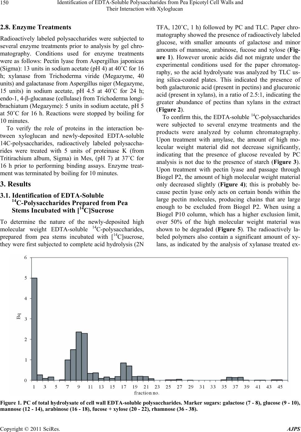 Identification of EDTA-Soluble Polysaccharides from Pea Epicotyl Cell Walls and Their Interaction with Xyloglucan Copyright © 2011 SciRes. AJPS 150 2.8. Enzyme Treatments Radioactively labeled polysaccharides were subjected to several enzyme treatments prior to analysis by gel chro- matography. Conditions used for enzyme treatments were as follows: Pectin lyase from Aspergillus japonicas (Sigma): 13 units in sodium acetate (pH 4) at 40˚C for 16 h; xylanase from Trichoderma viride (Megazyme, 40 units) and galactanase from Aspergillus niger (Megazyme, 15 units) in sodium acetate, pH 4.5 at 40˚C for 24 h; endo-1, 4-β-glucanase (cellulase) from Trichoderma longi- brachiatum (Megazyme): 5 units in sodium acetate, pH 5 at 50˚C for 16 h. Reactions were stopped by boiling for 10 minutes. To verify the role of proteins in the interaction be- tween xyloglucan and newly-deposited EDTA-soluble 14C-polysaccharides, radioactively labeled polysaccha- rides were treated with 5 units of proteinase K (from Tritirachium album, Sigma) in Mes, (pH 7) at 37˚C for 16 h prior to performing binding assays. Enzyme treat- ment was terminated by boiling for 10 minutes. 3. Results 3.1. Identification of EDTA-Soluble 14C-Polysaccharides Prepared from Pea Stems Incubated with [14C]Sucrose To determine the nature of the newly-deposited high molecular weight EDTA-soluble 14C-polysaccharides, prepared from pea stems incubated with [14C]sucrose, they were first subjected to complete acid hydrolysis (2N TFA, 120˚C, 1 h) followed by PC and TLC. Paper chro- matography showed the presence of radioactively labeled glucose, with smaller amounts of galactose and minor amounts of mannose, arabinose, fucose and xylose (Fig- ure 1). However uronic acids did not migrate under the experimental conditions used for the paper chromatog- raphy, so the acid hydrolysate was analyzed by TLC us- ing silica-coated plates. This indicated the presence of both galacturonic acid (present in pectins) and glucuronic acid (present in xylans), in a ratio of 2.5:1, indicating the greater abundance of pectins than xylans in the extract (Figure 2). To confirm this, the EDTA-soluble 14C-polysaccharides were subjected to several enzyme treatments and the products were analyzed by column chromatography. Upon treatment with amylase, the amount of high mo- lecular weight material did not decrease significantly, indicating that the presence of glucose revealed by PC analysis is not due to the presence of starch (Figure 3). Upon treatment with pectin lyase and passage through Biogel P2, the amount of high molecular weight material only decreased slightly (Figure 4); this is probably be- cause pectin lyase only acts on certain bonds within the large pectin molecules, producing chains that are large enough to be excluded from Biogel P2. When using a Biogel P10 column, which has a higher exclusion limit, over 50% of the high molecular weight material was shown to be degraded (Figure 5). The radioactively la- beled polymers also contain a significant amount of xy- lans, as indicated by the analysis of xylanase treated ex- Figure 1. PC of total hydrolysate of cell wall EDTA-soluble polysaccharides. Marker sugars: galactose (7 - 8), glucose (9 - 10), mannose (12 - 14), arabinose (16 - 18), fucose + xylose (20 - 22), rhamnose (36 - 38). 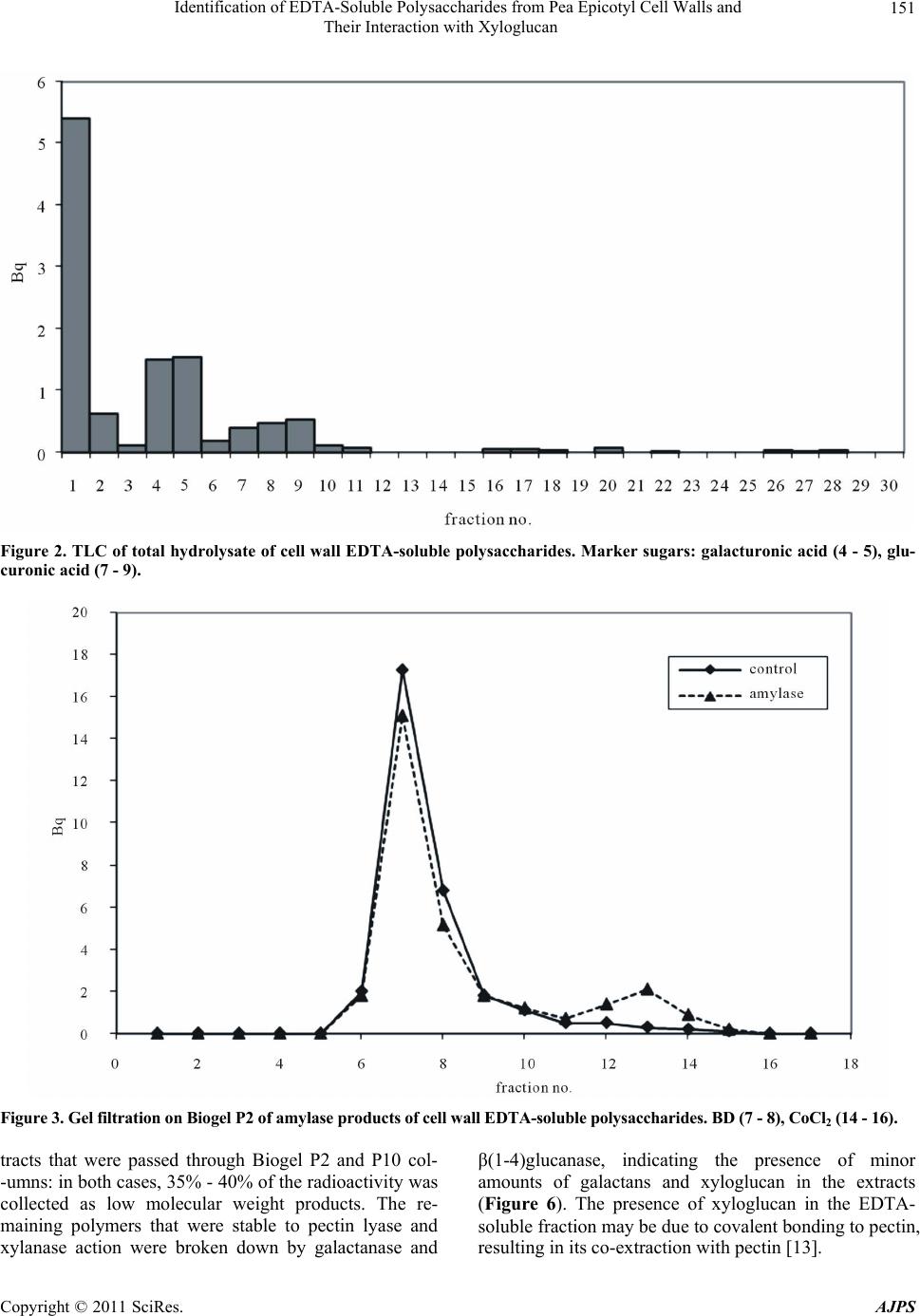 Identification of EDTA-Soluble Polysaccharides from Pea Epicotyl Cell Walls and 151 Their Interaction with Xyloglucan Figure 2. TLC of total hydrolysate of cell wall EDTA-soluble polysaccharides. Marker sugars: galacturonic acid (4 - 5), glu- curonic acid (7 - 9). Figure 3. Gel filtration on Biogel P2 of amylase products of cell wall EDTA-soluble polysaccharides. BD (7 - 8), CoCl2 (14 - 16). tracts that were passed through Biogel P2 and P10 col- -umns: in both cases, 35% - 40% of the radioactivity was collected as low molecular weight products. The re- maining polymers that were stable to pectin lyase and xylanase action were broken down by galactanase and β(1-4)glucanase, indicating the presence of minor amounts of galactans and xyloglucan in the extracts (Figure 6). The presence of xyloglucan in the EDTA- soluble fraction may be due to covalent bonding to pectin, esulting in its co-extraction with pectin [13]. r Copyright © 2011 SciRes. AJPS 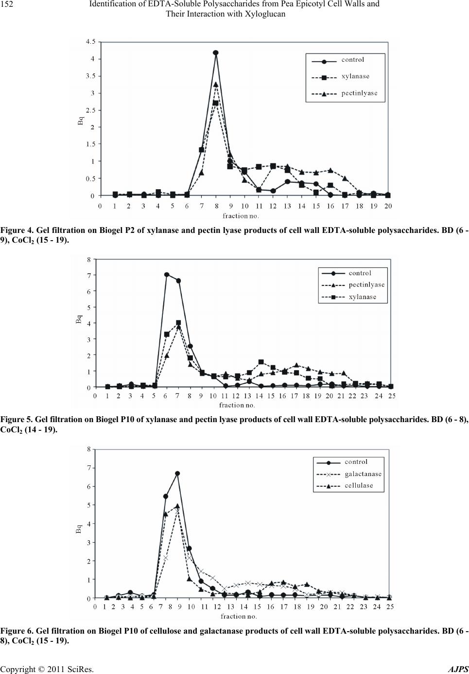 Identification of EDTA-Soluble Polysaccharides from Pea Epicotyl Cell Walls and 152 Their Interaction with Xyloglucan Figure 4. Gel filtration on Biogel P2 of xylanase and pectin lyase products of cell wall EDTA-soluble poly saccharides. BD (6 - 9), CoCl2 (15 - 19). Figure 5. Gel filtration on Biogel P10 of xylanase and pectin lyase products of cell wall EDTA-soluble polysaccharides. BD (6 - 8), CoCl2 (14 - 19). Figure 6. Gel filtration on Biogel P10 of cellulose and galactanase products of cell wall EDTA-soluble polysaccharides. BD (6 - ), CoCl2 (15 - 19). 8 Copyright © 2011 SciRes. AJPS 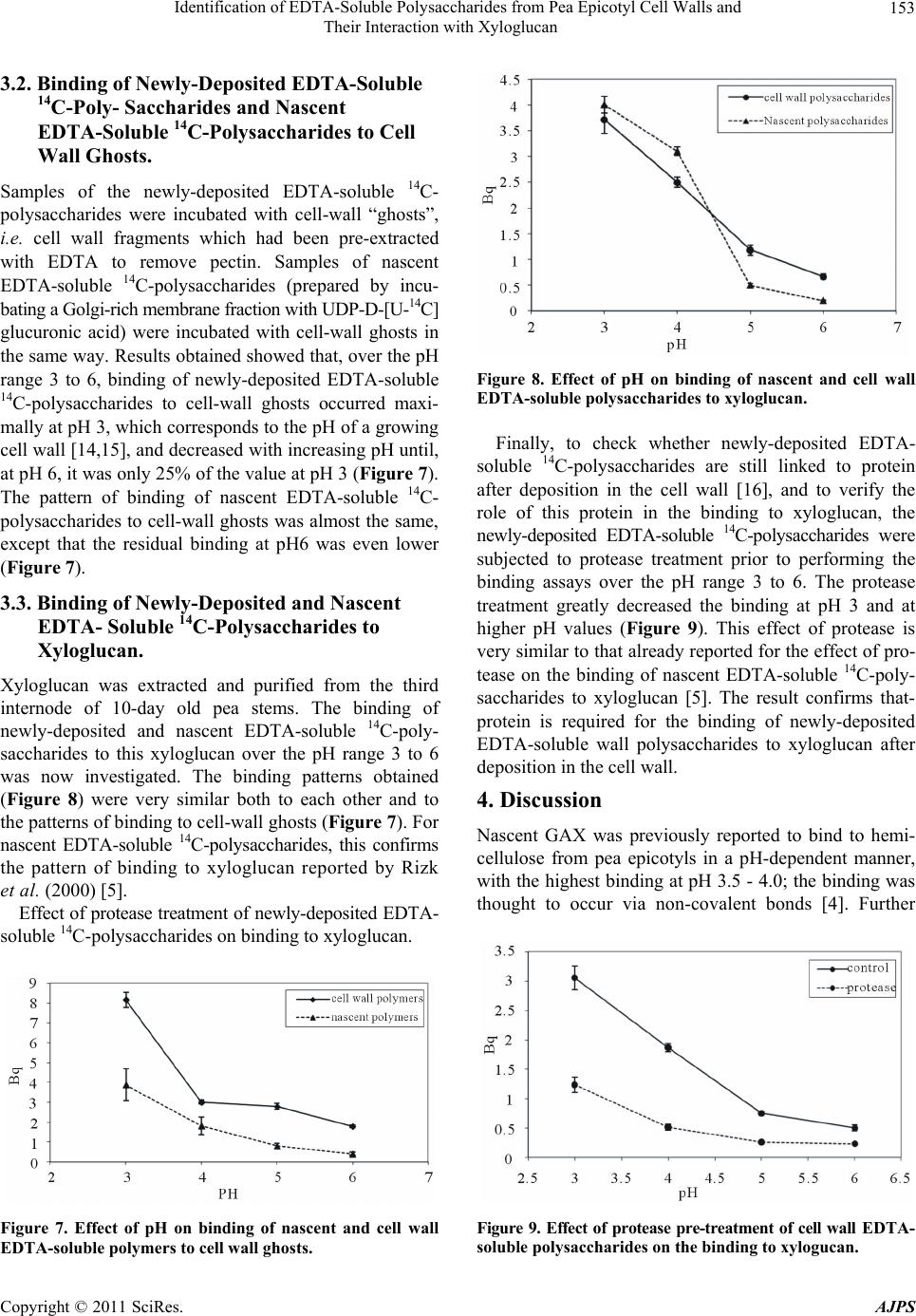 Identification of EDTA-Soluble Polysaccharides from Pea Epicotyl Cell Walls and 153 Their Interaction with Xyloglucan 3.2. Binding of Newly-Deposited EDTA-Soluble 14C-Poly- Saccharides and Nascent EDTA-Soluble 14C-Polysaccharides to Cell Wall Ghosts. Samples of the newly-deposited EDTA-soluble 14C- polysaccharides were incubated with cell-wall “ghosts”, i.e. cell wall fragments which had been pre-extracted with EDTA to remove pectin. Samples of nascent EDTA-soluble 14C-polysaccharides (prepared by incu- bating a Golgi-rich membrane fraction with UDP-D-[U-14C] glucuronic acid) were incubated with cell-wall ghosts in the same way. Results obtained showed that, over the pH range 3 to 6, binding of newly-deposited EDTA-soluble 14C-polysaccharides to cell-wall ghosts occurred maxi- mally at pH 3, which corresponds to the pH of a growing cell wall [14,15], and decreased with increasing pH until, at pH 6, it was only 25% of the value at pH 3 (Figure 7). The pattern of binding of nascent EDTA-soluble 14C- polysaccharides to cell-wall ghosts was almost the same, except that the residual binding at pH6 was even lower (Figure 7). 3.3. Binding of Newly-Deposited and Nascent EDTA- Soluble 14C-Polysaccharides to Xyloglucan. Xyloglucan was extracted and purified from the third internode of 10-day old pea stems. The binding of newly-deposited and nascent EDTA-soluble 14C-poly- saccharides to this xyloglucan over the pH range 3 to 6 was now investigated. The binding patterns obtained (Figure 8) were very similar both to each other and to the patterns of binding to cell-wall ghosts (Figure 7). For nascent EDTA-soluble 14C-polysaccharides, this confirms the pattern of binding to xyloglucan reported by Rizk et al. (2000) [5]. Effect of protease treatment of newly-deposited EDTA- soluble 14C-polysaccharides on binding to xyloglucan. Figure 7. Effect of pH on binding of nascent and cell wall EDTA-soluble polymers to cell wall ghosts. Figure 8. Effect of pH on binding of nascent and cell wall EDTA-soluble polysaccharides to xyloglucan. Finally, to check whether newly-deposited EDTA- soluble 14C-polysaccharides are still linked to protein after deposition in the cell wall [16], and to verify the role of this protein in the binding to xyloglucan, the newly-deposited EDTA-soluble 14C-polysaccharides were subjected to protease treatment prior to performing the binding assays over the pH range 3 to 6. The protease treatment greatly decreased the binding at pH 3 and at higher pH values (Figure 9). This effect of protease is very similar to that already reported for the effect of pro- tease on the binding of nascent EDTA-soluble 14C-poly- saccharides to xyloglucan [5]. The result confirms that- protein is required for the binding of newly-deposited EDTA-soluble wall polysaccharides to xyloglucan after deposition in the cell wall. 4. Discussion Nascent GAX was previously reported to bind to hemi- cellulose from pea epicotyls in a pH-dependent manner, with the highest binding at pH 3.5 - 4.0; the binding was thought to occur via non-covalent bonds [4]. Further Figure 9. Effect of protease pre-treatment of cell wall EDTA- soluble polysaccharides on the binding to xyloguc a n. Copyright © 2011 SciRes. AJPS 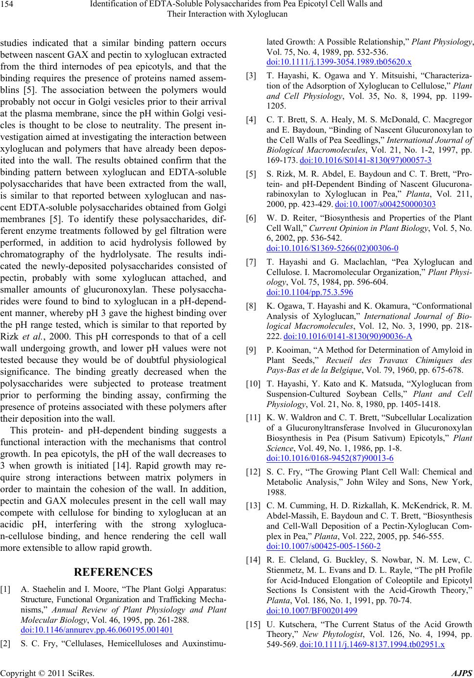 Identification of EDTA-Soluble Polysaccharides from Pea Epicotyl Cell Walls and 154 Their Interaction with Xyloglucan studies indicated that a similar binding pattern occurs between nascent GAX and pectin to xyloglucan extracted from the third internodes of pea epicotyls, and that the binding requires the presence of proteins named assem- blins [5]. The association between the polymers would probably not occur in Golgi vesicles prior to their arrival at the plasma membrane, since the pH within Golgi vesi- cles is thought to be close to neutrality. The present in- vestigation aimed at investigating the interaction between xyloglucan and polymers that have already been depos- ited into the wall. The results obtained confirm that the binding pattern between xyloglucan and EDTA-soluble polysaccharides that have been extracted from the wall, is similar to that reported between xyloglucan and nas- cent EDTA-soluble polysaccharides obtained from Golgi membranes [5]. To identify these polysaccharides, dif- ferent enzyme treatments followed by gel filtration were performed, in addition to acid hydrolysis followed by chromatography of the hydrlolysate. The results indi- cated the newly-deposited polysaccharides consisted of pectin, probably with some xyloglucan attached, and smaller amounts of glucuronoxylan. These polysaccha- rides were found to bind to xyloglucan in a pH-depend- ent manner, whereby pH 3 gave the highest binding over the pH range tested, which is similar to that reported by Rizk et al., 2000. This pH corresponds to that of a cell wall undergoing growth, and lower pH values were not tested because they would be of doubtful physiological significance. The binding greatly decreased when the polysaccharides were subjected to protease treatment prior to performing the binding assay, confirming the presence of proteins associated with these polymers after their deposition into the wall. This protein- and pH-dependent binding suggests a functional interaction with the mechanisms that control growth. In pea epicotyls, the pH of the wall decreases to 3 when growth is initiated [14]. Rapid growth may re- quire strong interactions between matrix polymers in order to maintain the cohesion of the wall. In addition, pectin and GAX molecules present in the cell wall may compete with cellulose for binding to xyloglucan at an acidic pH, interfering with the strong xylogluca- n-cellulose binding, and hence rendering the cell wall more extensible to allow rapid growth. REFERENCES [1] A. Staehelin and I. Moore, “The Plant Golgi Apparatus: Structure, Functional Organization and Trafficking Mecha- nisms,” Annual Review of Plant Physiology and Plant Molecular Bi ology, Vol. 46, 1995, pp. 261-288. doi:10.1146/annurev.pp.46.060195.001401 [2] S. C. Fry, “Cellulases, Hemicelluloses and Auxinstimu- lated Growth: A Possible Relationship,” Plant Physiology, Vol. 75, No. 4, 1989, pp. 532-536. doi:10.1111/j.1399-3054.1989.tb05620.x [3] T. Hayashi, K. Ogawa and Y. Mitsuishi, “Characteriza- tion of the Adsorption of Xyloglucan to Cellulose,” Plant and Cell Physiology, Vol. 35, No. 8, 1994, pp. 1199- 1205. [4] C. T. Brett, S. A. Healy, M. S. McDonald, C. Macgregor and E. Baydoun, “Binding of Nascent Glucuronoxylan to the Cell Walls of Pea Seedlings,” International Journal of Biological Macromolecules, Vol. 21, No. 1-2, 1997, pp. 169-173. doi:10.1016/S0141-8130(97)00057-3 [5] S. Rizk, M. R. Abdel, E. Baydoun and C. T. Brett, “Pro- tein- and pH-Dependent Binding of Nascent Glucurona- rabinoxylan to Xyloglucan in Pea,” Planta, Vol. 211, 2000, pp. 423-429. doi:10.1007/s004250000303 [6] W. D. Reiter, “Biosynthesis and Properties of the Plant Cell Wall,” Current Opinion in Plant Biology, Vol. 5, No. 6, 2002, pp. 536-542. doi:10.1016/S1369-5266(02)00306-0 [7] T. Hayashi and G. Maclachlan, “Pea Xyloglucan and Cellulose. I. Macromolecular Organization,” Plant Physi- ology, Vol. 75, 1984, pp. 596-604. doi:10.1104/pp.75.3.596 [8] K. Ogawa, T. Hayashi and K. Okamura, “Conformational Analysis of Xyloglucan,” International Journal of Bio- logical Macromolecules, Vol. 12, No. 3, 1990, pp. 218- 222. doi:10.1016/0141-8130(90)90036-A [9] P. Kooiman, “A Method for Determination of Amyloid in Plant Seeds,” Recueil des Travaux Chimiques des Pays-Bas et de la Belgique, Vol. 79, 1960, pp. 675-678. [10] T. Hayashi, Y. Kato and K. Matsuda, “Xyloglucan from Suspension-Cultured Soybean Cells,” Plant and Cell Physiology, Vol. 21, No. 8, 1980, pp. 1405-1418. [11] K. W. Waldron and C. T. Brett, “Subcellular Localization of a Glucuronyltransferase Involved in Glucuronoxylan Biosynthesis in Pea (Pisum Sativum) Epicotyls,” Plant Science, Vol. 49, No. 1, 1986, pp. 1-8. doi:10.1016/0168-9452(87)90013-6 [12] S. C. Fry, “The Growing Plant Cell Wall: Chemical and Metabolic Analysis,” John Wiley and Sons, New York, 1988. [13] C. M. Cumming, H. D. Rizkallah, K. McKendrick, R. M. Abdel-Massih, E. Baydoun and C. T. Brett, “Biosynthesis and Cell-Wall Deposition of a Pectin-Xyloglucan Com- plex in Pea,” Planta, Vol. 222, 2005, pp. 546-555. doi:10.1007/s00425-005-1560-2 [14] R. E. Cleland, G. Buckley, S. Nowbar, N. M. Lew, C. Stienmetz, M. L. Evans and D. L. Rayle, “The pH Profile for Acid-Induced Elongation of Coleoptile and Epicotyl Sections Is Consistent with the Acid-Growth Theory,” Planta, Vol. 186, No. 1, 1991, pp. 70-74. doi:10.1007/BF00201499 [15] U. Kutschera, “The Current Status of the Acid Growth Theory,” New Phytologist, Vol. 126, No. 4, 1994, pp. 549-569. doi:10.1111/j.1469-8137.1994.tb02951.x Copyright © 2011 SciRes. AJPS 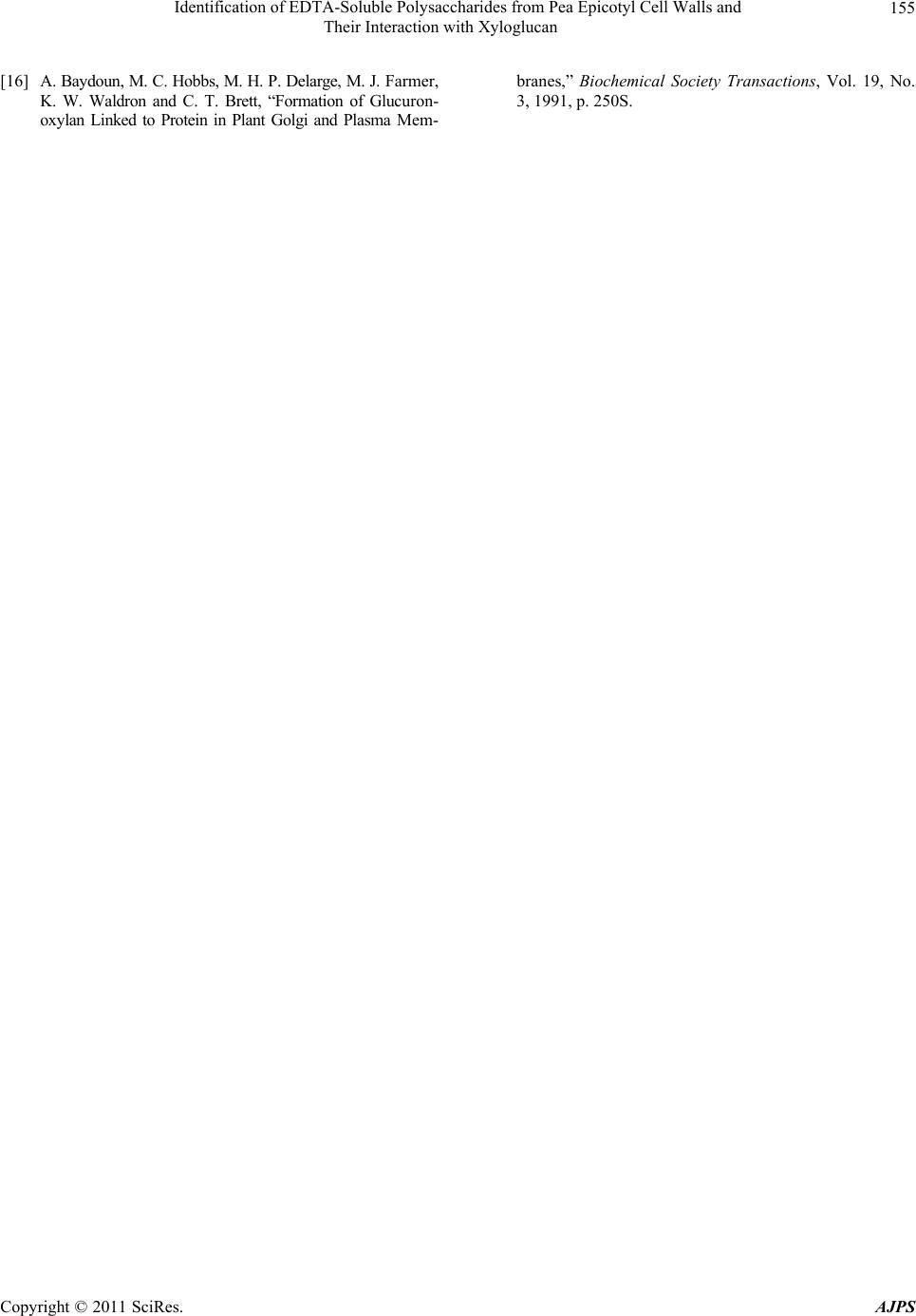 Identification of EDTA-Soluble Polysaccharides from Pea Epicotyl Cell Walls and Their Interaction with Xyloglucan Copyright © 2011 SciRes. AJPS 155 [16] A. Baydoun, M. C. Hobbs, M. H. P. Delarge, M. J. Farmer, K. W. Waldron and C. T. Brett, “Formation of Glucuron- oxylan Linked to Protein in Plant Golgi and Plasma Mem- branes,” Biochemical Society Transactions, Vol. 19, No. 3, 1991, p. 250S. |