

Paper Menu >>
Journal Menu >>
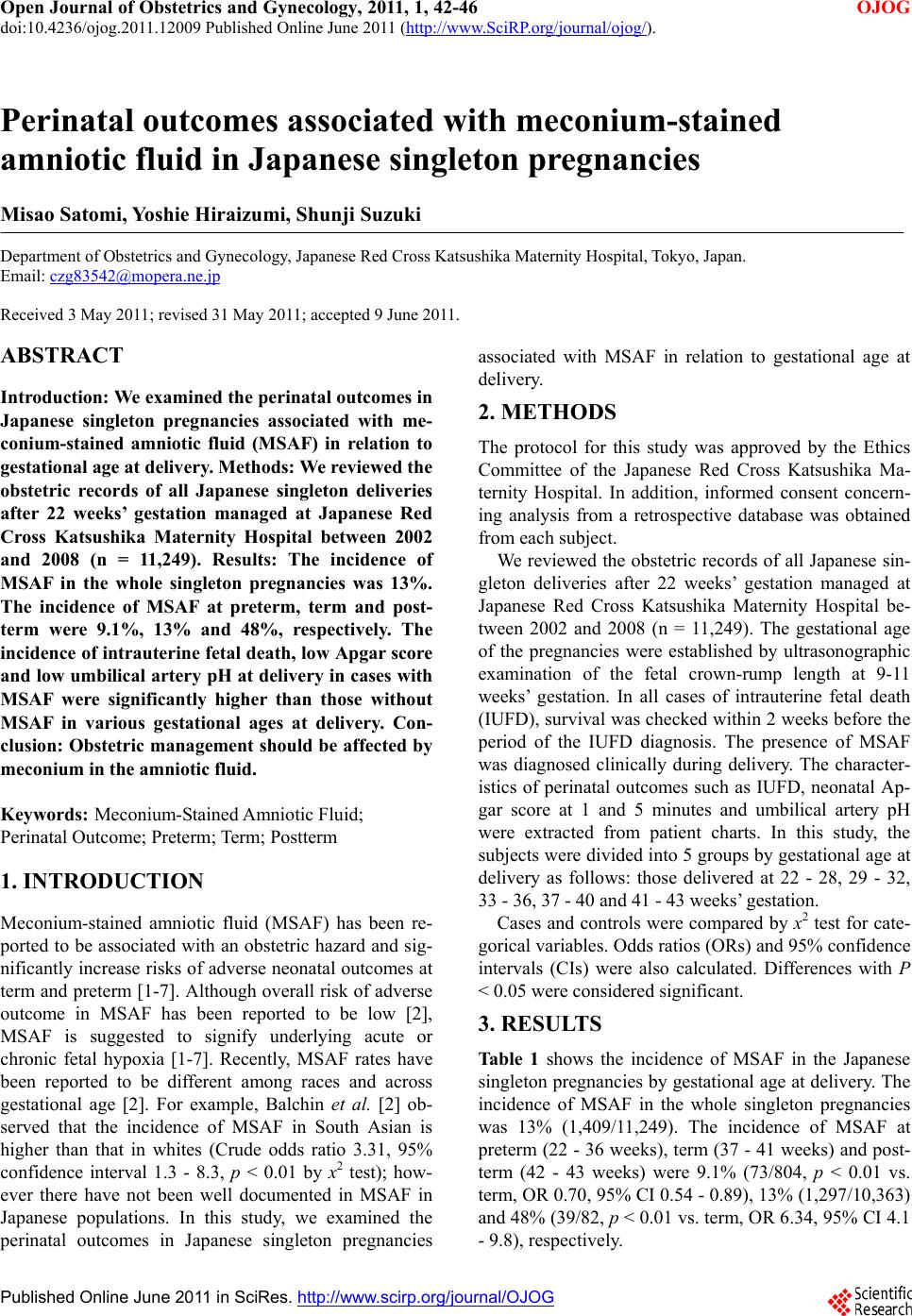 Open Journal of Obstetrics and Gynecology, 2011, 1, 42-46 doi:10.4236/ojog.2011.12009 Published Online June 2011 (http://www.SciRP.org/journal/ojog/ OJOG ). Published Online June 2011 in SciRes. http://www.scirp.org/journal/OJOG Perinatal outcomes associated with meconium-stained amniotic fluid in Japanese singleton pregnancies Misao Satomi, Yoshie Hiraizumi, Shunji Suzuki Department of Obstetrics and Gynecology, Japanese Red Cross Katsushika Maternity Hospital, Tokyo, Japan. Email: [email protected] Received 3 May 2011; revised 31 May 2011; accepted 9 June 2011. ABSTRACT Introduction: We examined the perinatal outcomes in Japanese singleton pregnancies associated with me- conium-stained amniotic fluid (MSAF) in relation to gestational age at delivery. Methods: We reviewed the obstetric records of all Japanese singleton deliveries after 22 weeks’ gestation managed at Japanese Red Cross Katsushika Maternity Hospital between 2002 and 2008 (n = 11,249). Results: The incidence of MSAF in the whole singleton pregnancies was 13%. The incidence of MSAF at preterm, term and post- term were 9.1%, 13% and 48%, respectively. The incidence of intrauterine fetal death, low Apgar score and low umbilical artery pH at delivery in cases with MSAF were significantly higher than those without MSAF in various gestational ages at delivery. Con- clusio n: Obstetric management should be affected by meconium in the amniotic fluid. Keywords: Meconium-Stained Amniotic Fluid; Perinatal Outcome; Preterm; Term; Postterm 1. INTRODUCTION Meconium-stained amniotic fluid (MSAF) has been re- ported to be associated with an obstetric hazard and sig- nificantly increase risks of adverse neonatal outcomes at term and preterm [1-7]. Although overall risk of adverse outcome in MSAF has been reported to be low [2], MSAF is suggested to signify underlying acute or chronic fetal hypoxia [1-7]. Recently, MSAF rates have been reported to be different among races and across gestational age [2]. For example, Balchin et al. [2] ob- served that the incidence of MSAF in South Asian is higher than that in whites (Crude odds ratio 3.31, 95% confidence interval 1.3 - 8.3, p < 0.01 by x2 test); how- ever there have not been well documented in MSAF in Japanese populations. In this study, we examined the perinatal outcomes in Japanese singleton pregnancies associated with MSAF in relation to gestational age at delivery. 2. METHODS The protocol for this study was approved by the Ethics Committee of the Japanese Red Cross Katsushika Ma- ternity Hospital. In addition, informed consent concern- ing analysis from a retrospective database was obtained from each subject. We reviewed the obstetric records of all Japanese sin- gleton deliveries after 22 weeks’ gestation managed at Japanese Red Cross Katsushika Maternity Hospital be- tween 2002 and 2008 (n = 11,249). The gestational age of the pregnancies were established by ultrasonographic examination of the fetal crown-rump length at 9-11 weeks’ gestation. In all cases of intrauterine fetal death (IUFD), survival was checked within 2 weeks before the period of the IUFD diagnosis. The presence of MSAF was diagnosed clinically during delivery. The character- istics of perinatal outcomes such as IUFD, neonatal Ap- gar score at 1 and 5 minutes and umbilical artery pH were extracted from patient charts. In this study, the subjects were divided into 5 groups by gestational age at delivery as follows: those delivered at 22 - 28, 29 - 32, 33 - 36, 37 - 40 and 41 - 43 weeks’ gestation. Cases and controls were compared by x2 test for cate- gorical variables. Odds ratios (ORs) and 95% confidence intervals (CIs) were also calculated. Differences with P < 0.05 were considered significant. 3. RESULTS Table 1 shows the incidence of MSAF in the Japanese singleton pregnancies by gestational age at delivery. The incidence of MSAF in the whole singleton pregnancies was 13% (1,409/11,249). The incidence of MSAF at preterm (22 - 36 weeks), term (37 - 41 weeks) and post- term (42 - 43 weeks) were 9.1% (73/804, p < 0.01 vs. term, OR 0.70, 95% CI 0.54 - 0.89), 13% (1,297/10,363) and 48% (39/82, p < 0.01 vs. term, OR 6.34, 95% CI 4.1 - 9.8), respectively. 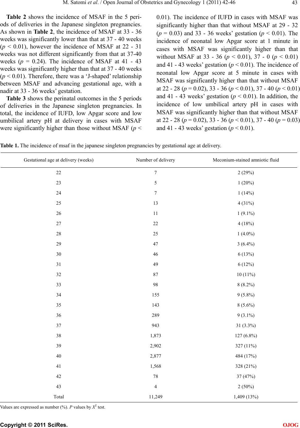 M. Satomi et al. / Open Journal of Obstetrics and Gynecology 1 (2011) 42-46 43 Ta ble 2 shows the incidence of MSAF in the 5 peri- ods of deliveries in the Japanese singleton pregnancies. As shown in Table 2, the incidence of MSAF at 33 - 36 weeks was significantly lower than that at 37 - 40 weeks (p < 0.01), however the incidence of MSAF at 22 - 31 weeks was not different significantly from that at 37-40 weeks (p = 0.24). The incidence of MSAF at 41 - 43 weeks was significantly higher than that at 37 - 40 weeks (p < 0.01). Therefore, there was a ‘J-shaped’ relationship between MSAF and advancing gestational age, with a nadir at 33 - 36 weeks’ gestation. Table 3 shows the perinatal outcomes in the 5 periods of deliveries in the Japanese singleton pregnancies. In total, the incidence of IUFD, low Apgar score and low umbilical artery pH at delivery in cases with MSAF were significantly higher than those without MSAF (p < 0.01). The incidence of IUFD in cases with MSAF was significantly higher than that without MSAF at 29 - 32 (p = 0.03) and 33 - 36 weeks’ gestation (p < 0.01). The incidence of neonatal low Apgar score at 1 minute in cases with MSAF was significantly higher than that without MSAF at 33 - 36 (p < 0.01), 37 - 0 (p < 0.01) and 41 - 43 weeks’ gestation (p < 0.01). The incidence of neonatal low Apgar score at 5 minute in cases with MSAF was significantly higher than that without MSAF at 22 - 28 (p = 0.02), 33 - 36 (p < 0.01), 37 - 40 (p < 0.01) and 41 - 43 weeks’ gestation (p < 0.01). In addition, the incidence of low umbilical artery pH in cases with MSAF was significantly higher than that without MSAF at 22 - 28 (p = 0.02), 33 - 36 (p < 0.01), 37 - 40 (p = 0.03) and 41 - 43 weeks’ gestation (p < 0.01). Table 1. The incidence of msaf in the japanese singleton pregnancies by gestational age at delivery. Gestational age at delivery (weeks) Number of delivery Meconium-stained amniotic fluid 22 7 2 (29%) 23 5 1 (20%) 24 7 1 (14%) 25 13 4 (31%) 26 11 1 (9.1%) 27 22 4 (18%) 28 25 1 (4.0%) 29 47 3 (6.4%) 30 46 6 (13%) 31 49 6 (12%) 32 87 10 (11%) 33 98 8 (8.2%) 34 155 9 (5.8%) 35 143 8 (5.6%) 36 289 9 (3.1%) 37 943 31 (3.3%) 38 1,873 127 (6.8%) 39 2,902 327 (11%) 40 2,877 484 (17%) 41 1,568 328 (21%) 42 78 37 (47%) 43 4 2 (50%) Total 11,249 1,409 (13%) Values are expressed as number (%). P values by Χ2 test. C opyright © 2011 SciRes. OJOG 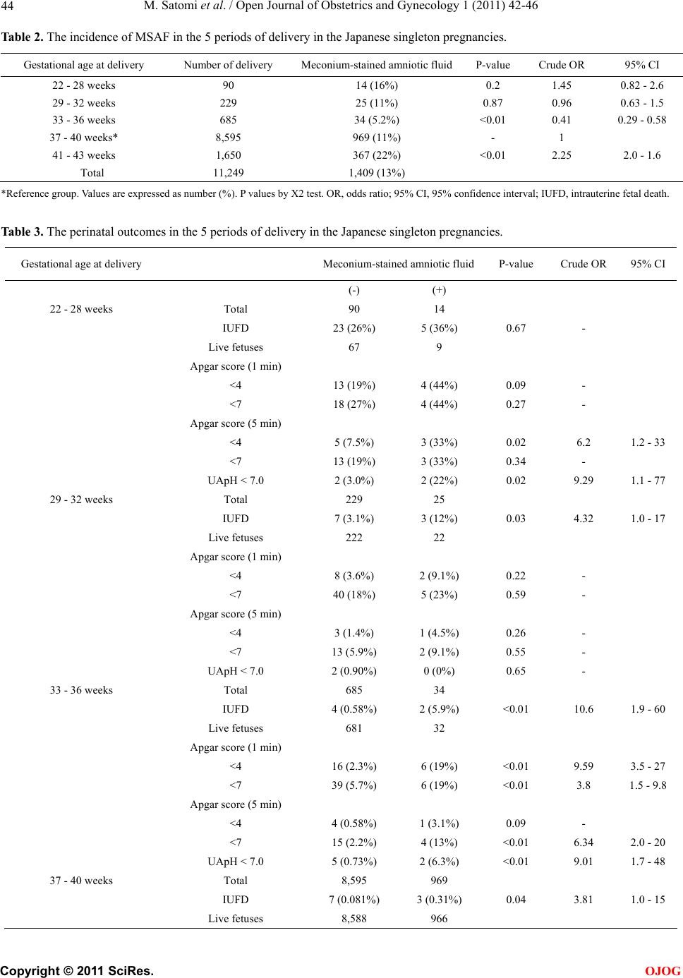 M. Satomi et al. / Open Journal of Obstetrics and Gynecology 1 (2011) 42-46 44 Table 2. The incidence of MSAF in the 5 periods of delivery in the Japanese singleton pregnancies. Gestational age at delivery Number of delivery Meconium-stained amniotic fluid P-value Crude OR 95% CI 22 - 28 weeks 90 14 (16%) 0.2 1.45 0.82 - 2.6 29 - 32 weeks 229 25 (11%) 0.87 0.96 0.63 - 1.5 33 - 36 weeks 685 34 (5.2%) <0.01 0.41 0.29 - 0.58 37 - 40 weeks* 8,595 969 (11%) - 1 41 - 43 weeks 1,650 367 (22%) <0.01 2.25 2.0 - 1.6 Total 11,249 1,409 (13%) *Reference group. Values are expressed as number (%). P values by Χ2 test. OR, odds ratio; 95% CI, 95% confidence interval; IUFD, intrauterine fetal death. Table 3. The perinatal outcomes in the 5 periods of delivery in the Japanese singleton pregnancies. Gestational age at delivery Meconium-stained amniotic fluidP-value Crude OR 95% CI (-) (+) 22 - 28 weeks Total 90 14 IUFD 23 (26%) 5 (36%) 0.67 - Live fetuses 67 9 Apgar score (1 min) <4 13 (19%) 4 (44%) 0.09 - <7 18 (27%) 4 (44%) 0.27 - Apgar score (5 min) <4 5 (7.5%) 3 (33%) 0.02 6.2 1.2 - 33 <7 13 (19%) 3 (33%) 0.34 - UApH < 7.0 2 (3.0%) 2 (22%) 0.02 9.29 1.1 - 77 29 - 32 weeks Total 229 25 IUFD 7 (3.1%) 3 (12%) 0.03 4.32 1.0 - 17 Live fetuses 222 22 Apgar score (1 min) <4 8 (3.6%) 2 (9.1%) 0.22 - <7 40 (18%) 5 (23%) 0.59 - Apgar score (5 min) <4 3 (1.4%) 1 (4.5%) 0.26 - <7 13 (5.9%) 2 (9.1%) 0.55 - UApH < 7.0 2 (0.90%) 0 (0%) 0.65 - 33 - 36 weeks Total 685 34 IUFD 4 (0.58%) 2 (5.9%) <0.01 10.6 1.9 - 60 Live fetuses 681 32 Apgar score (1 min) <4 16 (2.3%) 6 (19%) <0.01 9.59 3.5 - 27 <7 39 (5.7%) 6 (19%) <0.01 3.8 1.5 - 9.8 Apgar score (5 min) <4 4 (0.58%) 1 (3.1%) 0.09 - <7 15 (2.2%) 4 (13%) <0.01 6.34 2.0 - 20 UApH < 7.0 5 (0.73%) 2 (6.3%) <0.01 9.01 1.7 - 48 37 - 40 weeks Total 8,595 969 IUFD 7 (0.081%) 3 (0.31%) 0.04 3.81 1.0 - 15 Live fetuses 8,588 966 C opyright © 2011 SciRes. OJOG 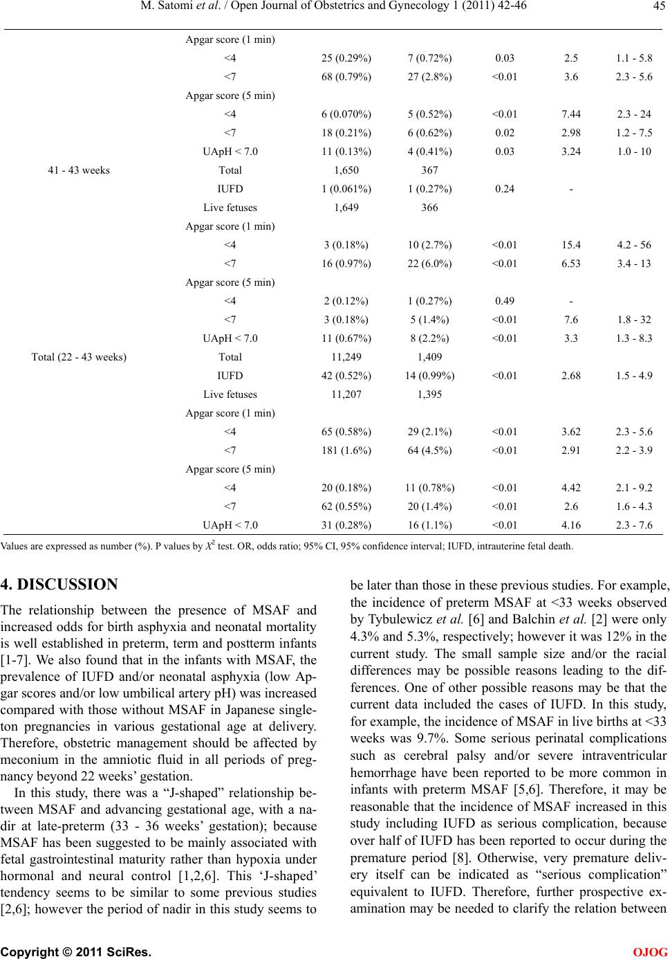 M. Satomi et al. / Open Journal of Obstetrics and Gynecology 1 (2011) 42-46 Copyright © 2011 SciRes. 45 OJOG Apgar score (1 min) <4 25 (0.29%) 7 (0.72%) 0.03 2.5 1.1 - 5.8 <7 68 (0.79%) 27 (2.8%) <0.01 3.6 2.3 - 5.6 Apgar score (5 min) <4 6 (0.070%) 5 (0.52%) <0.01 7.44 2.3 - 24 <7 18 (0.21%) 6 (0.62%) 0.02 2.98 1.2 - 7.5 UApH < 7.0 11 (0.13%) 4 (0.41%) 0.03 3.24 1.0 - 10 41 - 43 weeks Total 1,650 367 IUFD 1 (0.061%) 1 (0.27%) 0.24 - Live fetuses 1,649 366 Apgar score (1 min) <4 3 (0.18%) 10 (2.7%) <0.01 15.4 4.2 - 56 <7 16 (0.97%) 22 (6.0%) <0.01 6.53 3.4 - 13 Apgar score (5 min) <4 2 (0.12%) 1 (0.27%) 0.49 - <7 3 (0.18%) 5 (1.4%) <0.01 7.6 1.8 - 32 UApH < 7.0 11 (0.67%) 8 (2.2%) <0.01 3.3 1.3 - 8.3 Total (22 - 43 weeks) Total 11,249 1,409 IUFD 42 (0.52%) 14 (0.99%) <0.01 2.68 1.5 - 4.9 Live fetuses 11,207 1,395 Apgar score (1 min) <4 65 (0.58%) 29 (2.1%) <0.01 3.62 2.3 - 5.6 <7 181 (1.6%) 64 (4.5%) <0.01 2.91 2.2 - 3.9 Apgar score (5 min) <4 20 (0.18%) 11 (0.78%) <0.01 4.42 2.1 - 9.2 <7 62 (0.55%) 20 (1.4%) <0.01 2.6 1.6 - 4.3 UApH < 7.0 31 (0.28%) 16 (1.1%) <0.01 4.16 2.3 - 7.6 Values are expressed as number (%). P values by Χ2 test. OR, odds ratio; 95% CI, 95% confidence interval; IUFD, intrauterine fetal death. 4. DISCUSSION The relationship between the presence of MSAF and increased odds for birth asphyxia and neonatal mortality is well established in preterm, term and postterm infants [1-7]. We also found that in the infants with MSAF, the prevalence of IUFD and/or neonatal asphyxia (low Ap- gar scores and/or low umbilical artery pH) was increased compared with those without MSAF in Japanese single- ton pregnancies in various gestational age at delivery. Therefore, obstetric management should be affected by meconium in the amniotic fluid in all periods of preg- nancy beyond 22 weeks’ gestation. In this study, there was a “J-shaped” relationship be- tween MSAF and advancing gestational age, with a na- dir at late-preterm (33 - 36 weeks’ gestation); because MSAF has been suggested to be mainly associated with fetal gastrointestinal maturity rather than hypoxia under hormonal and neural control [1,2,6]. This ‘J-shaped’ tendency seems to be similar to some previous studies [2,6]; however the period of nadir in this study seems to be later than those in these previous studies. For example, the incidence of preterm MSAF at <33 weeks observed by Tybulewicz et al. [6] and Balchin et al. [2] were only 4.3% and 5.3%, respectively; however it was 12% in the current study. The small sample size and/or the racial differences may be possible reasons leading to the dif- ferences. One of other possible reasons may be that the current data included the cases of IUFD. In this study, for example, the incidence of MSAF in live births at <33 weeks was 9.7%. Some serious perinatal complications such as cerebral palsy and/or severe intraventricular hemorrhage have been reported to be more common in infants with preterm MSAF [5,6]. Therefore, it may be reasonable that the incidence of MSAF increased in this study including IUFD as serious complication, because over half of IUFD has been reported to occur during the premature period [8]. Otherwise, very premature deliv- ery itself can be indicated as “serious complication” equivalent to IUFD. Therefore, further prospective ex- amination may be needed to clarify the relation between  M. Satomi et al. / Open Journal of Obstetrics and Gynecology 1 (2011) 42-46 46 MSAF and perinatal outcomes at very preterm. At 41 - 43 weeks’ gestation, on the other hand, the in- cidence of low Apgar score and low umbilical artery pH in cases with MSAF were also significantly higher than those without MSAF. Postterm gestation itself has been suggested to be associated with the increased risk of perinatal mortality [9]. The higher rate of perinatal mor- bidity at postterm gestation may be due to hypoxia/ acidemia associated with “relative placental insuffi- ciency” where the placenta can no longer keep up the demands of the fetus [10]. These conditions may be also associated with the presence of MSAF. In addition, when MSAF is superimposed on fetal acidemia, there is an increased risk of meconium aspiration syndrome [4]. Therefore, the greater risk of adverse neonatal outcomes at ≥41 weeks in the current study support these previous suggestions especially in cases with MSAF [4,10]. In conclusion, obstetric management should be af- fected by meconium in the amniotic fluid in various gestational ages at delivery. Therefore, management re- quires awareness of this potential risk, appropriate in- trapartum care and a combined obstetric-neonatal ap- proach in cases with MSAF. REFERENCES [1] Ash, A.K. (2000) Managing patients with meconium- stained amniotic fluid. Hospital Medicine, 61, 844-848. [2] Balchin, I., Whittaker, J.C., Lamont, R.F. and Steer, P.J. (2011) Maternal and fetal characteristics associated with meconium-stained amniotic fluid. Obstetrics & Gyne- cology, 117, 828-835. doi:10.1097/AOG.0b013e3182117a26 [3] Natah, L., Leveno, K.J., Carmody III, T.J., Kelly, M.A. and Sherman, M.L. (1994) Meconium: A 1990s perspec- tive on an old obstetric hazard. Obstetrics & Gynecology, 83, 329-332. [4] Ramin, K.D., Leveno, K.J., Kelly, M.A. and Carmody, T.J. (1996) Amniotic fluid meconium: A fetal environ- mental hazard. Obstetrics & Gynecology, 87, 181-184. doi:10.1016/0029-7844(95)00403-3 [5] Spinillo, A., Fazzi, E., Capuzzo, E., Stronati, M., Piazzi, G. and Ferrari, A. (1997) Meconium-stained amniotic fluid and risk for cerebral palsy in preterm infants. Ob- stetrics & Gynecology, 90, 519-523. doi:10.1016/S0029-7844(97)00308-6 [6] Tybulewicz, A.T., Clegg, S.K., Fonfe, G.J. and Stenson, B.J. (2004) Preterm meconium staining of the amniotic fluid: associated findings and risk of adverse clinical outcome. Archives of Disease in Childhood—Fetal & Neonatal Edition, 89, F328-F330. doi:10.1136/adc.2002.021949 [7] Ziadeh, S.M. and Sunna, E. (2000) Obstetric and perina- tal outcome of pregnancies with term labour and me- conium-stained amniotic fluid. Archives of Gynecology and Obstetrics, 264, 84-87. doi:10.1007/s004040000088 [8] Gardosi, J., Mul, T., Mongelli, M. and Fagan, D. (1998) Analysis of birthweight and gestational age in antepar- tum stillbirths. British Journal of Obstetrics and Gyne- cology, 105, 524-530. doi:10.1111/j.1471-0528.1998.tb10153.x [9] Suzuki, S. (2009) Post-term pregnancy as a risk factor for intrapartum fetal distress. Journal of Perinatal Medicine, 37, 420-421. doi:10.1515/JPM.2009.055 [10] Volherr, H. (1975) Placental insufficiency in relation to postterm pregnancy and fetal postmaturity: Evaluation of fetoplacental function; management of the postterm gra- vida. American Journal of Obstetrics & Gynecology, 123, 67-103. C opyright © 2011 SciRes. OJOG |