

Paper Menu >>
Journal Menu >>
 Open Journal of Obstetrics and Gynecology, 2011, 1, 55-63 doi:10.4236/ojog.2011.12012 Published Online June 2011 (http://www.SciRP.org/journal/ojog/ OJOG ). Published Online June 2011 in SciRes. http://www.scirp.org/journal/OJOG Preoperative evaluation of P53 and bcl-2 over expression in clinical stage 1 endometrial carcinoma and their correlation with surgico-pathological data and prognosis of patients Emad A. Fyallah1, Reda A. Hemida1, Kamal I. An war 1, Nada A. Nadia2, Lotfy S. Sherif1, Mohammad T. Sayed-Ahmed1 1Departments of Obstetrics and Gynecology, Mansoura University, Nile Delta, Egypt; 2Pathology, Faculty of Medicine, Mansoura University, Nile Delta, Egypt. Email: [email protected] Received 29 April 2011; revised 26 May 2011; accepted 3 June 2011. ABSTRACT Introduction: P53 and bcl-2 over expression was re- ported to affect the biology and prognosis of patients endometrial carcinoma. Methods: This prospective study included 38 patients with histologically con- firmed endometrial carcinoma and staged clinically as stage I. Immunohistochemical staining of the tu- mor specimens obtained by dilatation and curettage (D&C) with P53 an d bcl-2 mono clonal antibodies was done. The surgical, pathological and follow up data of all patients were studied. Results: There were 18 cases (47.4%) with positive P53 over expression. P53 over expression was found to be associated with in- creasing grade of differentiation, advanced stage, and non endometrioid type (significant), and increased depth of myometrial invasion (non-significant). It was associated with more recurrence rate (22% ver- sus 15%) and shorter mean survival time (33.9 versus 29.2 months). Bcl-2 expression was present in24 cases (63.2%) of the studied group. There was significant decrease in bcl-2expression with poorly differentiated, advanced stage, increased depth of myometrial inva- sion, and non-endometrioid type. It was not signifi- cantly correlated with recurrence rate and mean sur- vival time. Conclusion: P53 over expression in the D&C specimens was associated with adverse surgi- copathological criteria, increased mortality rate, and shorter survival time in patients with endometrial carcinoma. A significant decrease in bcl-2 expression was associated with adverse surgicopathological cri- teria, but it was not significantly correlated with prognosis of the patients. Keywords: Endometrial Carcinoma; Prognosis; P53; Bcl-2 1. INTRODUCTION Endometrial carcinoma is the most common malignancy of the female genital tract accounting for almost half of all gynecologic cancers in western world. It represents the fourth most common cancer after breast, lungs and bowel cancers, and the seventh leading cause of death from malignancy in women [1]. Mariani et al. [2], reported that in the pretreatment curettage specimen, the presence of unfavorable level of P53 and bcl-2 or non endometrioid histological feature or combination of those, can significantly predict lymph node status in patients with endometrial carcinoma. P53 gene mutation was found to affect the biological features of endometrial carcinoma, as it was found to be associated with more aggressive histologic subtypes than endometrioid carcinoma. Strong expression of p53 cor- relates with advanced stage and high grade of the tumor and was detected more frequently in endometrial cancer with lympho-vascular space invasion [3]. Bcl-2 belongs to a family of apoptosis-regulatory genes which may either promote cell survival or en- courage cell death [4]. Expression of bcl-2 does not only contribute to oncogenesis but also to chemotherapy re- sistance in variety of tumors by inhibiting apoptosis [5]. Bcl-2 expression was found to be increased in grade 1 and 2 endometrioid adenocarcinoma, while in the serous papillary endometrial cancer showed immuno-nega- tivity to bcl-2 [6]. Bcl-2 may have an importance in the progression of endometrial carcinoma [7]. To the best of our knowledge, there are few reported studies to evaluate the correlation of pre-operative test- ing of P53 and bcl-2 expression with the surgico-patho-  E. A. Fyallah et al. / Open Journal of Obstetrics and Gynecology 1 (2011) 55-63 56 logical data and prognosis of patients in clinical stage 1 endometrial carcinoma. We tried to evaluate th is point in this prospective clinical study. 2. PATIENTS AND METHODS This prospective clinical study was conducted at the de- partments of Gynecology and Pathology, Faculty of Medicine, during the period from April 2005 to October 2008. The study included 38 patients with histologically confirmed endometrial carcinoma and staged clinically as stage I. The routine pre-operative work up of these patients was done. Dilatation and curettage was done, a part of the biopsy was prepared in paraffin sections for his- tologic examination and another part was used for im- munohistochemical staining with P53 and bcl-2 mono- clonal antibodies. P53 immune staining: Immunohistochemical reaction was carried out for detection of P53 protein over expres- sion in endometrial tumor tissue using monoclonal mouse anti-human P53 protein, clone; DO-7, supplied by DAKO corporation, USA. Positive staining was de- fined as homogenous pattern of nuclear staining (brow- nish coloration) that involved more than 5% of the cells. Bcl-2 immunohistochem ical staining: The primary an- tibody used was the second generation monoclonal mouse anti-bcl-2 protein (Biogenex, Cat no. Am 287). Using the high power field, the immunoreactivity (posi- tive cases) for bcl-2 was determined by the percentage of tumor cells showing cytoplasmic (brown) staining that involved more than 5% of the cells. After that, patients were subjected to surgical treat- ment, in which peritoneal cytology, extra-fascial hyster- ectomy, bilateral salpingo-oophrectomy and pelvic lymph node sampling were done. The use of adjuvant radiotherapy was individualized in the tumor board meetings .Candidates of postopera- tive radiotherapy were suggested by use of prognostic factors such as surgical staging, depth of myometrial invasion, histologic type and tumor grading. Follow up: Patients were followed up every three months, by history, physical examinations, and ultra- sound. MRI was done every 6 months. The surgico- pa- thological data of all patients were studied. Overall sur- vival, recurrence rate and duration were estimated. 2.1. Statistical Analysis Statistical analysis was done by using SPSS (Statistical package of social science) program version 10, 1999. The data were parametric by using Kolmogrov-Smirnov test. The qualitative data were presented in the form of means, standard deviation and range. Student test was used for comparison of the two groups. One way ANO- VA test was used to compare more than two groups. Man-Whitney test was used to compare the non-para- metric data. Sensitivity, specificity and accuracy were calculated. Kaplan-Meier survival analysis was done to calculate the cumulative survival. Significance was con- sidered when P value is less than 0.05. 2.2. Ethical Considerations The research was approved by The Ethical Review Committee of our Faculty of Medicine. Formal and written consents were taken from all patients. 3. RESULTS This study included 38 cases of histologically confirmed endometrial carcinoma, clinical stage I, during the pe- riod from April 2005 to October 2008. The mean follow up duration from the treatment was 21.5 month, (range 6 - 40 months). After surgical staging (according to FIGO staging 1988), there were 30 cases with stage I, 4 cases with stage II and 4 cases with stage III. Pretreatment study of P53 over expression by “Im- muno-histochemical stain” was done for the studied group; there were 18 cases (47.4%) with positive P53 over expression. P53 over expression was found to be associated with increasing grade of differentiation, advanced stage, and non endometrioid type (significant), and increased depth of myometrial invasion (non-significant) as can be con- cluded fr om Table 1. The relation between P53 over expression and recur- rence of the disease in the study group: Although the recurrence of the disease in patients with negative P53 over expression was 15% (3 of 20 cases), in the po sitive group it was 22 % (4 of 18 cases) but this d ifference was not statistically significant (P value 0.576). The survival time was prolonged in P53 negative pa- tients, as the mean survival time in p53 negative group was 33.9 months while it was 29.21 months in p53 posi- tive group (Table 2). Figure 1: Kaplan-Meier estimates the influence of P53 over expression in the recurrence of the studied group. There was difference in the mean recurrence du- ration in both groups. The relation between P53 over expression and mortal- ity of the studied group is shown in Table 3, the mortal- ity between P53 po s itive patients was higher 16.6% (3 of 18 cases) in comparison to the mortality between nega- tive patients, which was 5% (1 of 20 cases) the differ- ence is significant (P value 0.026). Figure 2: Kaplan-Meier estimates the influence of P53 over expression in the mortality duration of the stu- died group: There was difference in the mean mortality duration in both groups. It was shorter in positive group. Bcl-2 expression was studied also, bcl-2 expression C opyright © 2011 SciRes. OJOG 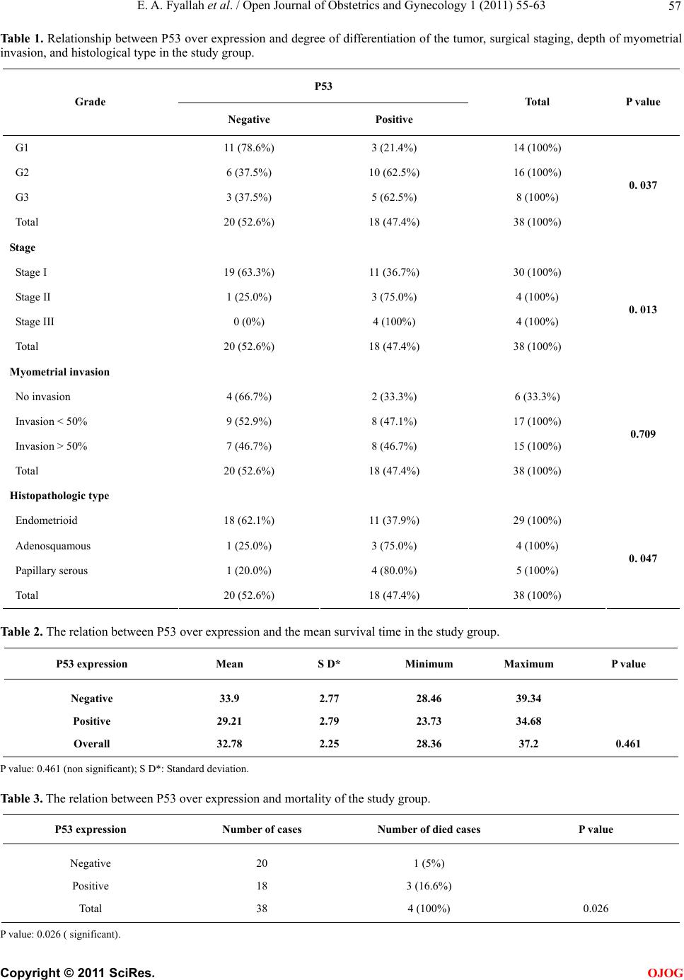 E. A. Fyallah et al. / Open Journal of Obstetrics and Gynecology 1 (2011) 55-63 Copyright © 2011 SciRes. 57 OJOG Table 1 . Relationship between P53 over expression and degree of differentiation of the tumor, surgical staging, depth of myometrial invasion, and histological type in the study group. P53 Grade Negative Positive Total P value G1 11 (78.6%) 3 (21.4%) 14 (100%) G2 6 (37.5%) 10 (62.5%) 16 (100%) G3 3 (37.5%) 5 (62.5%) 8 ( 10 0%) Total 20 (52.6%) 18 (47.4%) 38 (100%) 0. 037 Stage Stage I 19 (63.3%) 11 (36.7%) 30 (100%) Stage II 1 (25.0%) 3 (75.0%) 4 (100%) Stage III 0 (0%) 4 (100%) 4 (100%) Total 20 (52.6%) 18 (47.4%) 38 (100%) 0. 013 Myometria l invasion No invasion 4 (66.7%) 2 (33.3%) 6 (33.3%) Invasion < 50% 9 (52.9%) 8 (47.1%) 17 (100%) Invasion > 50% 7 (46.7%) 8 (46.7%) 15 (100%) Total 20 (52.6%) 18 (47.4%) 38 (100%) 0.709 Histopathologic type Endometrioid 18 (62.1%) 11 (37.9%) 29 (100%) Adenosquamous 1 (25.0%) 3 (75.0%) 4 (100%) Papillary serous 1 (20.0%) 4 (80.0%) 5 ( 1 00 % ) Total 20 (52.6%) 18 (47.4%) 38 (100%) 0. 047 Table 2. The relation between P53 over expression and the mean survival time in the study group. P53 expression Mean S D* Minimum Maximum P value Negative Positive Overall 33.9 29.21 32.78 2.77 2.79 2.25 28.46 23.73 28.36 39.34 34.68 37.2 0.461 P value: 0.461 (non significant); S D*: St andard deviation. Table 3. The relation between P53 over expression and mortality of the study group. P53 expression Number of cases Number of died cases P value Negative Positive Total 20 18 38 1 (5%) 3 (16.6%) 4 (100%) 0.026 P value: 0.026 ( significant). 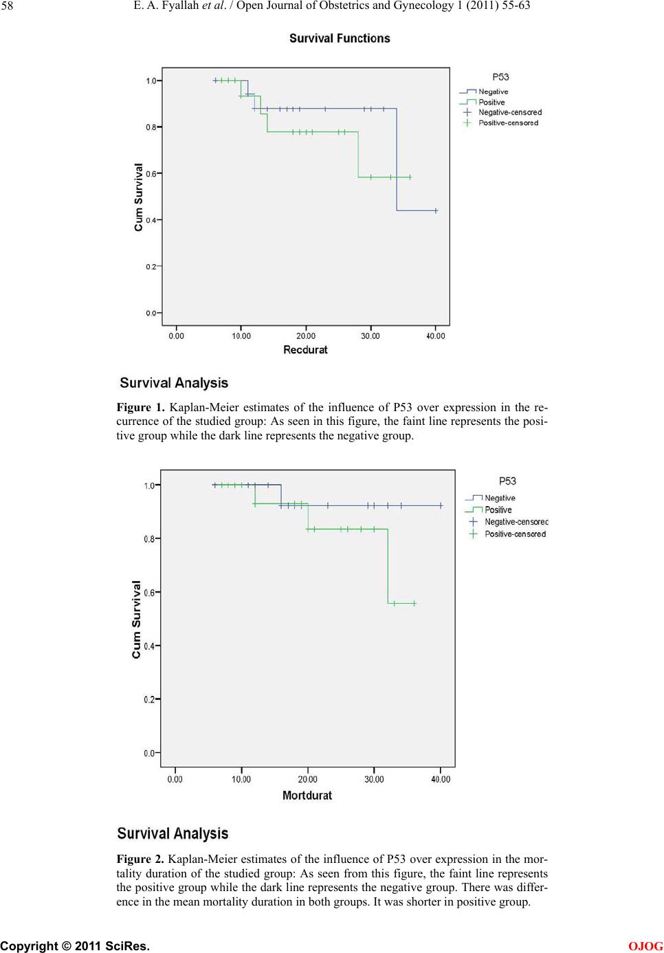 E. A. Fyallah et al. / Open Journal of Obstetrics and Gynecology 1 (2011) 55-63 58 Figure 1. Kaplan-Meier estimates of the influence of P53 over expression in the re- currence of the studied group: As seen in this figure, the faint line represents the posi- tive group while the dark line represents the negative group. Figure 2. Kaplan-Meier estimates of the influence of P53 over expression in the mor- tality duration of the studied group: As seen from this figure, the faint line represents the positive group while the dark line represents the negative group. There was differ- ence in the mean mortality duration in both groups. It was shorter in positive group. C opyright © 2011 SciRes. OJOG 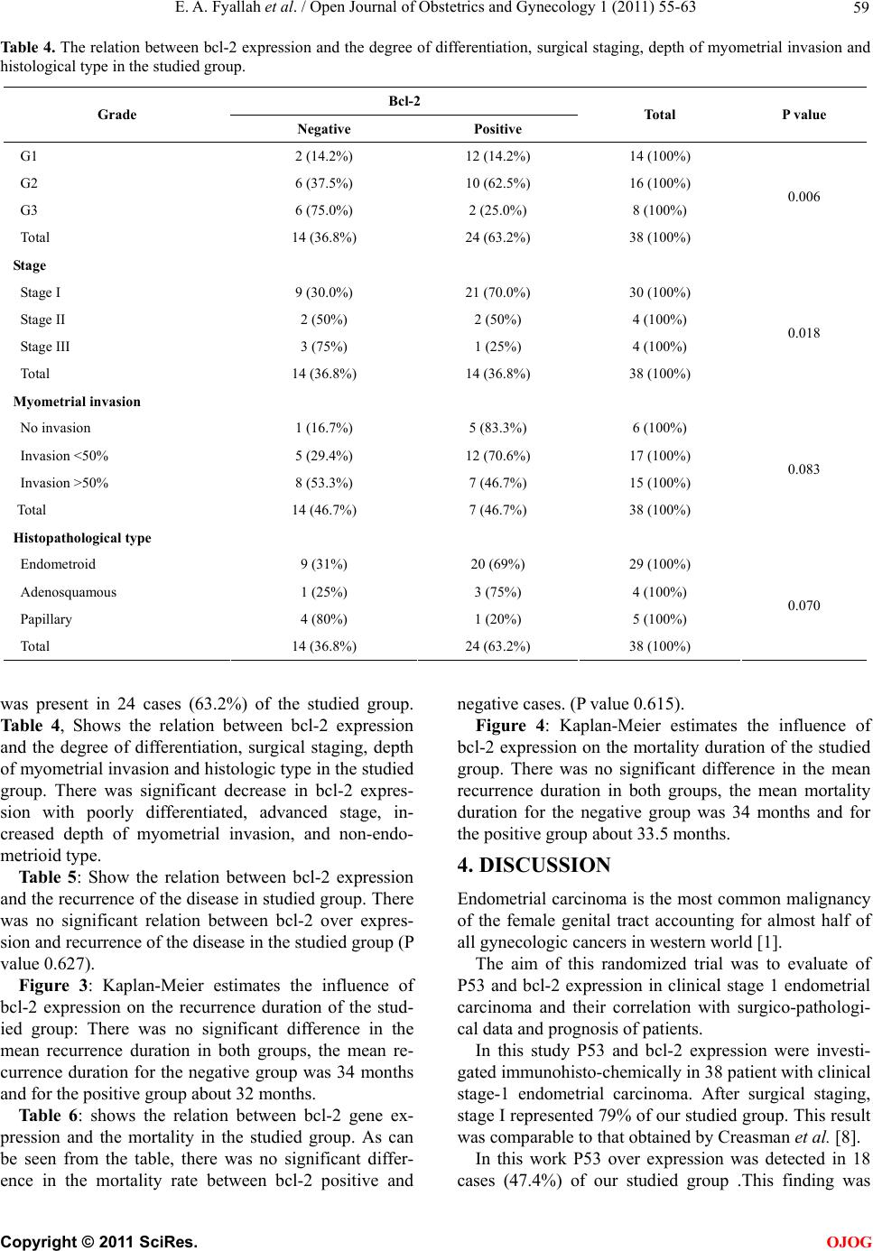 E. A. Fyallah et al. / Open Journal of Obstetrics and Gynecology 1 (2011) 55-63 Copyright © 2011 SciRes. 59 OJOG Table 4. The relation between bcl-2 expression and the degree of differentiation, surgical staging, depth of myometrial invasion and histological type in the studied group. Bcl-2 Grade Negative Positive Total P value G1 2 (14.2%) 12 (14.2%) 14 (100%) G2 6 (37.5%) 10 (62.5%) 16 (100%) G3 6 (75.0%) 2 (25.0%) 8 (100%) Total 14 (36.8%) 24 (63.2%) 38 (100%) 0.006 Stage Stage I 9 (30.0%) 21 (70.0%) 30 (100%) Stage II 2 (50%) 2 (50%) 4 (100%) Stage III 3 (75%) 1 (25%) 4 (100%) Total 14 (36.8%) 14 (36.8%) 38 (100%) 0.018 Myometria l invasion No invasion 1 (16.7%) 5 (83.3%) 6 (100%) Invasion <50% 5 (29.4%) 12 (70.6%) 17 (100%) Invasion >50% 8 (53.3%) 7 (46.7%) 15 (100%) Total 14 (46.7%) 7 (46.7%) 38 (100%) 0.083 Histopathological type Endometroid 9 (31%) 20 (69%) 29 (100%) Adenosquamous 1 (25%) 3 (75%) 4 (100%) Papillary 4 (80%) 1 (20%) 5 (100%) Total 14 (36.8%) 24 (63.2%) 38 (100%) 0.070 was present in 24 cases (63.2%) of the studied group. Table 4, Shows the relation between bcl-2 expression and the degree of differentiation, surgical staging, depth of myometrial invasion and histologic type in the studied group. There was significant decrease in bcl-2 expres- sion with poorly differentiated, advanced stage, in- creased depth of myometrial invasion, and non-endo- metrioid type. Ta ble 5 : Show the relation between bcl-2 expression and the recurrence of the disease in studied group. There was no significant relation between bcl-2 over expres- sion and recurren ce of the disease in th e stud ied group (P value 0.627). Figure 3: Kaplan-Meier estimates the influence of bcl-2 expression on the recurrence duration of the stud- ied group: There was no significant difference in the mean recurrence duration in both groups, the mean re- currence duration for the negative group was 34 months and for the positive group about 32 months. Table 6: shows the relation between bcl-2 gene ex- pression and the mortality in the studied group. As can be seen from the table, there was no significant differ- ence in the mortality rate between bcl-2 positive and negative cases. (P value 0.615). Figure 4: Kaplan-Meier estimates the influence of bcl-2 expression on the mortality dur ation of the studied group. There was no significant difference in the mean recurrence duration in both groups, the mean mortality duration for the negative group was 34 months and for the positive group about 33.5 months. 4. DISCUSSION Endometrial carcinoma is the most common malignancy of the female genital tract accounting for almost half of all gynecologic cancers in western world [1]. The aim of this randomized trial was to evaluate of P53 and bcl-2 expression in clinical stage 1 endometrial carcinoma and their correlation with surgico-pathologi- cal data and prognosis of patients. In this study P53 and bcl-2 expression were investi- gated immunohisto-chemically in 38 patient with clinical stage-1 endometrial carcinoma. After surgical staging, stage I represented 79% of our studied group. This result was comparable to that obtained by Creasman et al. [8]. In this work P53 over expression was detected in 18 cases (47.4%) of our studied group .This finding was 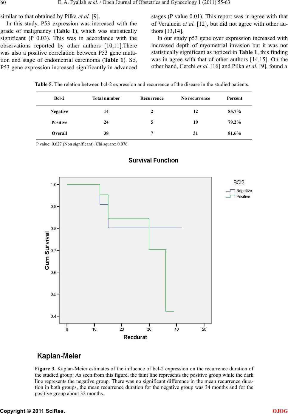 E. A. Fyallah et al. / Open Journal of Obstetrics and Gynecology 1 (2011) 55-63 60 similar to that obtained by Pilka et al. [9]. In this study, P53 expression was increased with the grade of malignancy (Table 1), which was statistically significant (P 0.03). This was in accordance with the observations reported by other authors [10,11].There was also a positive correlation between P53 gene muta- tion and stage of endometrial carcinoma (Ta ble 1 ). So, P53 gene expression increased significantly in advanced stages (P value 0.01). This report was in agree with that of Veralucia et al. [12], but did not agree with other au- thors [13,14]. In our study p53 gene over expression increased with increased depth of myometrial invasion but it was not statistically significant as noticed in Table 1, this finding was in agree with that of other authors [14,15]. On the other hand, Cerchi et al. [16] and Pilka et al. [9], found a Table 5. The relation between bcl-2 expression and recurrence of the disease in the studied patients. Bcl-2 Total number Recurrence No recurrence Percent Negative Positive Overall 14 24 38 2 5 7 12 19 31 85.7% 79.2% 81.6% P value: 0.627 (Non significant). Chi square: 0.076 Figure 3. Kaplan-Meier estimates of the influence of bcl-2 expression on the recurrence duration of the studied group: As seen from this figure, the faint line represents the positive group while the dark line represents the negative group. There was no significant difference in the mean recurrence dura- tion in both groups, the mean recurrence duration for the negative group was 34 months and for the positive group about 32 months. C opyright © 2011 SciRes. OJOG 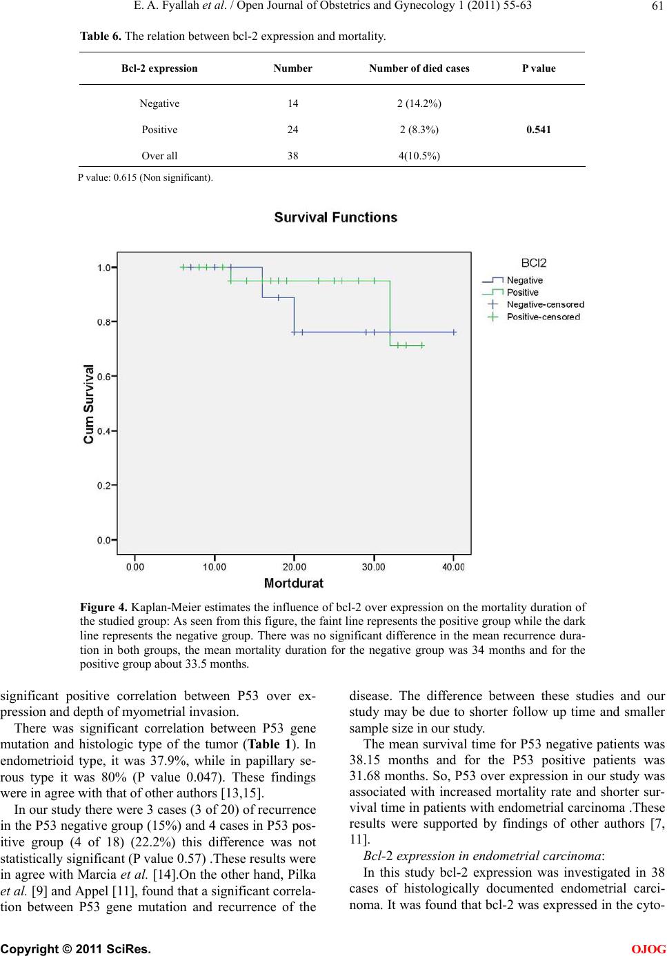 E. A. Fyallah et al. / Open Journal of Obstetrics and Gynecology 1 (2011) 55-63 61 Table 6. The relation between bcl-2 expression and mortality. Bcl-2 expre ssion Number Number of died cases P value Negative Positive Over all 14 24 38 2 (14.2%) 2 (8.3%) 4(10.5%) 0.541 P value: 0.615 (Non significant). Figure 4. Kaplan-Meier estimates the influence of bcl-2 over expression on the mortality duration of the studied group: As seen from this figure, the faint line represents the positive group while the dark line represents the negative group. There was no significant difference in the mean recurrence dura- tion in both groups, the mean mortality duration for the negative group was 34 months and for the positive group about 33.5 months. significant positive correlation between P53 over ex- pression and depth of myometrial invasion. There was significant correlation between P53 gene mutation and histologic type of the tumor (Table 1). In endometrioid type, it was 37.9%, while in papillary se- rous type it was 80% (P value 0.047). These findings were in agree with that of other authors [1 3,15]. In our study there were 3 cases (3 of 20) of recurrence in the P53 negative gr oup (15%) and 4 cases in P53 pos- itive group (4 of 18) (22.2%) this difference was not statistically significant (P value 0.57) .These results were in agree with Marcia et al. [14].On the other hand, Pilka et al. [9] and Appel [11], found that a significant correla- tion between P53 gene mutation and recurrence of the disease. The difference between these studies and our study may be due to shorter follow up time and smaller sample size in our study. The mean survival time for P53 negative patients was 38.15 months and for the P53 positive patients was 31.68 months. So, P53 over expression in our study was associated with increased mortality rate and shorter sur- vival time in patients with endometrial carcinoma .These results were supported by findings of other authors [7, 11]. Bcl-2 expression in endometrial carcinoma: In this study bcl-2 expression was investigated in 38 cases of histologically documented endometrial carci- noma. It was found that bcl-2 was expressed in the cyto- C opyright © 2011 SciRes. OJOG 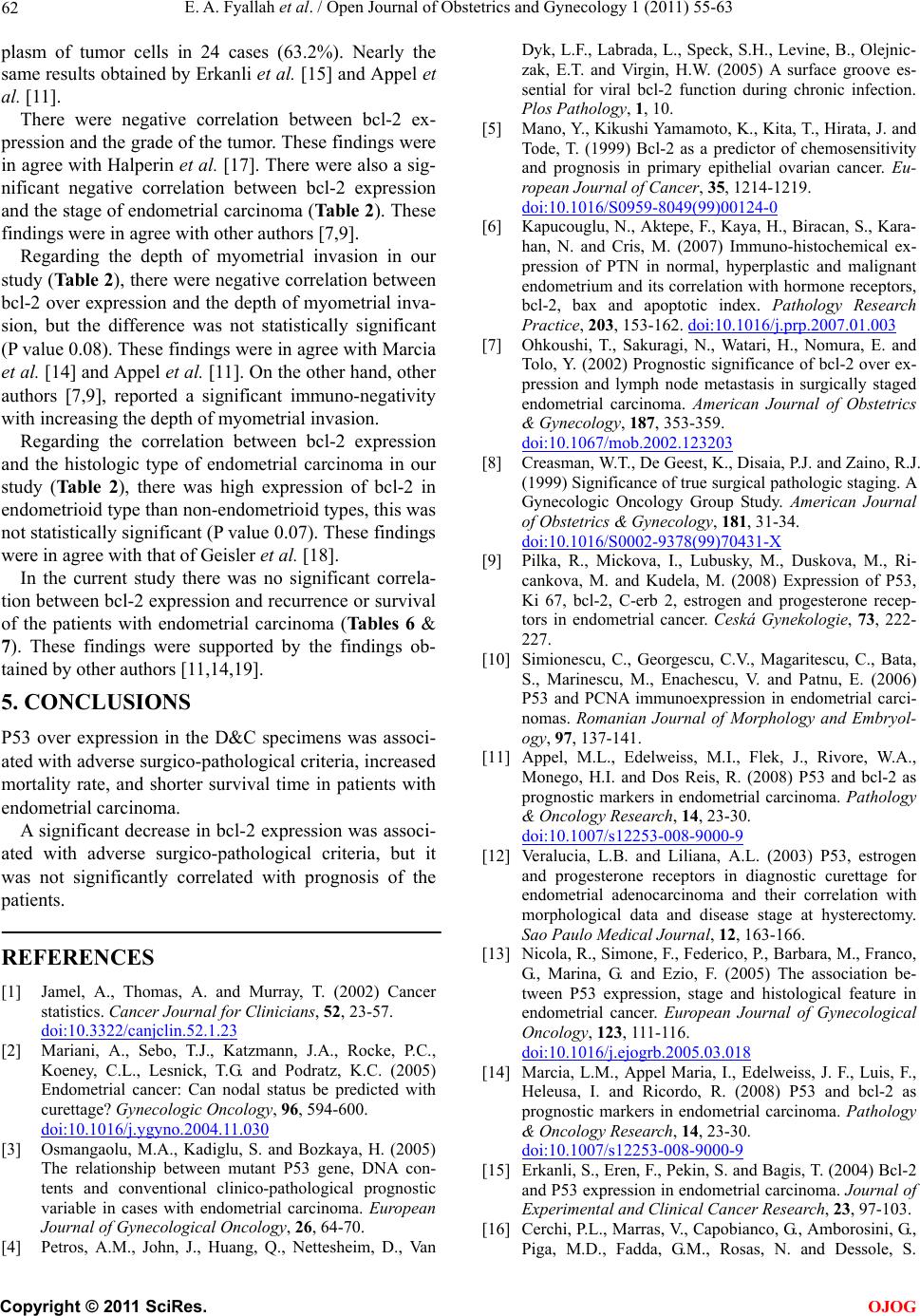 E. A. Fyallah et al. / Open Journal of Obstetrics and Gynecology 1 (2011) 55-63 62 plasm of tumor cells in 24 cases (63.2%). Nearly the same results obtained by Erkanli et al. [15] and Appel et al. [11]. There were negative correlation between bcl-2 ex- pression and the grade of the tumor. These findings were in agree with Halperin et al. [17]. There were also a sig- nificant negative correlation between bcl-2 expression and the stage of endometrial carcinoma (Table 2). These findings were in agree with other authors [7,9]. Regarding the depth of myometrial invasion in our study (Table 2), there were negative correlation between bcl-2 over expression and the depth of myometrial inva- sion, but the difference was not statistically significant (P value 0.08). These findings were in agree with Marcia et al. [14] and Appel et al. [11]. On the other hand, other authors [7,9], reported a significant immuno-negativity with increasing the depth of myometrial invasion. Regarding the correlation between bcl-2 expression and the histologic type of endometrial carcinoma in our study (Table 2), there was high expression of bcl-2 in endometrioid type than non-endometrioid typ es, this was not statistically significant (P value 0.07). These findings were in agree with that of Geisler et al. [18]. In the current study there was no significant correla- tion between bcl-2 expression and recurrence or survival of the patients with endometrial carcinoma (Ta b le s 6 & 7). These findings were supported by the findings ob- tained by other authors [11,14,19]. 5. CONCLUSIONS P53 over expression in the D&C specimens was associ- ated with adverse surgico-pathological criteria, increased mortality rate, and shorter survival time in patients with endometrial carcinoma. A significant decrease in bcl-2 expression was associ- ated with adverse surgico-pathological criteria, but it was not significantly correlated with prognosis of the patients. REFERENCES [1] Jamel, A., Thomas, A. and Murray, T. (2002) Cancer statistics. Cancer Journal for Clinicians, 52, 23-57. doi:10.3322/canjclin.52.1.23 [2] Mariani, A., Sebo, T.J., Katzmann, J.A., Rocke, P.C., Koeney, C.L., Lesnick, T.G. and Podratz, K.C. (2005) Endometrial cancer: Can nodal status be predicted with curettage? Gynecologic Oncology, 96, 594-600. doi:10.1016/j.ygyno.2004.11.030 [3] Osmangaolu, M.A., Kadiglu, S. and Bozkaya, H. (2005) The relationship between mutant P53 gene, DNA con- tents and conventional clinico-pathological prognostic variable in cases with endometrial carcinoma. European Journal of Gynecological Oncology, 26, 64-70. [4] Petros, A.M., John, J., Huang, Q., Nettesheim, D., Van Dyk, L.F., Labrada, L., Speck, S.H., Levine, B., Olejnic- zak, E.T. and Virgin, H.W. (2005) A surface groove es- sential for viral bcl-2 function during chronic infection. Plos Pathology, 1, 10. [5] Mano, Y., Kikushi Yamamoto, K., Kita, T., Hirata, J. and Tode, T. (1999) Bcl-2 as a predictor of chemosensitivity and prognosis in primary epithelial ovarian cancer. Eu- ropean Journal of Cancer, 35, 1214-1219. doi:10.1016/S0959-8049(99)00124-0 [6] Kapucouglu, N., Aktepe, F., Kaya, H., Biracan, S., Kara- han, N. and Cris, M. (2007) Immuno-histochemical ex- pression of PTN in normal, hyperplastic and malignant endometrium and its correlation with hormone receptors, bcl-2, bax and apoptotic index. Pathology Research Practice, 203, 153-162. doi:10.1016/j.prp.2007.01.003 [7] Ohkoushi, T., Sakuragi, N., Watari, H., Nomura, E. and Tolo, Y. (2002) Prognostic significance of bcl-2 over ex- pression and lymph node metastasis in surgically staged endometrial carcinoma. American Journal of Obstetrics & Gynecology, 187, 353-359. doi:10.1067/mob.2002.123203 [8] Creasman, W.T., De Geest, K., Disaia, P.J. and Zaino, R.J. (1999) Significance of true surgical pathologic staging. A Gynecologic Oncology Group Study. American Journal of Obstetrics & Gynecology, 181, 31-34. doi:10.1016/S0002-9378(99)70431-X [9] Pilka, R., Mickova, I., Lubusky, M., Duskova, M., Ri- cankova, M. and Kudela, M. (2008) Expression of P53, Ki 67, bcl-2, C-erb 2, estrogen and progesterone recep- tors in endometrial cancer. Ceská Gynekologie, 73, 222- 227. [10] Simionescu, C., Georgescu, C.V., Magaritescu, C., Bata, S., Marinescu, M., Enachescu, V. and Patnu, E. (2006) P53 and PCNA immunoexpression in endometrial carci- nomas. Romanian Journal of Morphology and Embryol- ogy, 97, 137-141. [11] Appel, M.L., Edelweiss, M.I., Flek, J., Rivore, W.A., Monego, H.I. and Dos Reis, R. (2008) P53 and bcl-2 as prognostic markers in endometrial carcinoma. Pathology & Oncology Research, 14, 23-30. doi:10.1007/s12253-008-9000-9 [12] Veralucia, L.B. and Liliana, A.L. (2003) P53, estrogen and progesterone receptors in diagnostic curettage for endometrial adenocarcinoma and their correlation with morphological data and disease stage at hysterectomy. Sao Paulo Medical Journal, 12, 163-166. [13] Nicola, R., Simone, F., Federico, P., Barbara, M., Franco, G., Marina, G. and Ezio, F. (2005) The association be- tween P53 expression, stage and histological feature in endometrial cancer. European Journal of Gynecological Oncology, 123, 111-116. doi:10.1016/j.ejogrb.2005.03.018 [14] Marcia, L.M., Appel Maria, I., Edelweiss, J. F., Luis, F., Heleusa, I. and Ricordo, R. (2008) P53 and bcl-2 as prognostic markers in endometrial carcinoma. Pathology & Oncology Research, 14, 23-30. doi:10.1007/s12253-008-9000-9 [15] Erkanli, S., Eren, F., Pekin, S. and Bagis, T. (2004) Bcl-2 and P53 expression in endometrial carcinoma. Journal of Experimental and Clinical Cancer Research, 23, 97-103. [16] Cerchi, P.L., Marras, V., Capobianco, G., Amborosini, G., Piga, M.D., Fadda, G.M., Rosas, N. and Dessole, S. C opyright © 2011 SciRes. OJOG  E. A. Fyallah et al. / Open Journal of Obstetrics and Gynecology 1 (2011) 55-63 Copyright © 2011 SciRes. 63 OJOG (2001) Prognostic value of P53, C-erb-B2 and MIB-1 in endometrial carcinoma. European Journal of Gyneco- logical Oncology, 22, 451-453. [17] Halperin, R., Zehavi, S., Habler, E. and Bukovsky, I. (2001) Comparative immunohistochemical study of en- dometroid and serous papillary carcinoma of the endo- metrium. European Journal of Gynecological Oncology, 22, 122-126. [18] Geisler, J.P., Geisler, H.E., Wiemann, M.C., Zhan, Z., Miller, G.A. and Crabtree, W. (1998) Lack of bcl-2 per- sistence: An independent prognostic indicator of poor prognosis in endometrial carcinoma. Gynecologic On- cology, 71, 305-307. doi:10.1006/gyno.1998.5192 [19] Fanning, J., Brown, S., Phibbs, G., Kramer, T. and Zaher, A. (2002) Immunohistochemical evaluation is not prog- nostic for recurrence in fully staged high-risk endo- metrial cancer. International Journal of Gynecological Cancer, 12, 286-289. doi:10.1046/j.1525-1438.2002.t01-1-01103.x |