

Paper Menu >>
Journal Menu >>
 World Journal of AIDS, 2011, 1, 31-36 doi:10.4236/wja.2011.12006 Published Online June 2011 (http://www.SciRP.org/journal/wja) Copyright © 2011 SciRes. WJA 31 Immunovirological and Biochemical Changes in Nigerian Patients with Hepatitis B Coinfection on Antiretroviral Therapy Jesse Abiodun Otegbayo1, Titilola Stella Akingbola2, Joshua Odunayo Akinyemi3, Kayode Solomon Adedapo4, Georgina Njideka Odaibo5, Yetunde Adebisi Aken Óva2, David Olufemi Olaleye5, Isaac Folorunsho Adewole7, Robert Murphy8, Phylis Kanki9 1Departments of Medicine, University of Ibadan, Ibadan, Nigeria; 2Department of Haematology, University of Ibadan, Ibadan, Nige- ria; 3Department of Epidemiology and Biostatistics, University of Ibadan, Ibadan, Nigeria; 4Department of Chemical Pathology, University of Ibadan, Ibadan, Nigeria; 5Department of Virology, University of Ibadan, Ibadan, Nigeria; 6Department of Haematology, University of Ibadan, Ibadan, Nigeria; 7Department of Obstetrics and Gynaecology, University of Ibadan, Ibadan, Nigeria; 8Department of Infectious Diseases, North-western University Feinberg School of Medicine, Chicago, USA; 9Department of cancer Biology, Harvard School of Public Health, Harvard University, Boston, USA. Email: [email protected] Received April 3rd, 2011; Revised May 3rd, 2011; Accepted May 24th, 2011. ABSTRACT Hepatitis B virus (HBV) co-infection with HIV is high among Nigerians. Some studies have suggested impaired CD4 recovery among coinfected patients compared to the HIV mono infected. This retrospective study of treatment-naïve HIV infected patients was aimed at determining the trend of changes in CD4+ counts, HIV-RNA, renal and liver func- tion tests in response to combined antiretroviral therapy (CART). A questionnaire was utilised to extract clinical and laboratory da ta of HBV co infected HIV /AIDS patients before treatment and at six, twelve and eighteen months of ther- apy with CART. Findings were compared to those of HIV mono infected. Relevant statistical instruments were used to analyse for comparisons of means of Log10 HIV viral load and CD4 count using SPSS package 15.0. All levels of sig- nificance were at 5%. Two thousand five hundred and sixty two patients were analysed. Of these, 354 (13.8%) were HBsAg positive. Majority (63.1%) were females. Most of the recruited patients were on combivir and nevirapine. The median CD4 count fo r the HBsAg negative was 104 cells/(mm3 IQR 34-171) and it was significantly higher than those of the positive (91 cells/mm3) (p < 0.05). ALT and AST were higher among HBsAg positives, while urea and creatinine levels were similar. The median change in CD4 count from baseline and during the course of therapy were similar in the two groups. S imilarly, virological responses were not different in the two g roups at the various time points. In con- clusion no significant difference in the rate of CD4 recovery and HIV-RNA decline in among coinfected and monoin- fected HIV patients at different stages of therapy. Keywords: Hepatitis B, Coinfection, CD4, Viral Load, Antiretroviral, Nigeria 1. Introduction Hepatitis B virus (HBV) is a common major endemic infection in Nigeria [1,2] and its association with com- mon major liver diseases is well established in our envi- ronment [3,4] as in other parts of the world. HIV/AIDS continue to receive significant attention and a sizeable proportion of the health care budget because of the yet to be conquered pandemic. Previous studies in Nigeria have shown a significant hepatitis B and C virus co-infection with HIV infection [5,6]. These studies however showed that hepatitis C, either as mono-infection or as co- infection with HIV infection is relatively low compared with HBV. Many of the antiretroviral drugs in current use are associated with hepatotoxicity among other side-effects. Hepatotoxicity of antiretroviral drugs espe- cially co-infected patients with hepatitis viruses is a ma- jor concern, with increasing incidence of liver lidease [7]. Some studies have addressed the immunological (CD4+) and viral (HIV-RNA) responses in HIV/AIDS patients on combined antiretroviral therapy (CART). Micheloud et al. in their study among HCV-coinfected paediatric patients 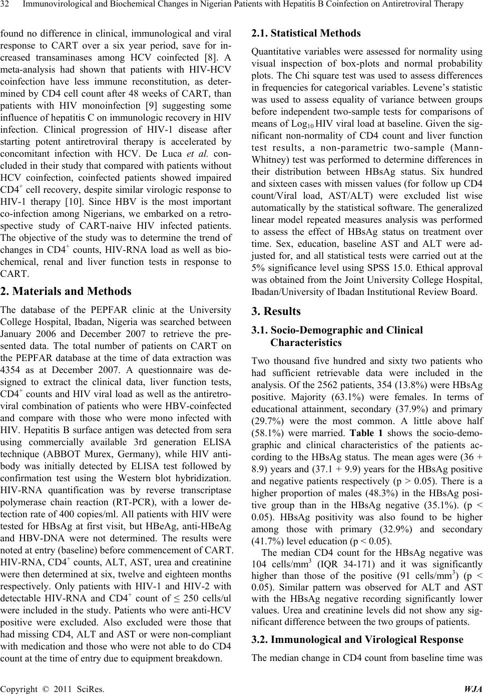 32 Immunovirological and Biochemical Changes in Nigerian Patients with Hepatitis B Coinfection on Antiretroviral Therapy found no difference in clinical, immunological and viral response to CART over a six year period, save for in- creased transaminases among HCV coinfected [8]. A meta-analysis had shown that patients with HIV-HCV coinfection have less immune reconstitution, as deter- mined by CD4 cell count after 48 weeks of CART, than patients with HIV monoinfection [9] suggesting some influence of hepatitis C on immunologic recovery in HIV infection. Clinical progression of HIV-1 disease after starting potent antiretroviral therapy is accelerated by concomitant infection with HCV. De Luca et al. con- cluded in their study that compared with patients without HCV coinfection, coinfected patients showed impaired CD4+ cell recovery, despite similar virologic response to HIV-1 therapy [10]. Since HBV is the most important co-infection among Nigerians, we embarked on a retro- spective study of CART-naive HIV infected patients. The objective of the study was to determine the trend of changes in CD4+ counts, HIV-RNA load as well as bio- chemical, renal and liver function tests in response to CART. 2. Materials and Methods The database of the PEPFAR clinic at the University College Hospital, Ibadan, Nigeria was searched between January 2006 and December 2007 to retrieve the pre- sented data. The total number of patients on CART on the PEPFAR database at the time of data extraction was 4354 as at December 2007. A questionnaire was de- signed to extract the clinical data, liver function tests, CD4+ counts and HIV viral load as well as the antiretro- viral combination of patients who were HBV-coinfected and compare with those who were mono infected with HIV. Hepatitis B surface antigen was detected from sera using commercially available 3rd generation ELISA technique (ABBOT Murex, Germany), while HIV anti- body was initially detected by ELISA test followed by confirmation test using the Western blot hybridization. HIV-RNA quantification was by reverse transcriptase polymerase chain reaction (RT-PCR), with a lower de- tection rate of 400 copies/ml. All patients with HIV were tested for HBsAg at first visit, but HBeAg, anti-HBeAg and HBV-DNA were not determined. The results were noted at entry (baseline) before commencement of CART. HIV-RNA, CD4+ counts, ALT, AST, urea and creatinine were then determined at six, twelve and eighteen months respectively. Only patients with HIV-1 and HIV-2 with detectable HIV-RNA and CD4+ count of ≤ 250 cells/ul were included in the study. Patients who were anti-HCV positive were excluded. Also excluded were those that had missing CD4, ALT and AST or were non-compliant with medication and those who were not able to do CD4 count at the time of entry due to equipment breakdown. 2.1. Statistical Methods Quantitative variables were assessed for normality using visual inspection of box-plots and normal probability plots. The Chi square test was used to assess differences in frequencies for categorical variables. Levene’s statistic was used to assess equality of variance between groups before independent two-sample tests for comparisons of means of Log10 HIV viral load at baseline. Given the sig- nificant non-normality of CD4 count and liver function test results, a non-parametric two-sample (Mann- Whitney) test was performed to determine differences in their distribution between HBsAg status. Six hundred and sixteen cases with missen values (for follow up CD4 count/Viral load, AST/ALT) were excluded list wise automatically by the statistical software. The generalized linear model repeated measures analysis was performed to assess the effect of HBsAg status on treatment over time. Sex, education, baseline AST and ALT were ad- justed for, and all statistical tests were carried out at the 5% significance level using SPSS 15.0. Ethical approval was obtained from the Joint University College Hospital, Ibadan/University of Ibadan Institutional Review Board. 3. Results 3.1. Socio-Demographic and Clinical Characteristics Two thousand five hundred and sixty two patients who had sufficient retrievable data were included in the analysis. Of the 2562 patients, 354 (13.8%) were HBsAg positive. Majority (63.1%) were females. In terms of educational attainment, secondary (37.9%) and primary (29.7%) were the most common. A little above half (58.1%) were married. Table 1 shows the socio- demo- graphic and clinical characteristics of the patients ac- cording to the HBsAg status. The mean ages were (36 + 8.9) years and (37.1 + 9.9) years for the HBsAg positive and negative patients respectively (p > 0.05). There is a higher proportion of males (48.3%) in the HBsAg posi- tive group than in the HBsAg negative (35.1%). (p < 0.05). HBsAg positivity was also found to be higher among those with primary (32.9%) and secondary (41.7%) level education (p < 0.05). The median CD4 count for the HBsAg negative was 104 cells/mm3 (IQR 34-171) and it was significantly higher than those of the positive (91 cells/mm3) (p < 0.05). Similar pattern was observed for ALT and AST with the HBsAg negative recording significantly lower values. Urea and creatinine levels did not show any sig- nificant difference between the two groups of patients. 3.2. Immunological and Virological Response The median change in CD4 count from baseline time was Copyright © 2011 SciRes. WJA 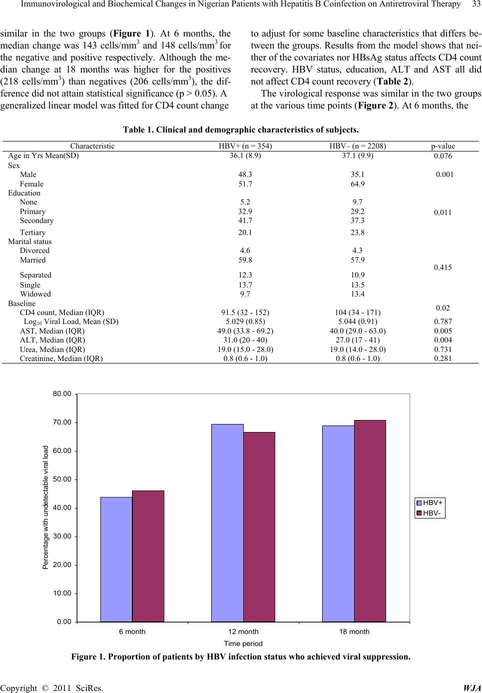 Immunovirological and Biochemical Changes in Nigerian Patients with Hepatitis B Coinfection on Antiretroviral Therapy Copyright © 2011 SciRes. WJA 33 similar in the two groups (Figure 1). At 6 months, the median change was 143 cells/mm3 and 148 cells/mm3 for the negative and positive respectively. Although the me- to adjust for some baseline characteristics that differs be- tween the groups. Results from the model shows that nei- ther of the covariates nor HBsAg status affects CD4 count recovery. HBV status, education, ALT and AST all did not affect CD4 count recovery (Table 2). dian change at 18 months was higher for the positives (218 cells/mm3) than negatives (206 cells/mm3), the dif- ference did not attain statistical significance (p > 0.05). A The virological response was similar in the two groups at the various time points (Figure 2). At 6 months, the generalized linear model was fitted for CD4 count change Table 1. Clinical and demographic characteristics of subjects. Characteristic HBV+ (n = 354) HBV– (n = 2208) p-value Age in Yrs Mean(SD) 36.1 (8.9) 37.1 (9.9) 0.076 Sex Male 48.3 35.1 Female 51.7 64.9 0.001 Education None 5.2 9.7 Primary 32.9 29.2 Secondary 41.7 37.3 Tertiary 20.1 23.8 0.011 Marital status Divorced 4.6 4.3 Married 59.8 57.9 Separated 12.3 10.9 Single 13.7 13.5 Widowed 9.7 13.4 0.415 Baseline CD4 count, Median (IQR) 91.5 (32 - 152) 104 (34 - 171) 0.02 Log 10 Viral Load, Mean (SD) 5.029 (0.85) 5.044 (0.91) 0.787 AST, Median (IQR) 49.0 (33.8 - 69.2) 40.0 (29.0 - 63.0) 0.005 ALT, Median (IQR) 31.0 (20 - 40) 27.0 (17 - 41) 0.004 Urea, Median (IQR) 19.0 (15.0 - 28.0) 19.0 (14.0 - 28.0) 0.731 Creatinine, Median (IQR) 0.8 (0.6 - 1.0) 0.8 (0.6 - 1.0) 0.281 0.00 10.00 20.00 30.00 40.00 50.00 60.00 70.00 80.00 6 month12 month18 month Time period Percentage with undetectable viral load HBV+ HBV- Figure 1. Proportion of patients by HBV infection status who achieved viral suppression. 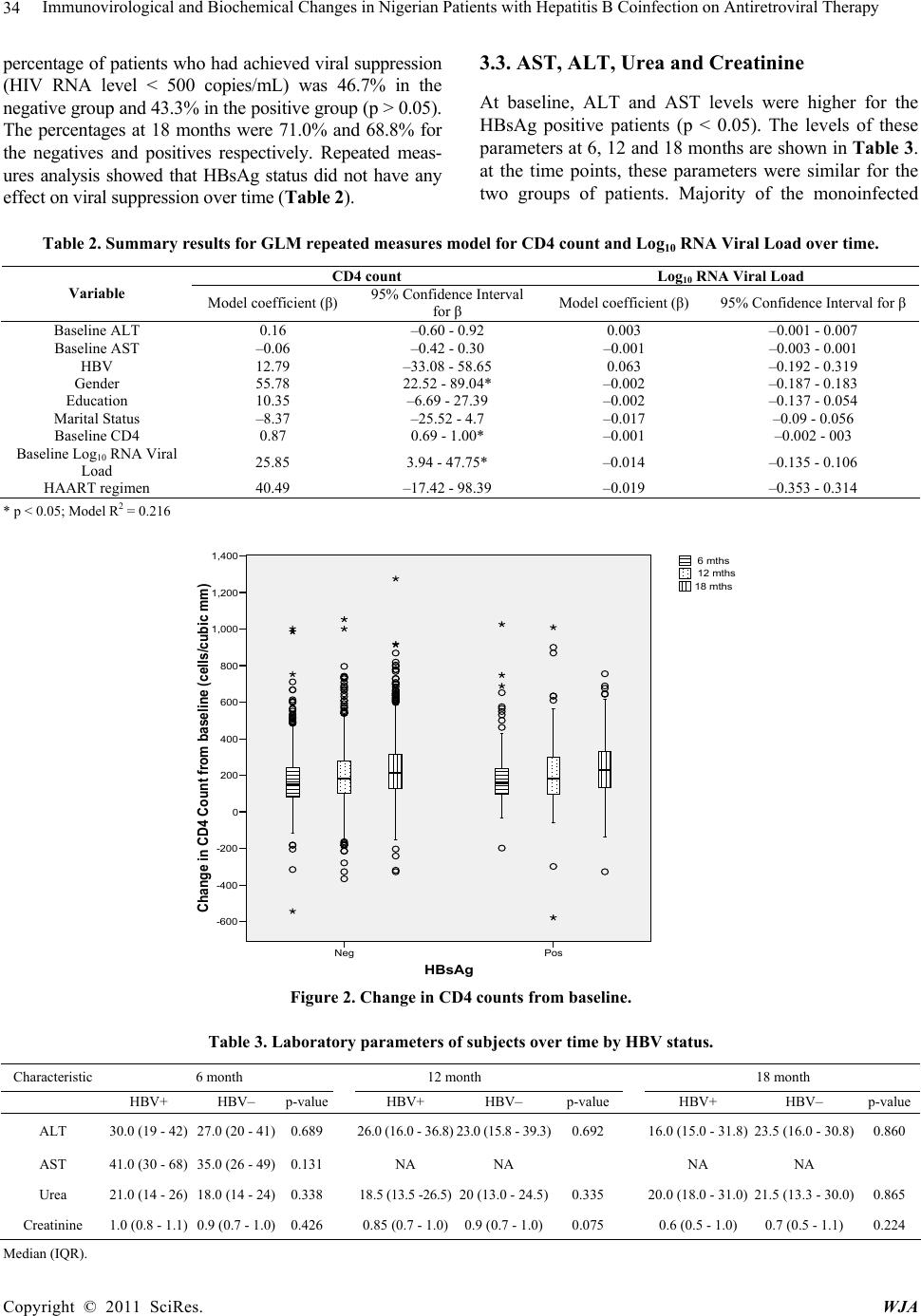 Immunovirological and Biochemical Changes in Nigerian Patients with Hepatitis B Coinfection on Antiretroviral Therapy 34 percentage of patients who had achieved viral suppression (HIV RNA level < 500 copies/mL) was 46.7% in the negative group and 43.3% in the positive group (p > 0.05). The percentages at 18 months were 71.0% and 68.8% for the negatives and positives respectively. Repeated meas- ures analysis showed that HBsAg status did not have any effect on viral suppression over time (Table 2). 3.3. AST, ALT, Urea and Creatinine At baseline, ALT and AST levels were higher for the HBsAg positive patients (p < 0.05). The levels of these parameters at 6, 12 and 18 months are shown in Table 3. at the time points, these parameters were similar for the two groups of patients. Majority of the monoinfected Table 2. Summary results for GLM repeated measures model for CD4 count and Log10 RNA Viral Load over time. CD4 count Log10 RNA Viral Load Variable Model coefficient (β) 95% Confidence Interval for β Model coefficient (β) 95% Confidence Interval for β Baseline ALT 0.16 –0.60 - 0.92 0.003 –0.001 - 0.007 Baseline AST –0.06 –0.42 - 0.30 –0.001 –0.003 - 0.001 HBV 12.79 –33.08 - 58.65 0.063 –0.192 - 0.319 Gender 55.78 22.52 - 89.04* –0.002 –0.187 - 0.183 Education 10.35 –6.69 - 27.39 –0.002 –0.137 - 0.054 Marital Status –8.37 –25.52 - 4.7 –0.017 –0.09 - 0.056 Baseline CD4 0.87 0.69 - 1.00* –0.001 –0.002 - 003 Baseline Log10 RNA Viral Load 25.85 3.94 - 47.75* –0.014 –0.135 - 0.106 HAART regimen 40.49 –17.42 - 98.39 –0.019 –0.353 - 0.314 * p < 0.05; Model R2 = 0.216 HBsAg PosNeg Change in CD4 Count from baseline (cells/cubic mm) 1,400 1,200 1,000 800 600 400 200 0 -200 -400 -600 18 mths 12 mths 6 mths Figure 2. Change in CD4 counts from baseline. Table 3. Laboratory parameters of subjects over time by HBV status. Characteristic 6 month 12 month 18 month HBV+ HBV– p-value HBV+ HBV– p-valueHBV+ HBV– p-value ALT 30.0 (19 - 42) 27.0 (20 - 41) 0.689 26.0 (16.0 - 36.8)23.0 (15.8 - 39.3)0.692 16.0 (15.0 - 31.8) 23.5 (16.0 - 30.8)0.860 AST 41.0 (30 - 68) 35.0 (26 - 49) 0.131 NA NA NA NA Urea 21.0 (14 - 26) 18.0 (14 - 24) 0.338 18.5 (13.5 -26.5)20 (13.0 - 24.5)0.335 20.0 (18.0 - 31.0) 21.5 (13.3 - 30.0)0.865 Creatinine 1.0 (0.8 - 1.1) 0.9 (0.7 - 1.0) 0.426 0.85 (0.7 - 1.0)0.9 (0.7 - 1.0)0.075 0.6 (0.5 - 1.0) 0.7 (0.5 - 1.1) 0.224 Median (IQR). Copyright © 2011 SciRes. WJA  Immunovirological and Biochemical Changes in Nigerian Patients with Hepatitis B Coinfection on Antiretroviral Therapy 35 patients were on combivir and nevirapine, while the coin- fected patients were mainly on truvada and nevirapine. 4. Discussion This study has further shown a high HBV coinfection rate among our patient cohorts as previously noted by us and other authors in Africa [6,11,12] but higher than values obtained in the Western world with lower inci- dence of HBV [13,14]. The preponderance of the female gender had been suggested to be due to the fact that women may be more sensitive to changes in their health and probably socioculturally conditioned to seek health care more than men [6]. The significantly higher coinfec- tion among men compared with women in spite of a predominant female gender affected with HIV is likely a reflection of HBV prevalence among the general popula- tion which has been noted to be higher amongst men [15]. The lower prevalence of coinfection among the better educated individuals generally could be due to the level of awareness that may be associated with education and therefore behaviours and practices that may reduce HBV transmission [16]. It is however surprising that no dif- ference in HBV prevalence was found across the differ- ent marital status. It would have been expected, arguably though, that divorcees and the unmarried would be more exposed to transmission of HBV sexually. This phe- nomenon might be confirming the well known fact that HBV transmission is mainly horizontal in childhood among Africans. Ola et al. however found out that HIV monoinfection is commoner among married people (70.4%), suggesting heterosexual transmission of HIV in our environment as previously documented [16]. It would appear that the immunological status of HBV coinfection and monoinfection is similar, as evidenced by the similar CD4 counts in HIV monoinfected and HIV-coinfected at the start of CART. Similar observa- tions were made Idoko et al. in the Northern part of Ni- geria [17]. It is evident from our study that CART has no influence on CD4 recovery regardless of HBV status with time and duration of therapy. Idoko et al. however found an association with Hepatitis B e antigen (HBeAg) and CD4 T-cell recovery and HIV-RNA clearance [17]. Apart from the higher baseline values of transaminases among HBV coinfected patients, no temporal difference was observed in the normalization of ALT, urea and creatinine among coinfected and monoinfected patients, suggesting that HBV status did not significantly influ- ence the immunovirological response to CART, renal function and transaminase levels. The retrospective na- ture of this study and our inability to determine the date of acquisition of both HIV and HBV (acute or chronic), as well as unavailability of HBV DNA are some of the limitations of our study. The exclusion of subjects with missen data may also affect the result as it is unclear whether such cases were missing for reasons connected with the study outcome. REFERENCES [1] J. A. Otegbayo, F. A. Fasola and A. Abja, “Prevalence of Hepatitis B Surface and E Antigens, Risk Factors for Vi- ral Acquisition and Serum Transaminase among Blood Donors in Ibadan, Nigeria,” Trop Gastroenterol, Vol. 24, No. 4, 2003, pp. 196-197. [2] N. D. Sirisena, M. O. Njoku, J. A. Idoko, et al., “Carriage Rate of Hepatitis B Surface Antigen in an Urban Com- munity in Jos Plateau State, Nigeria,” The Nigerian Post- graduate Medical Journal, Vol. 9, No. 1, 2002, pp. 7-10. [3] S. O. Ola, J. A. Otegbayo, G. N. Odaibo, O. D. Olaleye and I. O. Olubuyide, “Serum Anti-HCV and Hepatitis B Surface Antigenaemia in Nigerian Patients with Acute Icteric Hepatitis,” West African Journal of Medicine, Vol. 21, Vol. 3, No. 2, 2002, pp. 215-217. [4] I. O. Olubuyide, B. Aliyu, O. A. Olalelye, S. O. Ola, F. Olawuyi, U. H. Malabu, S. O. Odemuyiwa, G. N. Odaibo and G. C. Cook, “Hepatitis B and C Virus and Hepato- cellular Carcinoma,” Transactions of the Royal Society of Tropical Medicine, Vol. 91, No. 1, 1997, pp. 38-41. doi:10.1016/S0035-9203(97)90387-4 [5] C. J. Uneke, O. Ogbu, P. U. Inyama, G. I. Anyanwu, M. O. Njoku and J. H. Idoko, “Prevalence of Hepatitis-B Surface Antigen among Blood Donors and Human Im- munodeficiency Virus-Infected Patients in Jos, Nigeria,” Memórias do Instituto Oswaldo Cruz, Vol. 100, No. 1, 2005, pp. 13-16. doi:10.1590/S0074-02762005000100002 [6] J. A. Otegbayo, B. O. Taiwo, T. S. Akingbola, G. N. Odaibo, K. S. Adedapo, S. Penugonda, I. F. Adewole, D. O. Olaleye, R. Murphy and P. Kanki, “Prevalence of Hepatitis B and C Seropositivity in a Nigerian Cohort of HIV-Infected Patients,” Annals of Hepatol, Vol. 7, No. 2, 2008, pp. 152-156. [7] D. L. Thomas, “Growing Importance of Liver Disease in HIV-Infected Persons,” Hepatology, Vol. 43, No. 2, 2006, pp. S221-S229. doi:10.1002/hep.21033 [8] D. Micheloud, J. Jensen, J. M. Bellón, R. Gonzalez, M. J. Mellado, M. L. Navarro, M. A. Muñoz-Fernández and S. Resino, “Spanish Group of Pediatric HIV Infection. Long-Term Response to Highly Active Antiretroviral Therapy in Human Immunodeficiency Virus and Hepati- tis C Virus Coinfected Children: 6 Years of Follow-up,” The Pediatric Infectious Disease Journal, Vol. 26, No. 11, 2007, pp. 1061-1064. doi:10.1097/INF.0b013e31812e531c [9] M. F. Miller, C. Haley, M. J. Koziel and C. F. Rowley, “Impact of Hepatitis C Virus on Immune Restoration in HIV-Infected Patients Who Start Highly Active Antiret- roviral Therapy: A Meta-Analysis,” Clinical Infectious Diseases, Vol. 41, No. 5, 2005, pp. 713-720. doi:10.1086/432618 [10] A.de Luca, R. Bugarini, A. C. Lepri, M. Puoti, E. Girardi, Copyright © 2011 SciRes. WJA  36 Immunovirological and Biochemical Changes in Nigerian Patients with Hepatitis B Coinfection on Antiretroviral Therapy Copyright © 2011 SciRes. WJA A. Antinori, A. Poggio, G. Pagano, G. Tositti, G. Cadeo, A. Macor, M. Toti and A. D’Arminio Monforte, “Italian Cohort Naive Antiretrovirals Study Group. Coinfection with Hepatitis Viruses and Outcome of Initial Antiretro- viral Regimens in Previously Naive HIV-Infected Sub- jects,” Archives of Internal Medicine, Vol. 162, No. 18, 2002, pp. 2125-2132. [11] J. C. Forbi, S. Gabadi, R. Alabi, H. O. Iperepolu, C. R. Pam, P. E. Entonu and S. M. Agwale, “The Role of Triple Infection with Hepatitis B Virus, Hepatitis C Virus, and Human Immunodeficiency Virus (HIV) Type-1 on CD4+ Lymphocyte Levels in the Highly HIV Infected Popula- tion of North-Central Nigeria,” Memórias do Instituto Oswaldo Cruz, Vol. 102, No. 4, 2007, pp. 535-537. doi:10.1590/S0074-02762007005000025 [12] A. M. di Bisceglie, M. Maskew, D. Schulze, A. Reyneke, L. McNamara and C. Firnhaber, “HIV-HBV Coinfection among South African Patients Receiving Antiretroviral Therapy,” Antiviral Therapy, Vol. 15, No. 3, 2010, pp. 499-503. doi:10.3851/IMP1494 [13] P. R. Spradling, J. T. Richardson, K. Buchacz, A. C. Moorman and J. T. Brooks, “The HIV Outpatient Study (HOPS) Investigators*. Prevalence of Chronic Hepatitis B Virus Infection among Patients in the HIV Outpatient Study, 1996-2007,” Journal of Viral Hepatitis, Vol. 17, No. 12, 2010, pp. 879-886. doi:10.1111/j.1365-2893.2009.01249.x [14] C. L. Thio and B. Hepatitis, “Human Immunodeficiency Virus Coinfection,” Hepatology, Vol. 49, No. 5, 2009, pp. S138-145. doi:10.1002/hep.22883 [15] M. P. Adoga, S. D. Gyar, S. Pechulano, O. D. Bashayi, S. E. Emiasegen, T. Zungwe, O. H. Iperepolu, C. Agupugo and S. M. Agwale, “Hepatitis B Virus Infections in Ap- parently Healthy Urban Nigerians: Data from Pre- Vac- cination Tests,” The Journal of Infection in Developing Countries, Vol. 4, No. 6, 2010, pp. 397-400. [16] S. O. Ola, M. M. Ladipo, J. A. Otegbayo, G. N. Odaibo, E. A. Bamgboye, O. G. Nwaorgu, S. W. Shokunbi and O. D. Olaleye, “Demographic Factors in HIV Infected Pa- tients Seen at UCH, Ibadan, Nigeria,” African Journal of Medicine & Medical Sciences, Vol. 34, No. 3, 2005, pp. 297-301. [17] J. Idoko, S. Meloni, M. Muazu, L. Nimzing, B. Badung, C. Hawkins, J. L. Sankalé, E. Ekong, R. Murphy, P. Kanki and C. L. Thio, “Impact of Hepatitis B virus Infec- tion on Human Immunodeficiency Virus Response to Antiretroviral Therapy in Nigeria,” Clinical Infectious Diseases, Vol. 49, No. 8, 2009, pp. 1268-1273. doi:10.1086/605675 |