

Paper Menu >>
Journal Menu >>
 Open Journal of Obstetrics and Gynecology, 2011, 1, 21-24 doi:10.4236/ojog.2011.12005 Published Online June 2011 (http://www.SciRP.org/journal/ojog/ OJOG ). Published Online June 2011 in SciRes. http://www.scirp.org/journal/OJOG Evaluation of a public expense-covered gynecologic screening program in Japan 2005-2009 Hiroshi Takagi, Satoshi Ichigo, Kazutoshi Matsunami, Atsushi Imai* Department of Obstetrics and Gynecology, Matsunami General Hospital, Kasamatsu, Gifu, Japan. Email: *[email protected] Received 14 April 2011; revised 31 May 2011; accepted 8 June 2011. ABSTRACT Introduction: In Japan, there is free physical check- up programs of cancer screening, by which asymp- tomatic participants undergo a medical examination at public expense. The present study aims to describe their gynecologic findings and compare them with the literatures reported from general hospitals and self- paid check-up programs. Methods: Medical records of Japanese women, who underwent gynecological examinations at public expense between 2005 and 2009, were retrospectively reviewed. Results: Of the cervical smears from 2850 women aged 21 - 82 years, 33 (1.1%) were classified as dysplastic and malignant changes: 28 of low-grade squamous intraepithelial lesion (LSIL), 3 high-grade squamous intraepithelial lesion (HSIL), 2 atypical squamous cells of unde- terimined significance (ASC-US). No case of cervical squamous cell carcinoma or adenocarcinoma was found. Ultrasonographic examination detected uterus enlargements and ovary tumors in less than 1% of cases. Most of participants (98%) revealed no gyne- cologic abnormalities. Conclusion: Annual gyneco- logic screening and proper follow-up programs even against asymptomatic women may remarkably re- duce the probability of (pre)malignant disease. Keywords: Cervical Smear Screening; Transvaginal Sonography; Gynecologic Check-up; Oncology 1. INTRODUCTION Cervical intraepithelial neoplasia typically develops into invasive cancer over a 10-year period and apparent cases of rapidly progressive cervical cancer are likely to be among women who have escaped screening and proper follow-up [1-6]. The cervical smear (Papanicolaou, Pap smear) is a routine screening test used for the detection of early cervical abnormalities, namely precancerous dysplastic changes of the uterine cervix [1-6]. Organized screening programs for cervical cancer using the cervical smears have been shown to be effective in decreasing mortality and incidence from the disease [1,7]. The cer- vical screening is a relatively simple, low cost and non- invasive method. Together with transvaginal ultrasono- graphy for detection of ovarian and uterine tumors, a routine cervical screening reduces the probability of de- veloping gynecological m a li gnant di seases . In many countries, undergoing cancer screening is not mandatory but voluntary. The level of knowledge and attitude toward screening are related to multiple factors such as ethnicity, place of residence, income, and social- economic status [8-12]. In Japan, there are free physical check-up programs of cancer screening, by which as- ymptomatic participants undergo a medical examination at public expense. The present study aims to describe their gynecologic findings and compare them with the literatures reported from general hospitals and self-paid check-up programs. 2. METHODS Between January 2005 and December 2009, 2850 as- ymptomatic women, age 21-82, visited one of four Ka- samatsu City-agreed gynecologic physicians for their physical check-up. The cost was fully covered by the municipal corporation. Gynecologic examinations in- cluded routine cancer screening (Papanocolaou test), transvaginal ultrasonography, and pelvic examination by a gynecologist. Cervical and endometrial smears were taken using a speculum and brush and classified into 6 categories: normal, low-grade squamous intraepithelial lesions (LSIL), high-grade squamous intraepithelial le- sions (HSIL), atypical squamouse cells of undetermined significance (ASC-US), cervical carcinoma, cervical adenocarcinoma. When classified as inadequate, the par- ticipants were soon resubmitted to smear examination. Their records of gynecologic findings were retrospec- tively reviewed. 3. RESULTS Table 1 shows the cytologic and ultrasonografic f indings 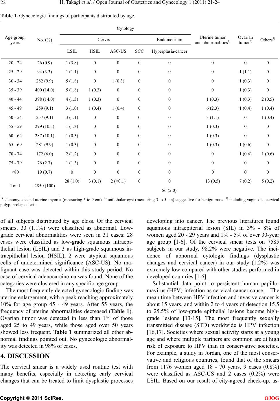 H. Takagi et al. / Open Journal of Obstetrics and Gynecology 1 (2011) 21-24 22 Table 1. Gynecologic findings of participants distributed by age. Cytology Cervix Endometrium Age group, years No. (%) LSIL HSIL ASC-USSCCHyperplasia/cancer Uterine tumor and abnormalities1) Ovarian tumor2) Others3) 20 - 24 26 (0.9) 1 (3.8) 0 0 0 0 0 0 0 25 - 29 94 (3.3) 1 (1.1) 0 0 0 0 0 1 (1.1) 0 30 - 34 282 (9.9) 5 (1.8) 0 1 (0.3) 0 0 0 1 (0.3) 0 35 - 39 400 (14.0) 5 (1.8) 1 (0.3) 0 0 0 0 1 (0.3) 0 40 - 44 398 (14.0) 4 (1.3) 1 (0.3) 0 0 0 1 (0.3) 1 (0.3) 2 (0.5) 45 - 49 259 (9.1) 3 (1.0) 1 (0.4) 1 (0.4) 0 0 6 (2.3) 1 (0.4) 1 (0.4) 50 - 54 257 (9.1) 3 (1.1) 0 0 0 0 3 (1.1) 0 1 (0.4) 55 - 59 299 (10.5) 1 (1.3) 0 0 0 0 1 (0.3) 0 0 60 - 64 287 (10.1) 1 (0.3) 0 0 0 0 1 (0.3) 0 0 65 - 69 281 (9.9) 1 (0.3) 0 0 0 0 1 (0.3) 1 (0.6) 0 70 - 74 172 (6.0) 2 (1.2) 0 0 0 0 0 1 (0.6) 1 (0.6) 75 - 79 76 (2.7) 1 (1.3) 0 0 0 0 0 0 0 <80 19 (0.7) 0 0 0 0 0 0 0 0 28 (1.0) 3 (0.1) 2 (<0.1) 0 0 13 (0.5) 7 (0.2) 5 (0.2) Total 2850 (100) 56 (2.0) 1) adenomyosis and uterine myoma (measuring 5 to 9 cm). 2) unilobular cyst (measuring 3 to 5 cm) suggestive for benign mass. 3) including vaginosis, cervical polyp, prolaps uteri. of all subjects distributed by age class. Of the cervical smears, 33 (1.1%) were classified as abnormal. Low- grade cervical abnormalities were seen in 31 cases: 28 cases were classified as low-grade squamous intraepi- thelial lesion (LSIL) and 3 as high-grade squamous in- traepithelial lesion (HSIL), 2 were atypical squamous cells of undetermined significance (ASC-US). No ma- lignant case was detected within this study period. No case of cervical adenocarcinoma was found. None of the categories were clustered in any specific age group. The most frequently detected gynecologic finding was uterine enlargement, with a peak reaching approximately 10% for age group 45 - 49 years. After 55 years, the frequency of uterine abnormalities decreased (Ta b l e 1 ). Ovarian tumor was detected in less than 1% of those aged 25 to 49 years, while those aged over 50 years showed less frequent. Ta b le 1 summarized all other ab- normal findings pointed out. No gynecologic abnormal- ity was detected in 98% of cases. 4. DISCUSSION The cervical smear is a widely used routine test with many benefits, especially in detecting early cervical changes that can be treated to limit dysplastic processes developing into cancer. The previous literatures found squamous intraepiterial lesion (SIL) in 3% - 8% of women aged 20 - 29 years and 1% - 5% of over 30-year age group [1-6]. Of the cervical smear tests on 7585 subjects in our study, 98.2% were negative. The inci- dence of abnormal cytologic findings (dysplastic changes and cervical cancer) in our study (1.2%) was extremely low compared with other studies performed in developed countries [1-6]. Substantial data point to persistent human papillo- mavirus (HPV) infection as cervical cancer cause. The mean time between HPV infection and invasive cancer is about 15 years, and within 2 to 4 years of detection 15.5 to 25.5% of low-grade epithelial lesions become high- grade lesions [13-15]. The most frequently sexually transmitted disease (STD) worldwide is HPV infection [16,17]. Societies where sexual activity starts at a young age and where multiple partners are common are at high risk of exposure to HPV than in conservative societies. For example, a study in Jordan, one of the most conser- vative and religious countries, found that of the smears from 1176 women aged 18 - 70 years, 9 cases (0.8%) were classified as ASC-US and 2 cases (0.2%) were LSIL. Based on our result of city-agreed check-up, as- C opyright © 2011 SciRes. OJOG  H. Takagi et al. / Open Journal of Obstetrics and Gynecology 1 (2011) 21-24 23 ymptomatic participants undergo a medical examination at public expense. The cultural tradition and high con- cern on check-up of our subjects restrict the likelihood of multiple sexual partners. This may explain why very low incidence of dysplastic changes and cervical cancer were found in our study group of women. Of pelvic mass, uterine myomas and/or adenomyosis are estimated to be present in 20% - 25% or reproduc- tive-age women, indicating that they are one of the most common human neoplasma [18-20]. A myoma does not necessarily produce symptoms, and even very large ones may go undetected by the patient, particularly if she is obese. Symptoms from myomas depend on their location, size and state of presentation; symptoms are present in 35% - 50% of patients with myomas. Ovarian tumors, cystic or solid, are also frequently asymptomatic and undetected by themselves. The diagnosis of these tumors is not usually difficult using ultrasonography at physical check-up. We observed lower frequency of uterine en- largement and ovarian tumors in our subjects. The present study based on symptom-free population suggested annual gynecologic screening and proper fol- low-up programs even against asymptomatic women may remarkably reduce the probability of (pre) maliga- nant disease. Since the study sample was shown to be representative populatio n of high-attitu de toward screen- ing but non-high income, most of our observations may have important implications in terms of public health. REFERENCES [1] Anttila, A., Ronco, G., Clifford, G., Bray, F., Hakama, M., Arbyn, M. and Weiderpass, E. (2004) Cervical cancer screening programmes and policies in 18 European countries. British Journal of Cancer, 91, 935-941. [2] Bray, F., Loos, A., McCarron, P., Weiderpass, E., Arbyn, M., Møller, H., Hakama, M. and Parkin, D. (2005) Trends in cervical squamous cell carcinoma incidence in 13 European countries: Changing risk and the effects of screening. Cancer Epidemiology, Biomarkers & Preven- tion, 14, 677-686. doi:10.1158/1055-9965.EPI-04-0569 [3] Greenlee, R., Hill-Harmon, M., Murray, T. and Thun, M. (2001) Cancer statistics, 2001. CA: A Cancer Journal for Clinicians, 51, 15-36. doi:10.3322/canjclin.51.1.15 [4] Hakama, M., Coleman, M., Alexe, D. and Auvinen, A. (2008) Cancer screening: Evidence and practice in Eu- rope 2008. European Journal of Cancer, 44, 1404-1413. doi:10.1016/j.ejca.2008.02.013 [5] Johannesson, G., Geirsson, G., Day, N. and Tulinius, H. (1982) Screening for cancer of the uterine cervix in Ice- land 1965-1978. Acta Obstetricia et Gynecologica Scan- dinavica, 61, 199-203. doi:10.3109/00016348209156556 [6] Mount, S. and Papillo, J. (1999) A study of 10,296 pedi- atric and adolescent Papanicolaou smear diagnoses in northern New England. Pediatrics, 103, 539-545. doi:10.1542/peds.103.3.539 [7] Nieminen, P., Kallio, M., Anttila, A. and Hakama, M. (1999) Organised vs. spontaneous Pap-smear screening for cervical cancer: A case-control study. International Journal of Cancer, 83, 55-58. doi:10.1002/(SICI)1097-0215(19990924)83:1<55::AID-I JC11>3.0.CO;2-U [8] Dietrich, A., Tobin, J., Cassells, A., Robinson, C., Greene, M., Sox, C., Beach, M., DuHamel, K. and Younge, R. (2006) Telephone care management to improve cancer screening among low-income women: A randomized, controlled trial. Annals of Internal Medicine, 144, 563- 571. [9] Lawson, H., Henson, R., Bobo, J. and Kaeser, M. (2000) Implementing recommendations for the early detection of breast and cervical cancer among low-income women. MMWR—Recommendations and Reports, 49, 37-55. [10] Ng, E., Wilkins, R., Fung, M. and Berthelot, J. (2004) Cervical cancer mortality by neighbourhood income in urban Canada from 1971 to 1996. Canadian Medical Association Journal, 170, 1545-1549. doi:10.1503/cmaj.1031528 [11] Schoenberg, N., Hopenhayn, C., Christ ian, A., Kn igh t, E. and Rubio, A. (2005) An in-depth and updated perspec- tive on determinants of cervical cancer screening among central Appal achian w omen. Women Health, 42, 89-105. doi:10.1300/J013v42n02_06 [12] Yabroff, K., Lawrence, W., King, J., Mangan, P., Wash- ington, K., Yi, B., Kerner, J. and Mandelblatt, J. (2005) Geographic disparities in cervical cancer mortality: What are the roles of risk factor prevalence, screening, and use of recommended treatment? The Journal of Rural Health, 21, 149-157. doi:10.1111/j.1748-0361.2005.tb00075.x [13] Muñoz, N., Bosch, F., de Sanjosé, S., Herrero, R., Cas- tellsagué, X., Shah, K., Snijders, P. and Meijer, C. (2003) Epidemiologic classification of human papillomavirus types associated with cervical cancer. The New England Journal of Medicine, 348, 518-527. doi:10.1056/NEJMoa021641 [14] Rocha-Zavaleta, L., Yescas, G., Cruz. R. and Cruz-Talo- nia, F. (2004) Human papillomavirus infection and cer- vical ectopy. International Journal of Gynecology & Ob- stetrics, 85, 259-266. doi:10.1016/j.ijgo.2003.10.002 [15] Tachezy, R., Saláková, M., Hamsíková, E., Kanka, J., Havránková, A. and Vonka, V. (2003) Prospective study on cervical neoplasia: Presence of HPV DNA in cyto- logical smears precedes the development of cervical neo- plastic lesions. Sexually Transmitted Infections, 79, 191- 196. doi:10.1136/sti.79.3.191 [16] Baseman, J. and Koutsky, L. (2005) The epidemiology of human papillomavirus infections. Journal of Clinical Vi- rology, 32, S16-24. doi:10.1016/j.jcv.2004.12.008 [17] Clavel, C., Masure, M., Bory, J., Putaud, I., Mangeonjean, C., Lorenzato, M., Nazeyrollas, P., Gabriel, R. and Que- reux, C., Birembaut, P. (2001) Human papillomavirus testing in primary screening for the detection of high- grade cervical lesions: A study of 7932 women. British Journal of Cancer, 84, 1616-1623. doi:10.1054/bjoc.2001.1845 [18] Levy, B. (2008) Modern management of uterine fibroids. Acta Obstetricia et Gynecologica Scandinavica, 87, 812- 823. doi:10.1080/00016340802146912 [19] Parker, W. (2007) Uterine myomas: Management. Fertil- C opyright © 2011 SciRes. OJOG  H. Takagi et al. / Open Journal of Obstetrics and Gynecology 1 (2011) 21-24 Copyright © 2011 SciRes. 24 OJOG ity and Sterility, 88, 255-271. doi:10.1016/j.fertnstert.2007.06.044 [20] Sankaran, S. and Manyonda, I. (2008) Medical manage- ment of fibroids. Best Practice & Research Clinical Ob- stetrics & Gynecology, 22, 655-667. doi:10.1016/j.bpobgyn.2008.03.001 |