

Paper Menu >>
Journal Menu >>
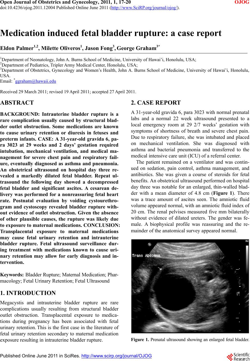 Open Journal of Obstetrics and Gynecology, 2011, 1, 17-20 doi:10.4236/ojog.2011.12004 Published Online June 2011 (http://www.SciRP.org/journal/ojog/ OJOG ). Published Online June 2011 in SciRes. http://www.scirp.org/journal/OJOG Medication induced fetal bladder rupture: a case report Eldon Palmer1,2, Milette Oliveros1, Jason Fong3, George Graham3* 1Department of Neonatology, John A. Burns School of Medicine, University of Hawai’i, Honolulu, USA; 2Department of Pediatrics, Tripler Army Medical Center, Honolulu, USA; 3Department of Obstetrics, Gynecology and Women’s Health, John A. Burns School of Medicine, University of Hawai’i, Honolulu, USA. Email: *[email protected] Received 29 March 2011; revised 19 April 2011; accepted 27 April 2011. ABSTRACT BACKGROUND: Intrauterine bladder rupture is a rare complication usually caused by structural blad- der outlet obstruction. Some medications are known to cause urinary retention or diuresis in fetuses and preterm infants. CASE: A 31-year-old gravida 6, pa- ra 3023 at 29 weeks and 2 days’ gestation required intubation, mechanical ventilation, and medical ma- nagement for severe chest pain and respiratory fail- ure, eventually diagnosed as asthma and pneumonia. An obstetrical ultrasound on hospital day three re- vealed a markedly dilated fetal bladder. Repeat ul- trasound the following day showed a decompressed fetal bladder and significant ascites. A cesarean de- livery was performed for a nonreassuring fetal heart rate. Postnatal evaluation by voiding cystourethro- gram and cystoscopy revealed bladder rupture with- out evidence of outlet obstruction. Given the absence of other plausible causes, the rupture was likely due to exposure to maternal medications. CONCLUSION: Transplacental exposure to maternal medications may cause fetal urinary retention and intrauterine bladder rupture. Fetal ultrasound surveillance dur- ing treatment with medications known to cause uri- nary retention may allow for early diagnosis and in- tervention. Keywords: Bladder Rupture; Maternal Medication; Phar- macology; Fetal Urinary Retention; Fetal Ultrasound 1. INTRODUCTION Megacystis and intrauterine bladder rupture are rare complications usually resulting from structural bladder outlet obstruction. Transplacental exposure to medica- tions during pregnancy has been associated with fetal urinary retention. Th is is the first case in the literature of fetal urinary retention secondary to maternal medication exposure resulting in intrauterine bladder rupture. 2. CASE REPORT A 31-year-old gravida 6, para 302 3 with normal pr enatal labs and a normal 22 week ultrasound presented to a local emergency room at 29 2/7 weeks’ gestation with symptoms of shortness of breath and severe chest pain. Due to respiratory failure, she was intubated and placed on mechanical ventilation. She was diagnosed with asthma and bacterial pneumonia and transferred to the medical intensive care unit (ICU) of a referral center. The patient remained on a ventilator and was contin- ued on sedation, pain control, asthma management, and antibiotics. She was given a course of steroids for fetal benefits. An obstetrical ultrasound performed o n hospital day three was notable for an enlarged, thin-walled blad- der with a mean diameter of 4.8 cm (Figure 1). There was a trace amount of ascites seen. The amniotic fluid volume appeared normal, with an amniotic fluid index of 20 cm. The renal pelvises measured five mm bilaterally without evidence of dilated ureters. The gender was fe- male. A biophysical profile was reassuring and the re- mainder of the anat omical survey appeared normal. Figure 1. Prenatal ultrasound showing an enlarged fetal bladder. 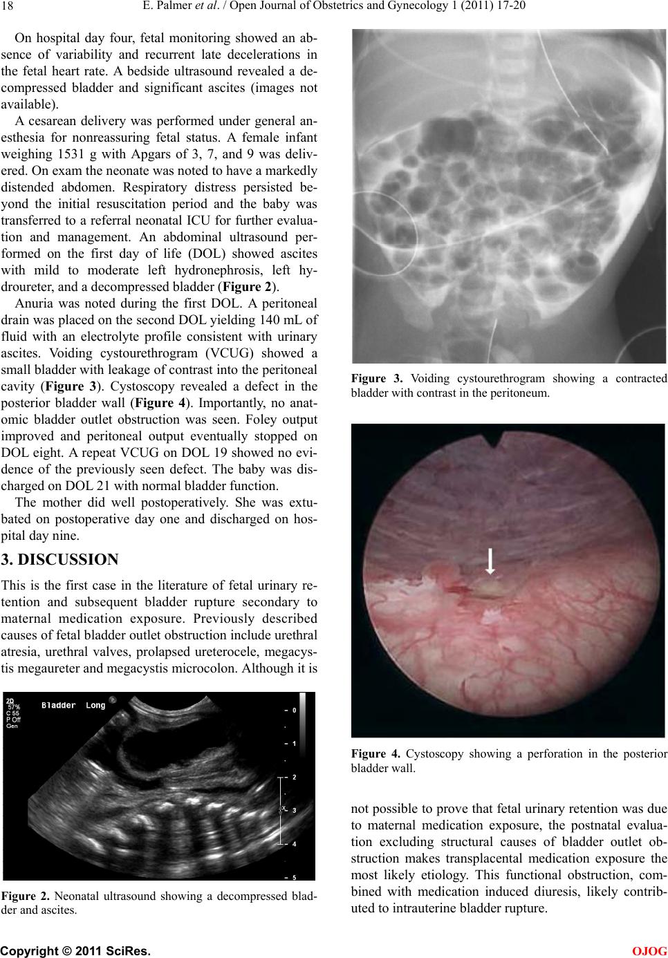 E. Palmer et al. / Open Journal of Obstetrics and Gynecology 1 (2011) 17-20 18 On hospital day four, fetal monitoring showed an ab- sence of variability and recurrent late decelerations in the fetal heart rate. A bedside ultrasound revealed a de- compressed bladder and significant ascites (images not available). A cesarean delivery was performed under general an- esthesia for nonreassuring fetal status. A female infant weighing 1531 g with Apgars of 3, 7, and 9 was deliv- ered. On exam the neonate was noted to have a markedly distended abdomen. Respiratory distress persisted be- yond the initial resuscitation period and the baby was transferred to a referral neonatal ICU for further evalua- tion and management. An abdominal ultrasound per- formed on the first day of life (DOL) showed ascites with mild to moderate left hydronephrosis, left hy- droureter, and a decompressed bladder (Figure 2). Anuria was noted during the first DOL. A peritoneal drain was placed on the second DOL yielding 140 mL of fluid with an electrolyte profile consistent with urinary ascites. Voiding cystourethrogram (VCUG) showed a small bladder with leakage of con trast into the peritoneal cavity (Figure 3). Cystoscopy revealed a defect in the posterior bladder wall (Figure 4). Importantly, no anat- omic bladder outlet obstruction was seen. Foley output improved and peritoneal output eventually stopped on DOL eight. A repeat VCUG on DOL 19 showed no evi- dence of the previously seen defect. The baby was dis- charged on DOL 21 wi th n or mal bladder functi on . The mother did well postoperatively. She was extu- bated on postoperative day one and discharged on hos- pital day nine. 3. DISCUSSION This is the first case in the literature of fetal urinary re- tention and subsequent bladder rupture secondary to maternal medication exposure. Previously described causes of fetal bladder outlet obstruction include urethral atresia, urethral valves, prolapsed ureterocele, megacys- tis megaureter and megacystis microcolon. Although it is Figure 2. Neonatal ultrasound showing a decompressed blad- der and ascites. Figure 3. Voiding cystourethrogram showing a contracted bladder with contrast in the peritoneum. Figure 4. Cystoscopy showing a perforation in the posterior bladder wall. not possible to prove that fetal urinary retention was due to maternal medication exposure, the postnatal evalua- tion excluding structural causes of bladder outlet ob- struction makes transplacental medication exposure the most likely etiology. This functional obstruction, com- bined with medication induced diuresis, likely contrib- uted to intrauterine bladder rupture. C opyright © 2011 SciRes. OJOG 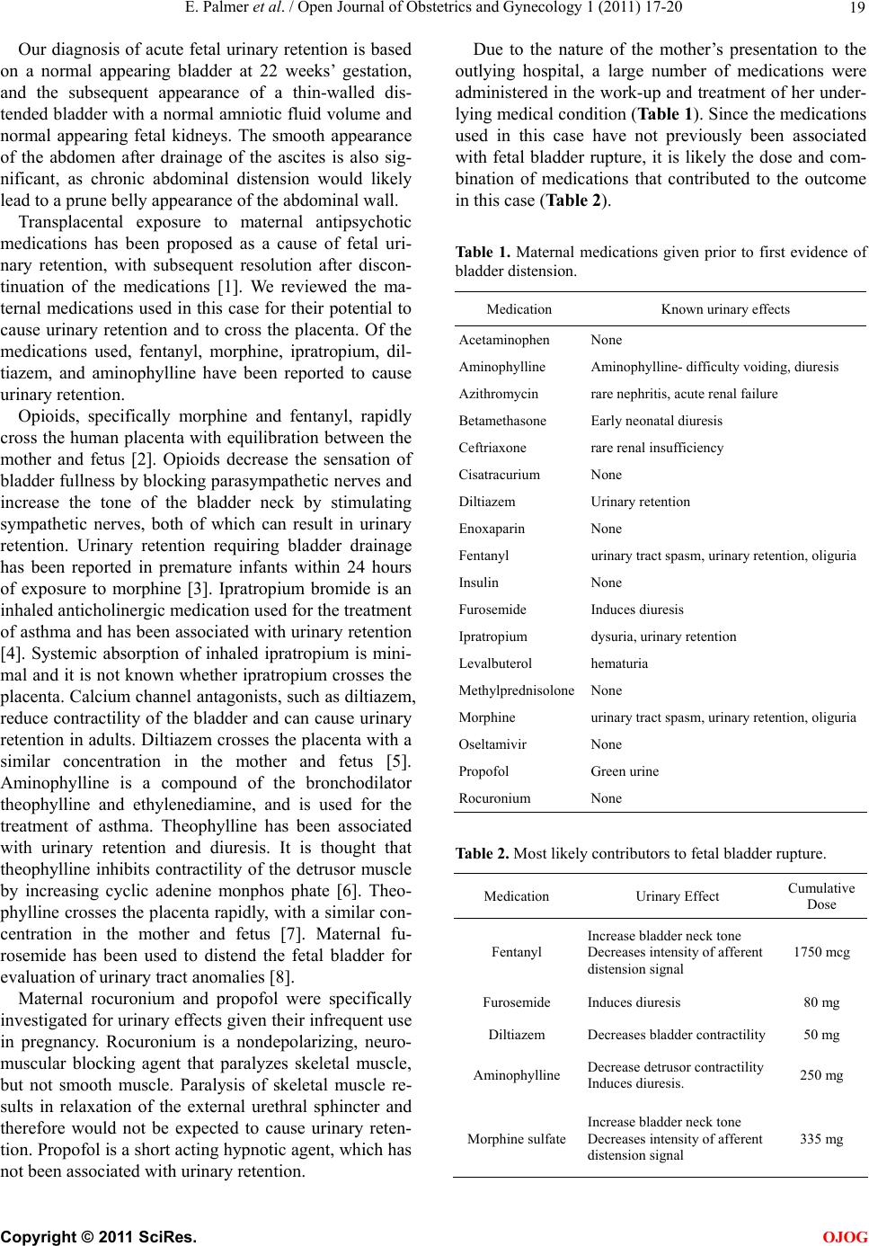 E. Palmer et al. / Open Journal of Obstetrics and Gynecology 1 (2011) 17-20 19 Our diagnosis of acute fetal urinary retention is based on a normal appearing bladder at 22 weeks’ gestation, and the subsequent appearance of a thin-walled dis- tended bladder with a normal amniotic fluid volume and normal appearing fetal kidneys. The smooth appearance of the abdomen after drainage of the ascites is also sig- nificant, as chronic abdominal distension would likely lead to a prune belly app earance of the abdominal wall. Transplacental exposure to maternal antipsychotic medications has been proposed as a cause of fetal uri- nary retention, with subsequent resolution after discon- tinuation of the medications [1]. We reviewed the ma- ternal medications used in this case for their potential to cause urinary retention and to cross the placenta. Of the medications used, fentanyl, morphine, ipratropium, dil- tiazem, and aminophylline have been reported to cause urinary retention. Opioids, specifically morphine and fentanyl, rapidly cross the human placenta with equilibration between the mother and fetus [2]. Opioids decrease the sensation of bladder fullness by blocking parasympathetic nerves and increase the tone of the bladder neck by stimulating sympathetic nerves, both of which can result in urinary retention. Urinary retention requiring bladder drainage has been reported in premature infants within 24 hours of exposure to morphine [3]. Ipratropium bromide is an inhaled anticho linergic medication us ed for th e treatment of asthma and has been associated with urinary retention [4]. Systemic absorption of inhaled ipratropium is mini- mal and it is not known whether ipratropium crosses the placenta. Calcium channel antagonists, such as diltiazem, reduce contractility o f the bladder and can cause urin ary retention in adults. Diltiazem crosses the placenta with a similar concentration in the mother and fetus [5]. Aminophylline is a compound of the bronchodilator theophylline and ethylenediamine, and is used for the treatment of asthma. Theophylline has been associated with urinary retention and diuresis. It is thought that theophylline inhibits contractility of the detrusor muscle by increasing cyclic adenine monphos phate [6]. Theo- phylline crosses the placenta rapidly, with a similar con- centration in the mother and fetus [7]. Maternal fu- rosemide has been used to distend the fetal bladder for evaluation of urinary tract anomalies [8]. Maternal rocuronium and propofol were specifically investigated for urinary effects given their infrequent use in pregnancy. Rocuronium is a nondepolarizing, neuro- muscular blocking agent that paralyzes skeletal muscle, but not smooth muscle. Paralysis of skeletal muscle re- sults in relaxation of the external urethral sphincter and therefore would not be expected to cause urinary reten- tion. Propofol is a short acting h ypnotic ag ent, which has not been associated with urinary retention. Due to the nature of the mother’s presentation to the outlying hospital, a large number of medications were administered in the work-up and treatment of her under- lying medical conditio n (Table 1). Since the medications used in this case have not previously been associated with fetal bladder rupture, it is likely the dose and com- bination of medications that contributed to the outcome in this case (Table 2). Ta b le 1 . Maternal medications given prior to first evidence of bladder distension. Medication Known urinary effect s Acetaminophen None Aminophylline Aminophylline- dif ficulty voiding, diuresis Azithromycin rare nephritis, acute renal failure Betamethasone Early neonatal diuresis Ceftriaxone rare renal insufficiency Cisatracurium None Diltiazem Urinary retention Enoxaparin None Fentanyl urinary tract spasm, urinary retention, oli guria Insulin None Furosemide Induces diuresis Ipratropium dysuria, urinary retention Levalbuterol hematuria MethylprednisoloneNone Morphine urinary tract spasm, urinary retention, oliguria Oseltamivir None Propofol Green urine Rocuronium None Table 2. Most likely contributors to fetal bladder rupture. Medication Urinary Effect Cumulative Dose Fentanyl Increase bladder neck tone Decreases intensity of afferent distension signal 1750 mcg Furosemide Induces diuresis 80 mg Diltiazem Decreases bladder contractility 50 mg Aminophylline Decrease detrusor contractility Induces diuresis. 250 mg Morphine sulfateIncrease bladder neck tone Decreases intensity of afferent distension signal 335 mg C opyright © 2011 SciRes. OJOG 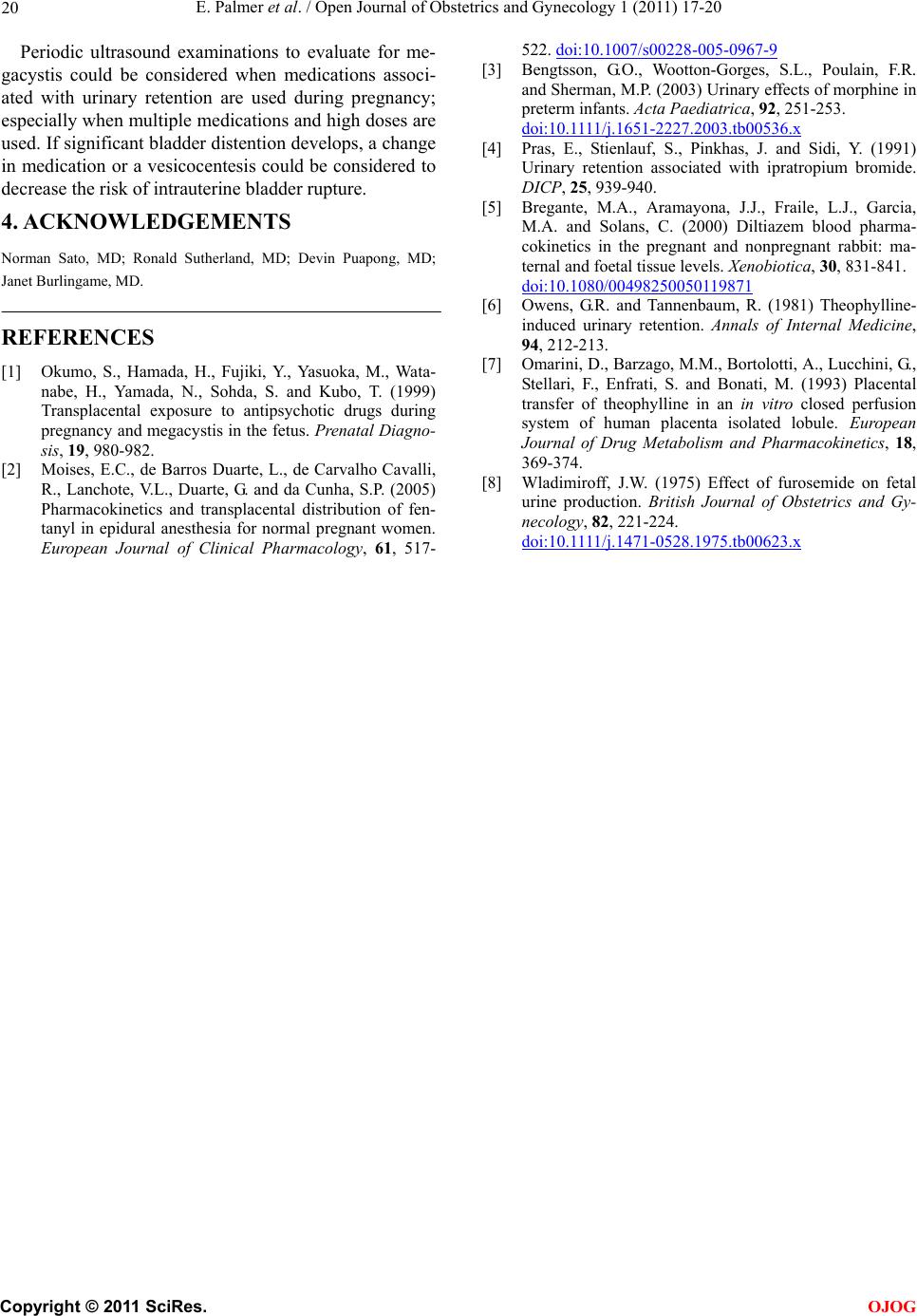 E. Palmer et al. / Open Journal of Obstetrics and Gynecology 1 (2011) 17-20 Copyright © 2011 SciRes. 20 Periodic ultrasound examinations to evaluate for me- gacystis could be considered when medications associ- ated with urinary retention are used during pregnancy; especially when multiple med ications and h igh doses are used. If significant bladder distention develops, a change in medication or a vesicocentesis could be considered to decrease the risk of intrauterine bladder rupture. OJOG 4. ACKNOWLEDGEMENTS Norman Sato, MD; Ronald Sutherland, MD; Devin Puapong, MD; Janet Burlingame, MD. REFERENCES [1] Okumo, S., Hamada, H., Fujiki, Y., Yasuoka, M., Wata- nabe, H., Yamada, N., Sohda, S. and Kubo, T. (1999) Transplacental exposure to antipsychotic drugs during pregnancy and megacystis in the fetus. Prenatal Diagno- sis, 19, 980-982. [2] Moises, E.C., de Barros Duarte, L., de Carvalho Cavalli, R., Lanchote, V.L., Duarte, G. and da Cunha, S.P. (2005) Pharmacokinetics and transplacental distribution of fen- tanyl in epidural anesthesia for normal pregnant women. European Journal of Clinical Pharmacology, 61, 517- 522. doi:10.1007/s00228-005-0967-9 [3] Bengtsson, G.O., Wootton-Gorges, S.L., Poulain, F.R. and Sherman, M.P. (2003) Urinary effects of morphine in preterm infants. Acta Paediatrica, 92, 251-253. d oi: 10 .1111 /j .1 651 -2227.2003.tb00536.x [4] Pras, E., Stienlauf, S., Pinkhas, J. and Sidi, Y. (1991) Urinary retention associated with ipratropium bromide. DICP, 25, 939-940. [5] Bregante, M.A., Aramayona, J.J., Fraile, L.J., Garcia, M.A. and Solans, C. (2000) Diltiazem blood pharma- cokinetics in the pregnant and nonpregnant rabbit: ma- ternal and foet al ti ssue levels. Xenobiotica, 30, 831-841. doi:10.1080/00498250050119871 [6] Owens, G.R. and Tannenbaum, R. (1981) Theophylline- induced urinary retention. Annals of Internal Medicine, 94, 212-213. [7] Omarini, D. , Barzago, M.M., Bortol otti, A., Lucchin i, G., Stellari, F., Enfrati, S. and Bonati, M. (1993) Placental transfer of theophylline in an in vitro closed perfusion system of human placenta isolated lobule. European Journal of Drug Metabolism and Pharmacokinetics, 18, 369-374. [8] Wladimiroff, J.W. (1975) Effect of furosemide on fetal urine production. British Journal of Obstetrics and Gy- necology, 82, 221-224. d oi: 10 .1111 /j .1 471 -0528.1975.tb00623.x |