

Paper Menu >>
Journal Menu >>
 Open Journal of Obstetrics and Gynecology, 2011, 1, 13-16 doi:10.4236/ojog.2011.12003 Published Online June 2011 (http://www.SciRP.org/journal/ojog/ OJOG ). Published Online June 2011 in SciRes. http://www.scirp.org/journal/OJOG Isolated proteinuria as an initial sign of severe preeclampsia Takahir o Ya mada, Takashi Ya mada, Mamoru Morikawa, Masamitsu Takeda, Ryutaro Nishida, Rina Akaishi, Hisanori Minakami Department of Obstetrics, Hokkaido University Graduate School of Medicine, Sapporo, Japan. Email: [email protected] Received 21 April 2011; revised 20 May 2011; accepted 27 May 2011. ABSTRACT Two pregnant women who initially developed pro- teinuria alone followed by serious preeclampsia are presented to emphasize that there is no adequate technical term to express the period of proteinuria alone based on the current criteria of pregnancy- induced hypertension. Case 1 exhibited a urinary protein concentration of 46 mg/dL in the absence of hypertension, and abdominal pain due to placental abruption with hypertension at gestational week (GW) 29–3/7 and 29–4/7, respectively. Case 2 exhibited a urinary protein/creatinine ratio of 2.67, developed hypertension, required cesarean section, and devel- oped posterior reversible encephalopathy syndrome at GW 28–1/7, 29–6/7, and 32–0/7, and on postpartum day 2, respectively. As women with proteinuria alone are not diagnosed as having preeclampsia and as a diagnosis of gestational proteinuria can be made only at 12 weeks postpartum, a prospective technical term applicable to the condition of proteinuria alone is needed to increase physicians’ attention to this co ndi- tion. Keywords: Posterior Reversible Encephalopathy Syndrome; Proteinuria; Placental Abruption; Pregnancy-Induced Hypertension 1. INTRODUCTION Based on the current criteria adopted in many countries, women with proteinuria alone are not diagnosed as hav- ing preeclampsia until they also exhibit hypertension [1]; in Japan, those who do not develop hypertension are diagnosed as having had gestational proteinuria. Thus, gestational proteinuria is a retrospective diagnosis. These criteria may have been based on the belief that clinical presentation of preeclampsia involves initial hyperten- sion and subsequent proteinuria. However, whether some pregnant women who initially exhibit proteinuria subsequently develop hypertension had not been exten- sively studied. The clinical outcomes of such women with gestational proteinuria, defined as those exhibiting transient proteinuria of ≥0.3 g/day appearing at or after 20 weeks of gestation and disappearing by 12 weeks postpartum, had been believed to be favorable, and pro- teinuria had been thought not to be an independent pre- dictor of an adverse outcome [2]. However, a recent report indicated that approximately 50% of women who develop proteinuria ≥0.3 g/day at and after 20 weeks of gestation in the absence of hyper- tension progress to preeclampsia [3]. If this is the case, it is misleading to suggest that the outcome of pregnancy in patients with isolated proteinuria is favorable. Here, we present two patients who showed proteinuria initially and subsequently developed hypertension, and finally experienced severe clinical conditions. We em- phasize that physicians have no adequate technical term to express the period of proteinuria alone in such pa- tients according to the current criteria of pregnancy-in- duced hypertension. 2. PATIENTS AND RESULTS This study was approved by the institutional review board of the Hokkaido University Hospital. Case 1: A 43-year-old nulliparous woman showed blood pressure (BP) of 124/75 mmHg and proteinuria (1 + on dipstick) at a regular antenatal visit at gestational week (GW) 28–3/7 in July 2008. She exhibited BP of 119/78 mmHg, urinary protein concentration of 46 mg/dL (Ta bl e 1 ), and weight gain of 1.7 kg in 1 week, and was diagnosed as having fetal growth restriction (FGR, estimated fetal body weight of 1082 g) the fol- lowing week (GW 29–3/7). Blood chemistry revealed anemia only. The patient requested treatment on an out- patient basis despite our recommendation of hospitaliza- tion. Thirty-four hours later, the patient presented with abdominal pain, hypertension (BP of 176/98 mmHg), and urinary protein concentration of 120 mg/dL. Placen- tal abruption was suspected based on ultrasonography findings and was confirmed by uneventful emergency cesarean section. A female infant with Apgar scores of 3 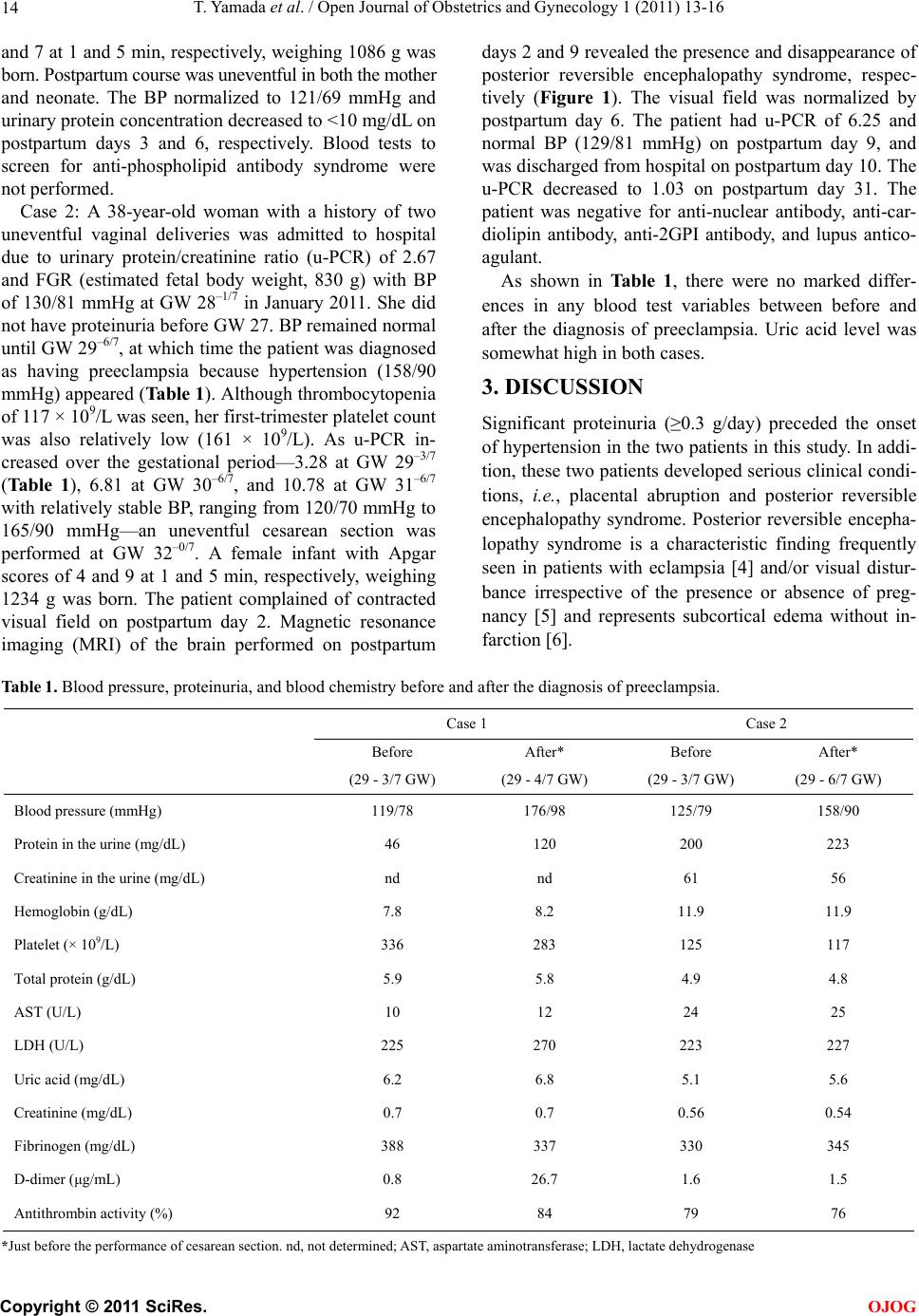 T. Yamada et al. / Open Journal of Obstetrics and Gynecology 1 (2011) 13-16 14 and 7 at 1 and 5 min, respectively, weighing 1086 g was born. Postpartum course was uneventful in both the mother and neonate. The BP normalized to 121/69 mmHg and urinary protein concentration decreased to <10 mg/dL on postpartum days 3 and 6, respectively. Blood tests to screen for anti-phospholipid antibody syndrome were not performed. Case 2: A 38-year-old woman with a history of two uneventful vaginal deliveries was admitted to hospital due to urinary protein/creatinine ratio (u-PCR) of 2.67 and FGR (estimated fetal body weight, 830 g) with BP of 130/81 mmHg at GW 28–1/7 in January 2011. She did not have proteinuria before GW 27. BP remained normal until GW 29–6/7, at which time the patient was diagnosed as having preeclampsia because hypertension (158/90 mmHg) appeared (Table 1). Although thrombocytopenia of 117 × 109/L was seen, her first-trimester platelet count was also relatively low (161 × 109/L). As u-PCR in- creased over the gestational period—3.28 at GW 29–3/7 (Table 1), 6.81 at GW 30–6/7, and 10.78 at GW 31–6/7 with relatively stable BP, ranging from 120/70 mmHg to 165/90 mmHg—an uneventful cesarean section was performed at GW 32–0/7. A female infant with Apgar scores of 4 and 9 at 1 and 5 min, respectively, weighing 1234 g was born. The patient complained of contracted visual field on postpartum day 2. Magnetic resonance imaging (MRI) of the brain performed on postpartum days 2 and 9 revealed the presence and disappearance of posterior reversible encephalopathy syndrome, respec- tively (Figure 1). The visual field was normalized by postpartum day 6. The patient had u-PCR of 6.25 and normal BP (129/81 mmHg) on postpartum day 9, and was discharged from hospital on postpartum day 10. The u-PCR decreased to 1.03 on postpartum day 31. The patient was negative for anti-nuclear antibody, anti-car- diolipin antibody, anti-2GPI antibody, and lupus antico- agulant. As shown in Table 1, there were no marked differ- ences in any blood test variables between before and after the diagnosis of preeclampsia. Uric acid level was somewhat high in both cases. 3. DISCUSSION Significant proteinuria (≥0.3 g/day) preceded the onset of hypertension in the two patients in this study. In addi- tion, these two patients developed serious clinical condi- tions, i.e., placental abruption and posterior reversible encephalopathy syndrome. Posterior reversible encepha- lopathy syndrome is a characteristic finding frequently seen in patients with eclampsia [4] and/or visual distur- bance irrespective of the presence or absence of preg- nancy [5] and represents subcortical edema without in- farction [6]. Table 1. Blood pressure, proteinuria, and blood chemistry before and after the diagnosis of preeclampsia. Case 1 Case 2 Before After* Before After* (29 - 3/7 GW) (29 - 4/7 GW) (29 - 3/7 GW) (29 - 6/7 GW) Blood pressure (mmHg) 119/78 176/98 125/79 158/90 Protein in the urine (mg/dL) 46 120 200 223 Creatinine in the urine (mg/dL) nd nd 61 56 Hemoglobin (g/dL) 7.8 8.2 11.9 11.9 Platelet (× 109/L) 336 283 125 117 Total protein (g/dL) 5.9 5.8 4.9 4.8 AST (U/L) 10 12 24 25 LDH (U/L) 225 270 223 227 Uric acid (mg/dL) 6.2 6.8 5.1 5.6 Creatinine (mg/dL) 0.7 0.7 0.56 0.54 Fibrinogen (mg/dL) 388 337 330 345 D-dimer (μg/mL) 0.8 26.7 1.6 1.5 Antithrombin activity (%) 92 84 79 76 *Just before the performance of cesarean section. nd, not determined; AST, aspartate aminotransferase; LDH, lactate dehydrogenase C opyright © 2011 SciRes. OJOG 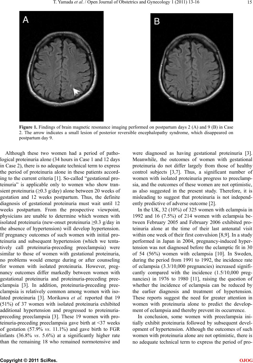 T. Yamada et al. / Open Journal of Obstetrics and Gynecology 1 (2011) 13-16 15 Figure 1. Findings of brain magnetic resonance imaging performed on postpartum days 2 (A) and 9 (B) in Case 2. The arrow indicates a small lesion of posterior reversible encephalopathy syndrome, which disappeared on postpartum day 9. Although these two women had a period of patho- logical proteinuria alone (34 hours in Case 1 and 12 days in Case 2), there is no adequate technical term to express the period of proteinuria alone in these patients accord- ing to the current criteria [1]. So-called “gestational pro- teinuria” is applicable only to women who show tran- sient proteinuria (≥0.3 g/day) alone between 20 weeks of gestation and 12 weeks postpartum. Thus, the definite diagnosis of gestational proteinuria must wait until 12 weeks postpartum. From the prospective viewpoint, physicians are unable to determine which women with isolated proteinuria (new-onset proteinuria ≥0.3 g/day in the absence of hypertension) will develop hypertension. If pregnancy outcomes of such women with initial pro- teinuria and subsequent hypertension (which we tenta- tively call proteinuria-preceding preeclampsia) were similar to those of women with gestational proteinuria, no problems would emerge during or after counseling for women with isolated proteinuria. However, preg- nancy outcomes differ markedly between women with gestational proteinuria and proteinuria-preceding pree- clampsia [3]. In addition, proteinuria-preceding pree- clampsia is relatively common among women with iso- lated proteinuria [3]. Morikawa et al. reported that 19 (51%) of 37 women with isolated proteinuria exhibited additional hypertension and progressed to proteinuria- preceding preeclampsia [3]. These 19 women with pro- teinuria-preceding preeclampsia gave birth at <37 weeks of gestation (57.9% vs. 11.1%) and gave birth to FGR infants (36.8% vs. 5.6%) at a significantly higher rate than the remaining 18 who remained normotensive and were diagnosed as having gestational proteinuria [3]. Meanwhile, the outcomes of women with gestational proteinuria do not differ largely from those of healthy control subjects [3,7]. Thus, a significant number of women with isolated proteinuria progress to preeclamp- sia, and the outcomes of these women are not optimistic, as also suggested in the present study. Therefore, it is misleading to suggest that proteinuria is not independ- ently predictive of adverse outcome [2]. In the UK, 32 (10%) of 325 women with eclampsia in 1992 and 16 (7.5%) of 214 women with eclampsia be- tween February 2005 and February 2006 exhibited pro- teinuria alone at the time of their last antenatal visit within one week of their first convulsion [8,9]. In a study performed in Japan in 2004, pregnancy-induced hyper- tension was not diagnosed before the eclamptic fit in 30 of 54 (56%) women with eclampsia [10]. In Sweden, during the period from 1991 to 1992, the incidence rate of eclampsia (3.3/10,000 pregnancies) increased signifi- cantly compared with the incidence (1.5/10,000 preg- nancies) in 1976 to 1980 [11], raising the question of whether the incidence of eclampsia can be reduced by the earlier diagnosis and treatment of hypertension. These reports suggest the need for greater attention in women with proteinuria alone to predict the develop- ment of eclampsia and thereby prevent its occurrence. In conclusion, some women with preeclampsia ini- tially exhibit proteinuria followed by subsequent devel- opment of hypertension. Although the outcomes of such women with proteinuria alone are not optimistic, there is no adequate technical term to express the period of pro- C opyright © 2011 SciRes. OJOG  T. Yamada et al. / Open Journal of Obstetrics and Gynecology 1 (2011) 13-16 16 teinuria alone according to the current criteria of preg- nancy-induced hypertension. The occurrence of women who develop eclampsia in the presence of proteinuria alone [8,9], in addition to the two women discussed in this report, indicates the need for a prospective technical term that is applicable to the clinical condition of pro- teinuria alone to increase physicians’ attention to preg- nant women with newly developed proteinuria in the absence of hypertension. REFERENCES [1] Report of the National High Blood Pressure Education Program Working Group on High Blood Pressure in Pregnancy. (2000) American Journal of Obstetrics & Gynecology, 183, S1-22. [2] Airoldi, J. and Weinstein, L. (2007) Clinical significance of proteinuria in pregnancy. Obstetrical & Gynecological Survey, 62, 117-124. doi:10.1097/01.ogx.0000253301.55009.ac [3] Morikawa, M., Yamada, T., Yamada, T., Cho, K., Yamada, H., Sakuragi, N. and Minakami, H. (2008) Pregnancy outcome of women who developed proteinuria in the ab- sence of hypertension after mid-gestation. Journal of Pe- rinatal Medicine, 36, 419-424. doi:10.1515/JPM.2008.062 [4] Thomas, S.V. (1998) Neurological aspects of eclampsia. Journal of the Neurological Sciences, 155, 37-43. doi:10.1016/S0022-510X(97)00274-8 [5] Nguyen-Lam, J. and Kiernan, M.C. (2008) Acute cortical blindness due to posterior reversible encephalopathy. Journal of Clinical Neuroscience, 15, 1182-1185. doi:10.1016/j.jocn.2007.04.025 [6] Hinchey, J., Chaves, C., Appignani, B., Breen, J., Pao, L., Wang, A., Pessin, M.S., Lamy, C., Mas, J.-L. and Caplan, L.R. (1996) A reversible leukoencephalopathy syndrome. The New England Journal of Medicine, 334, 494-500. doi:10.1056/NEJM199602223340803 [7] Holston, A.M., Qian, C., Yu, K.F., Epstein, F.H., Karu- manchi and S.A., Levine, R.J. (2009) Circulating angio- genic factors in gestational proteinuria without hyperten- sion. American Journal of Obstetrics & Gynecology, 200, e1-10. [8] Douglas, K.A. and Redman, C.W.G. (1994) Eclampsia in the United Kingdom. British Medical Journal, 309, 1395-1400. [9] Knight, M. (2007) Eclampsia in the United Kingdom 2005. British Journal of Obstetrics and Gynecology, 114 , 1072-1078. [10] Minakami, H., Kubo, T., Takeda, S., Kawabata, M., Ko- bayashi, T. and Nakabayashi, M. (2009) Placental abrup- tion, HELLP syndrome, and Eclampsia in Japan. Nippon Sanka-Fujinka Gakkai Zasshi, 61, 1559-1567. [11] Kullberg, G., Lindeberg, S. and Hanson, U. (2002) Ec- lampsia in Sweden. Hypertension in Pregnancy, 21, 13- 21. doi:10.1081/PRG-120002906 C opyright © 2011 SciRes. OJOG |