

Paper Menu >>
Journal Menu >>
 American Journal of Plant Sciences, 2011, 2, 276-281 doi:10.4236/ajps.2011.22030 Published Online June 2011 (http://www.SciRP.org/journal/ajps) Copyright © 2011 SciRes. AJPS Mango Malformation: I. Toxin Production Associated with Fusarium Pathogens Wafaa Haggag M.1, Hazza M.2, Sehab A.1, Abd El-Wahab M.1 1Department of Plant Pathology National Research Center, Dokki, Cairo, Egypt; 2 Science Faculty, Botany Department, Banha Uni- versity, Banha, Egypt. Email: [email protected] Received April 2nd, 2011; revised May 4th, 2011; accepted May 18th, 2011. ABSTRACT Eight Fusarium species i.e. F. subglutinans, F. solani, F. oxyspoum, F. sterilihyphosum, F. proliferatum, F. monili- forme, F. avena and F. chlamydspore isolated from mango malformed disease were tested for their ability to cause mango malformation disease and their production of moniliformin and total fumonisins (FB1 + FB2) using HPLC. A evaluated for moniliformin production, seven isolates were toxin producers, the production levels ranging from 0.51 to 8.90 µg/ml. The higher levels were produced by Fusarium subglutinans (8.51 µg/ml). Moderate concentrations of moniliformin was produced by F.moniliforme (6.90 µg/ml), F. oxysporum (6.30 µg/ml), F. proliferatum (4.10 μg/ml) and F. sterilihyphosum (1.10 μg/ml). Separation and identification of Fumonisin that was isolated from the pathogen- causing disease are made by (HPLC). A evaluated for total fumonisin production (FB1 + FB2), seven isolates were toxin producers, the production levels ranging from 0.10 to 8.30 µg/ml. The higher levels were produced by F. monili- forme (8.30 µg/ml. Moderate concentrations of fumonisin was produced by F .proliferatum (0.64 µg/ml) and F. subglu- tinans (0.50 µg/ml). Strong positive correlations between moniliformin and total fumonisins (FB1 + FB2) activities and malformation disease incidence by F. subglutinans, F. solani, F. oxyspoum, F. sterilihyphosum, F. proliferatum was observed. Keywords: Fusarium, Mango Malformation, Moniliformin and Fumonisins 1. Introduction Mango (Mangifera indica L.) is the most important fruit grown in tropical and subtropical region of the world. Mango (Mangifera indica L.) is the most important fruit crop in Egypt. Mango Malformation is one of the most destructive mango diseases [1,2]. Losses due to malfor- mation have not been accurately assessed because yield loss is not a linear function of disease severity [3]. Dur- ing a survey of mango plantations in Sindh for investi- gating the association of fungi with mango malformation disease (MMD), six fungal species viz., Fusarium nivale (Fr.) Ces, F. oxysporium, F. moniliforme, F. semitectum, Alternari alternata and Aspergillus niger were isolated and identified on the basis of their colony characteristics and conidial morphology [4]. F. sterilihyphosum and F. proliferatum are first report association with mango malformation in [5]. As noted by [6] mango malforma- tion probably involves two principles;1) the malforma- tion inducing principle (MIP) which works through im- balance in growth substance and in conditioning of cells; 2)The toxic principle (TP) which causes growth retardation and toxicity symptoms. Fuarium species, particularly F. moniliforme var subglutinans, are likely source of MIP and TP, and the causal agent of disease. Singh and Dhillon [7] proposed that ethylene might play role in mango malformation by suppression of apical dominance, causing more isodiametric growth of rachi- des and shortening and thickening of secondary branches of malformed panicles. Ram [8] stated that the most probable role of malformin in the causation of mango malformation might be mediated through alteration of membrane permeability leading to efflux of IAA or its metabolite. He indicates the possibility that malformin antagonizes IAA action through efflux action of auxins of the malformed cells causing loss of apical dominance at a very early stage of panicle development. Malformed mango panicles have been shown to contain reduced lev- els of auxin [7,9,10]. Strains of Fusarium proliferatum, F. subglutinans, F. anthophilum, F. annulatum, F. succisae, F. beomiforme, F. dlamini, F. napiforme, and F. nygamai from a variety of substrates and geographic areas were  Mango Malformation: I. Toxin Production Associated with Fusarium Pathogens277 tested for the production of fumonisin B1 in culture. None of the cultures of F. subglutinans, F. annulatum, F. succisae, or F. beomiforme produced fumonisin B1 in culture. Strains of F. proliferatum produced fumonisin B1 in amounts ranging from 155 to 2936 ppm, of the species tested, F. proliferatum is the most important producer of fumonisin B1 because of its association with corn and animal mycotoxicose such as porcine pulmo- nary edema. F. napiforme and F. nygamai also may be important because of their association with the food grains millet and sorghum [11]. Thus, objective of the present study is to study the pro- duction of toxins by pathogens involved in the causation of malformation. 2. Materials and Methods Fusarium species isolated from mango malformed dis- ease were tested for their ability to cause malformation. Mango seedlings cv. Sedekia (two years old) was inocu- lated with culture filtrate of Fusarium spp. by injection of the apical buds. Sterilized water was used as a control. Transplanted seedlings were monitored for development of malformation. At the end of the experiment (120 days), all surviving seedlings were examined for apical disease symptoms. Data were recorded on symptoms manifesta- tion as diseases incidence and severity (from 1 - 4 scale). 2.1. Determination of Moniliformin and Fumonisin Cultures were initially grown on agar slants for 7 - 10 days. A slant was macerated in 27 ml of sterile water. Aliquots of (2.5 ml) of the resulting suspension were added to 250 ml Erlenmeyer flasks containing 50 ml of inoculated medium made up of ultrapure water (1 L), NH4Cl (3 g), FeSO4·7H2O (0.2 g). MgSO4·7H2O (2 g). KH2PO4 (2 g) peptone (2 g), yeast extract (2 g) malt ex- tract (2 g ) and glucose (20 g) after 48 h of incubation in the dark at 28˚C on rotary shaker (220 rpm, 3.81 cm throw), the suspension was macerated and employed as moniliformin [12]. For production of moniliformin 2.5 ml aliquots were added to 250 ml Erlenmeyer flasks con- taining 50ml of production medium consisting of ul- trapure water, (NH4)2HPO4 (1 g). KH2PO4 (3 g), MgSO4- 7H2O (0.2 g) NaCL (5 g), Sucrose (40 g) and Glycerol (10 g), final pH (6.2). The flasks were covered in alumi- num foil to protect the toxin from the light and incubated for 10 days [13]. 2.1.1. Monilf ormin Analysis A 500 µl aliquots of culture filtrate was loaded onto a waters sep-pak®RP-18column pre-conditioned with wa- ter. The column was eluted with 2 ml of ion-pair solution (990:10 mix of 85:15 water/acetonitrile and 100:48:1.1 MKH2PO4/BU4N+OH [14]. A 100 µl aliquot was chro- matographed on a Lichrosorb®RP-18 250*4.6 MM HPLC with the ion—pair solution described above. The monili- formin was detected with the same detector as above and concentration evaluated by integration at 230 nm. Con- centrations were determined by reference to calibration curves established with standard provided by P. Scott of Health Canada [15]. 2.1.2. Fumoni si n Anal y s i s The cultured were filtered as above and analysis was performed on each replicate flask respectively as follows: A1 ml aliquot of filtrate was applied to abondElut Certify П® (200 mg, varian) column preconditioned by aspiring methanol (6 ml) and water (6 ml) under vacuum. The minicolumns were then washed with water (6 ml) and methanol (6 ml). Fumonisins were eluted with 0.1% trifluoroacetic acid (TFA) in methanol (3 ml). Fumonisin was quantified by HPLC as follows [15]. The TFA/ methanol/methanol fraction from the clean-up column was concentrated to dryness and taken up in 1ml of methanol. A20 µL aliquot was transferred to 2 ml vial and dried under stream of nitrogen. The residue was re- dissolved in 100 µL of 0.05 M sodium borate buffer, pH 8.3 (adjusted with 1NHCl). A freshly prepared solution of 4-fluoro-7-nitrobenzenofurazan (NBD-F) [100µL of 22 Mm NBD-F (Molecular probes Inc.) in 95% ethanol] was added. After heating for 70 Secs at 70˚C, the solu- tion was quenched in an ice bath and made up to 500 µL with a 1:1 mixture of HPLC mobile phases A(0.05 M NaH2PO4/Methanol adjusted to pH 6.3 with 2N NaoH, 1:1) and B (acetonitrile/ H2O, 8.2). A 20 µL aliquot was injected in duplicate onto aLichrosorb®5 µm RP-18250 MM × 4.6 MM column on a varian vista®5500 HPLC with a varian vista® CDS-401 data system. An 11 min linear geadient of о to 100% B followed by a 2 min pla- teau was run at a flow rate of 1 mL/min. the derivatized fumonisins were detected by their fluorescence at 490 nm after excitation at 450 nm (detector: water® model 420-E) are agent blank produced two main peaks at 6.3 min and 12.3 min. Concentrations were determined by refernce to calibration curves of fumonisin isolated in our laboratory (Miller et al., 1994). 2.2. HPLC Analysis and Method Development The HPLC system consisted of a ternary solvent pump (Gynkotek model 480), auto sampler (Gynkotek Gina 50), decade electrochemical detector with a glassy carbon electrode (Antec) and a diode array detector (Gynko- tek340S). Gynko soft software V5.60 was used to control the HPLC system and for data acquisition and analysis. The equipment was supplied by Dionex Softron (Idstein, Germany). Three columns, i.e. Multosphere C18 (3 μm; Copyright © 2011 SciRes. AJPS 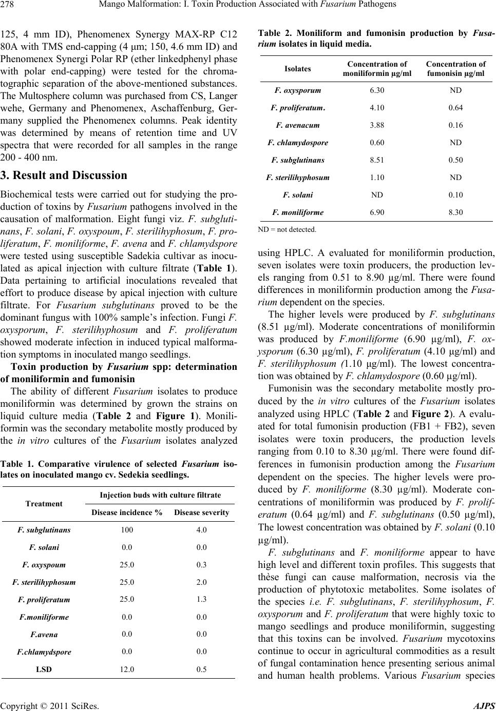 Mango Malformation: I. Toxin Production Associated with Fusarium Pathogens 278 125, 4 mm ID), Phenomenex Synergy MAX-RP C12 80A with TMS end-capping (4 μm; 150, 4.6 mm ID) and Phenomenex Synergi Polar RP (ether linkedphenyl phase with polar end-capping) were tested for the chroma- tographic separation of the above-mentioned substances. The Multosphere column was purchased from CS, Langer wehe, Germany and Phenomenex, Aschaffenburg, Ger- many supplied the Phenomenex columns. Peak identity was determined by means of retention time and UV spectra that were recorded for all samples in the range 200 - 400 nm. 3. Result and Discussion Biochemical tests were carried out for studying the pro- duction of toxins by Fusarium pathogens involved in the causation of malformation. Eight fungi viz. F. subgluti- nans, F. solani, F. oxyspoum, F. sterilihyphosum, F. pro- liferatum, F. moniliforme, F. avena and F. chlamydspore were tested using susceptible Sadekia cultivar as inocu- lated as apical injection with culture filtrate (Table 1). Data pertaining to artificial inoculations revealed that effort to produce disease by apical injection with culture filtrate. For Fusarium subglutinans proved to be the dominant fungus with 100% sample’s infection. Fungi F. oxysporum, F. sterilihyphosum and F. proliferatum showed moderate infection in induced typical malforma- tion symptoms in inoculated mango seedlings. Toxin production by Fusarium spp: determination of moniliformin and fumonisin The ability of different Fusarium isolates to produce moniliformin was determined by grown the strains on liquid culture media (Table 2 and Figure 1). Monili- formin was the secondary metabolite mostly produced by the in vitro cultures of the Fusarium isolates analyzed Table 1. Comparative virulence of selected Fusarium iso- lates on inoculated mango cv. Sedekia seedlings. Injection buds with culture filtrate Treatment Disease incidence % Disease severity F. subglutinans 100 4.0 F. solani 0.0 0.0 F. oxyspoum 25.0 0.3 F. sterilihyphosum 25.0 2.0 F. proliferatum 25.0 1.3 F.moniliforme 0.0 0.0 F.avena 0.0 0.0 F.chlamydspore 0.0 0.0 LSD 12.0 0.5 Table 2. Moniliform and fumonisin production by Fusa- rium isolates in liquid media. Concentration of fumonisin µg/ml Concentration of moniliformin µg/ml Isolates ND 6.30 F. oxysporum 0.64 4.10 F. proliferatum . 0.16 3.88 F. avenacum ND 0.60 F. chlamydospore 0.50 8.51 F. subglutinans ND 1.10 F. sterilihyphosum 0.10 ND F. solani 8.30 6.90 F. moniliforme ND = not detected. using HPLC. A evaluated for moniliformin production, seven isolates were toxin producers, the production lev- els ranging from 0.51 to 8.90 µg/ml. There were found differences in moniliformin production among the Fusa- rium dependent on the species. The higher levels were produced by F. subglutinans (8.51 µg/ml). Moderate concentrations of moniliformin was produced by F.moniliforme (6.90 µg/ml), F. ox- ysporum (6.30 µg/ml), F. proliferatum (4.10 μg/ml) and F. sterilihyphosum (1.10 μg/ml). The lowest concentra- tion was obtained by F. chlamydospore (0.60 µg/ml). Fumonisin was the secondary metabolite mostly pro- duced by the in vitro cultures of the Fusarium isolates analyzed using HPLC (Table 2 and Figure 2). A evalu- ated for total fumonisin production (FB1 + FB2), seven isolates were toxin producers, the production levels ranging from 0.10 to 8.30 µg/ml. There were found dif- ferences in fumonisin production among the Fusarium dependent on the species. The higher levels were pro- duced by F. moniliforme (8.30 µg/ml). Moderate con- centrations of moniliformin was produced by F. prolif- eratum (0.64 µg/ml) and F. subglutinans (0.50 µg/ml), The lowest concentration was obtained by F. solani (0.10 µg/ml). F. subglutinans and F. moniliforme appear to have high level and different toxin profiles. This suggests that thèse fungi can cause malformation, necrosis via the production of phytotoxic metabolites. Some isolates of the species i.e. F. subglutinans, F. sterilihyphosum, F. oxysporum and F. proliferatum that were highly toxic to mango seedlings and produce moniliformin, suggesting that this toxins can be involved. Fusarium mycotoxins continue to occur in agricultural commodities as a result of fungal contamination hence presenting serious animal and human health problems. Various Fusarium species Copyright © 2011 SciRes. AJPS  Mango Malformation: I. Toxin Production Associated with Fusarium Pathogens Copyright © 2011 SciRes. AJPS 279 have recently been found to produce several mycotoxins as moniliformin [9,11]. Moniliformin is formed in many cereals by a number of Fusarium species that include, besides F. moniliforme, F. avenaceum. F. subglutinans, F. proliferatum and others [4]. Malformin-like sub- stances are somehow involved in the causation of mal- formation, with malformin-stimulated ethylene produc- tion [10] causing a hormonal imbalance and conse- quently disturbed metabolism inducing malformation. Related, recently described species that have been Figure 1. HPLC of moniliformin production by Fusarium isolates in culture media. 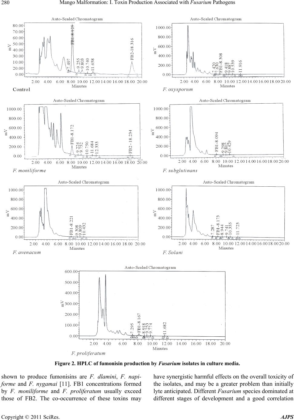 Mango Malformation: I. Toxin Production Associated with Fusarium Pathogens 280 Figure 2. HPLC of fumonisin production by Fusarium isolates in culture media. shown to produce fumonisins are F. dlamini, F. napi- forme and F. nygamai [11]. FB1 concentrations formed by F. moniliforme and F. proliferatum usually exceed those of FB2. The co-occurrence of these toxins may have synergistic harmful effects on the overall toxicity of the isolates, and may be a greater problem than initially lyte anticipated. Different Fusarium species dominated at different stages of development and a good correlation Copyright © 2011 SciRes. AJPS 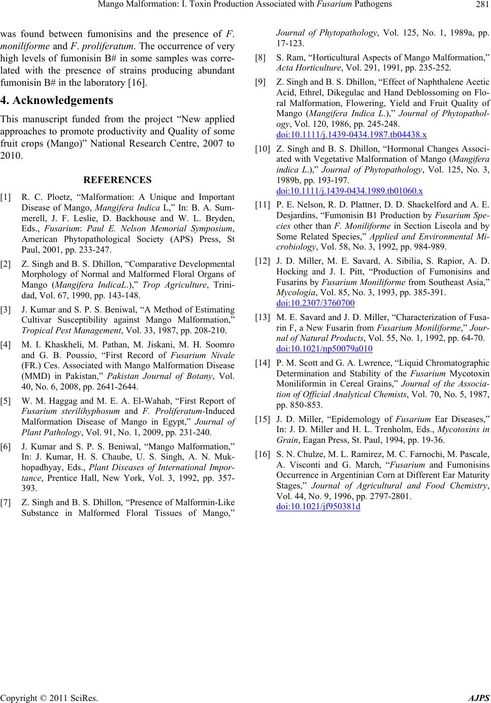 Mango Malformation: I. Toxin Production Associated with Fusarium Pathogens281 was found between fumonisins and the presence of F. moniliforme and F. proliferatum. The occurrence of very high levels of fumonisin B# in some samples was corre- lated with the presence of strains producing abundant fumonisin B# in the laboratory [16]. 4. Acknowledgements This manuscript funded from the project “New applied approaches to promote productivity and Quality of some fruit crops (Mango)” National Research Centre, 2007 to 2010. REFERENCES [1] R. C. Ploetz, “Malformation: A Unique and Important Disease of Mango, Mangifera Indica L,” In: B. A. Sum- merell, J. F. Leslie, D. Backhouse and W. L. Bryden, Eds., Fusarium: Paul E. Nelson Memorial Symposium, American Phytopathological Society (APS) Press, St Paul, 2001, pp. 233-247. [2] Z. Singh and B. S. Dhillon, “Comparative Developmental Morphology of Normal and Malformed Floral Organs of Mango (Mangifera IndicaL.),” Trop Agriculture, Trini- dad, Vol. 67, 1990, pp. 143-148. [3] J. Kumar and S. P. S. Beniwal, “A Method of Estimating Cultivar Susceptibility against Mango Malformation,” Tropical Pest Management, Vol. 33, 1987, pp. 208-210. [4] M. I. Khaskheli, M. Pathan, M. Jiskani, M. H. Soomro and G. B. Poussio, “First Record of Fusarium Nivale (FR.) Ces. Associated with Mango Malformation Disease (MMD) in Pakistan,” Pakistan Journal of Botany, Vol. 40, No. 6, 2008, pp. 2641-2644. [5] W. M. Haggag and M. E. A. El-Wahab, “First Report of Fusarium sterilihyphosum and F. Proliferatum-Induced Malformation Disease of Mango in Egypt,” Journal of Plant Pathology, Vol. 91, No. 1, 2009, pp. 231-240. [6] J. Kumar and S. P. S. Beniwal, “Mango Malformation,” In: J. Kumar, H. S. Chaube, U. S. Singh, A. N. Muk- hopadhyay, Eds., Plant Diseases of International Impor- tance, Prentice Hall, New York, Vol. 3, 1992, pp. 357- 393. [7] Z. Singh and B. S. Dhillon, “Presence of Malformin-Like Substance in Malformed Floral Tissues of Mango,” Journal of Phytopathology, Vol. 125, No. 1, 1989a, pp. 17-123. [8] S. Ram, “Horticultural Aspects of Mango Malformation,” Acta Horticulture, Vol. 291, 1991, pp. 235-252. [9] Z. Singh and B. S. Dhillon, “Effect of Naphthalene Acetic Acid, Ethrel, Dikegulac and Hand Deblossoming on Flo- ral Malformation, Flowering, Yield and Fruit Quality of Mango (Mangifera Indica L.),” Journal of Phytopathol- ogy, Vol. 120, 1986, pp. 245-248. doi:10.1111/j.1439-0434.1987.tb04438.x [10] Z. Singh and B. S. Dhillon, “Hormonal Changes Associ- ated with Vegetative Malformation of Mango (Mangifera indica L.),” Journal of Phytopathology, Vol. 125, No. 3, 1989b, pp. 193-197. doi:10.1111/j.1439-0434.1989.tb01060.x [11] P. E. Nelson, R. D. Plattner, D. D. Shackelford and A. E. Desjardins, “Fumonisin B1 Production by Fusarium Spe- cies other than F. Moniliforme in Section Liseola and by Some Related Species,” Applied and Environmental Mi- crobiology, Vol. 58, No. 3, 1992, pp. 984-989. [12] J. D. Miller, M. E. Savard, A. Sibilia, S. Rapior, A. D. Hocking and J. I. Pitt, “Production of Fumonisins and Fusarins by Fusarium Moniliforme from Southeast Asia,” Mycologia, Vol. 85, No. 3, 1993, pp. 385-391. doi:10.2307/3760700 [13] M. E. Savard and J. D. Miller, “Characterization of Fusa- rin F, a New Fusarin from Fusarium Moniliforme,” Jour- nal of Natural Products, Vol. 55, No. 1, 1992, pp. 64-70. doi:10.1021/np50079a010 [14] P. M. Scott and G. A. Lwrence, “Liquid Chromatographic Determination and Stability of the Fusarium Mycotoxin Moniliformin in Cereal Grains,” Journal of the Associa- tion of Official Analytical Chemists, Vol. 70, No. 5, 1987, pp. 850-853. [15] J. D. Miller, “Epidemology of Fusarium Ear Diseases,” In: J. D. Miller and H. L. Trenholm, Eds., Mycotoxins in Grain, Eagan Press, St. Paul, 1994, pp. 19-36. [16] S. N. Chulze, M. L. Ramirez, M. C. Farnochi, M. Pascale, A. Visconti and G. March, “Fusarium and Fumonisins Occurrence in Argentinian Corn at Different Ear Maturity Stages,” Journal of Agricultural and Food Chemistry, Vol. 44, No. 9, 1996, pp. 2797-2801. doi:10.1021/jf950381d Copyright © 2011 SciRes. AJPS |