

Paper Menu >>
Journal Menu >>
 American Journal of Plant Sciences, 2011, 2, 237-244 doi:10.4236/ajps.2011.22025 Published Online June 2011 (http://www.SciRP.org/journal/ajps) Copyright © 2011 SciRes. AJPS Direct Organogenesis from Cotyledons in Cultivars of Citrus clementina Hort. Ex Tan Giovanna Lombardo1, Roberta Alessandro2, Anna Scialabba1, Mariangela Sciandra3, Fabio De Pasquale4 1Department of Environmental Biology and Biodiversity, University of Palermo, Palermo, Italy; 2Department of Agro and Environ- mental Systems (S.Ag.A.), University of Palermo, Palermo, Italy; 3Department of Mathematical and Statistical Sciences “Silvio Vianelli”, University of Palermo, Palermo, Italy; 4Institute of Plant Genetics, National Council of Research, Research Division Pal- ermo, Palermo, Italy. Email: giolom[email protected] Received March 1st, 2011; revised April 23rd, 2011; accepted April 30th, 2011. ABSTRACT An efficient protocol to induce shoot buds regeneration in Citrus clementina cultivars (“Monreal”, “SRA 63” and “SRA 64”) by direct organogenesis has been developed using cotyledons as explants. Cotyledons transversely cut in three segments and entire ones were cultured on Murashige and Skoog (1962) solidified medium containing vitamins, 500 mg·l−1 malt extract, 50 g·l−1 sucrose and supplemented with three different concentrations of BAP (8.8, 13.2 and 17.6 μM). In all three cultivars the entire cotyledons showed more shoot morphogenic potential than transversely cut ones and after 60 incubation days the optimum BAP concentration was 17.6 μM in “Monreal” (50% ± 2.89% of frequency regeneration) and 13.2 μM in “SRA 63” (33.33% ± 3.33%) and “SRA 64” (25.93% ± 1.85%). In absence of BAP No morphogenesis occurred, demonstrating the absolute requirement of this hormone for shoots induction. The young shoots showed a regular growth in the culture tubes containing the basal medium without hormones, and the rooted plantlets survived after acclimatization. This protocol may find application in Citrus genetic improvement programs. Keywords: Citrus clementina, Cotyledons, Direct Organogenesis, Plant Regeneration, Tissue Culture 1. Introduction Citrus clementina Hort. ex Tanaka is a very important crop because of its excellent organoleptic and easy- peeling qualities. In C. clementina, as in Citrus spp., bud mutations arise spontaneously [1-4] and growers select them to improve horticultural traits of economically in- teresting species; there is also a high degree of inter- specific sexual compatibility, so clementine is highly used in genetic improvement programs as mother plant to obtain new genotypes through hybridization by con- trolled inbreeding. In vitro technique is a useful method to obtain true- to-type regenerated plants [5,6] as well as to induce so- matic mutations [7] affecting characters of agronomic interest in order to develop new and improved genotype lines. There are several studies on plant regeneration by organogenesis and embryogenesis from different types of explants in Citrus genus [8-13], but very little informa- tion is available about procedures for achieving regen- eration from clementine mandarin: embryos were in- duced from calli of ovular tissue [14], aberrant plants were obtained by direct and indirect somatic embryo- genesis from the nucellus of eight cultivars [7], and gy- nogenesis [15] and androgenesis [16-18] were studied in different cultivars. Cotyledons have high potential of regeneration [19,20] and represent a good source of tissue cultures. Organo- genesis from cotyledons was successfully obtained in Pongamia pinnata (L.) [21], Glicine max (L.) Merril [22], Dalbergia sissoo Roxb. [23], Capsicum annum L. [24] and Citrullus lanatus (Thunb.) Matsum. & Nakai [25]. Beyond, in the last decade cotyledons and cotyledonary nodal regions were used as target tissues for transforma- tion mediated by Agrobacterium [20]. The aim of this work was to induce in vitro plant re- generation in Citrus clementina to be used in Citrus ge- netic improvement programs. Three cultivars were inves- tigated in the experiments and cotyledons were used as explants. 2. Materials and Methods Ripe fruits of C. clementina “Monreal”, “SRA 63” and 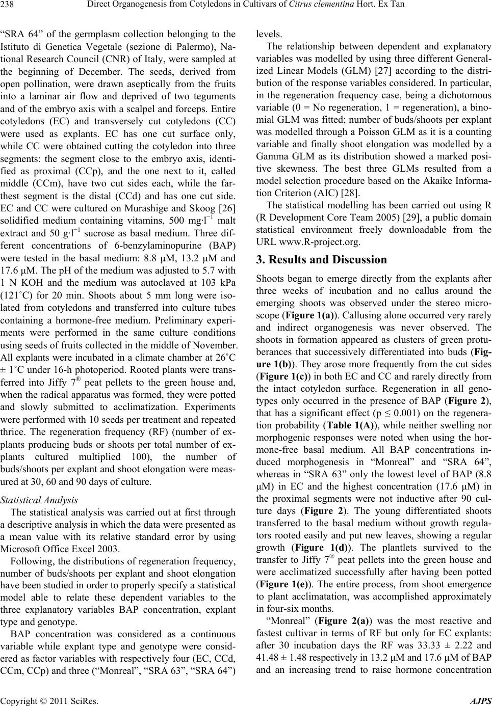 Direct Organogenesis from Cotyledons in Cultivars of Citrus clementina Hort. Ex Tan 238 “SRA 64” of the germplasm collection belonging to the Istituto di Genetica Vegetale (sezione di Palermo), Na- tional Research Council (CNR) of Italy, were sampled at the beginning of December. The seeds, derived from open pollination, were drawn aseptically from the fruits into a laminar air flow and deprived of two teguments and of the embryo axis with a scalpel and forceps. Entire cotyledons (EC) and transversely cut cotyledons (CC) were used as explants. EC has one cut surface only, while CC were obtained cutting the cotyledon into three segments: the segment close to the embryo axis, identi- fied as proximal (CCp), and the one next to it, called middle (CCm), have two cut sides each, while the far- thest segment is the distal (CCd) and has one cut side. EC and CC were cultured on Murashige and Skoog [26] solidified medium containing vitamins, 500 mg·l−1 malt extract and 50 g·l−1 sucrose as basal medium. Three dif- ferent concentrations of 6-benzylaminopurine (BAP) were tested in the basal medium: 8.8 μM, 13.2 μM and 17.6 μM. The pH of the medium was adjusted to 5.7 with 1 N KOH and the medium was autoclaved at 103 kPa (121˚C) for 20 min. Shoots about 5 mm long were iso- lated from cotyledons and transferred into culture tubes containing a hormone-free medium. Preliminary experi- ments were performed in the same culture conditions using seeds of fruits collected in the middle of November. All explants were incubated in a climate chamber at 26˚C ± 1˚C under 16-h photoperiod. Rooted plants were trans- ferred into Jiffy 7® peat pellets to the green house and, when the radical apparatus was formed, they were potted and slowly submitted to acclimatization. Experiments were performed with 10 seeds per treatment and repeated thrice. The regeneration frequency (RF) (number of ex- plants producing buds or shoots per total number of ex- plants cultured multiplied 100), the number of buds/shoots per explant and shoot elongation were meas- ured at 30, 60 and 90 days of culture. Statistical Analysis The statistical analysis was carried out at first through a descriptive analysis in which the data were presented as a mean value with its relative standard error by using Microsoft Office Excel 2003. Following, the distributions of regeneration frequency, number of buds/shoots per explant and shoot elongation have been studied in order to properly specify a statistical model able to relate these dependent variables to the three explanatory variables BAP concentration, explant type and genotype. BAP concentration was considered as a continuous variable while explant type and genotype were consid- ered as factor variables with respectively four (EC, CCd, CCm, CCp) and three (“Monreal”, “SRA 63”, “SRA 64”) levels. The relationship between dependent and explanatory variables was modelled by using three different General- ized Linear Models (GLM) [27] according to the distri- bution of the response variables considered. In particular, in the regeneration frequency case, being a dichotomous variable (0 = No regeneration, 1 = regeneration), a bino- mial GLM was fitted; number of buds/shoots per explant was modelled through a Poisson GLM as it is a counting variable and finally shoot elongation was modelled by a Gamma GLM as its distribution showed a marked posi- tive skewness. The best three GLMs resulted from a model selection procedure based on the Akaike Informa- tion Criterion (AIC) [28]. The statistical modelling has been carried out using R (R Development Core Team 2005) [29], a public domain statistical environment freely downloadable from the URL www.R-project.org. 3. Results and Discussion Shoots began to emerge directly from the explants after three weeks of incubation and no callus around the emerging shoots was observed under the stereo micro- scope (Figure 1(a)). Callusing alone occurred very rarely and indirect organogenesis was never observed. The shoots in formation appeared as clusters of green protu- berances that successively differentiated into buds (Fig- ure 1(b)). They arose more frequently from the cut sides (Figure 1(c)) in both EC and CC and rarely directly from the intact cotyledon surface. Regeneration in all geno- types only occurred in the presence of BAP (Figure 2), that has a significant effect (p ≤ 0.001) on the regenera- tion probability (Table 1(A)), while neither swelling nor morphogenic responses were noted when using the hor- mone-free basal medium. All BAP concentrations in- duced morphogenesis in “Monreal” and “SRA 64”, whereas in “SRA 63” only the lowest level of BAP (8.8 μM) in EC and the highest concentration (17.6 μM) in the proximal segments were not inductive after 90 cul- ture days (Figure 2). The young differentiated shoots transferred to the basal medium without growth regula- tors rooted easily and put new leaves, showing a regular growth (Figure 1(d)). The plantlets survived to the transfer to Jiffy 7® peat pellets into the green house and were acclimatized successfully after having been potted (Figure 1(e)). The entire process, from shoot emergence to plant acclimatation, was accomplished approximately in four-six months. “Monreal” (Figure 2(a)) was the most reactive and fastest cultivar in terms of RF but only for EC explants: after 30 incubation days the RF was 33.33 ± 2.22 and 41.48 ± 1.48 respectively in 13.2 μM and 17.6 μM of BAP and an increasing trend to raise hormone concentration Copyright © 2011 SciRes. AJPS 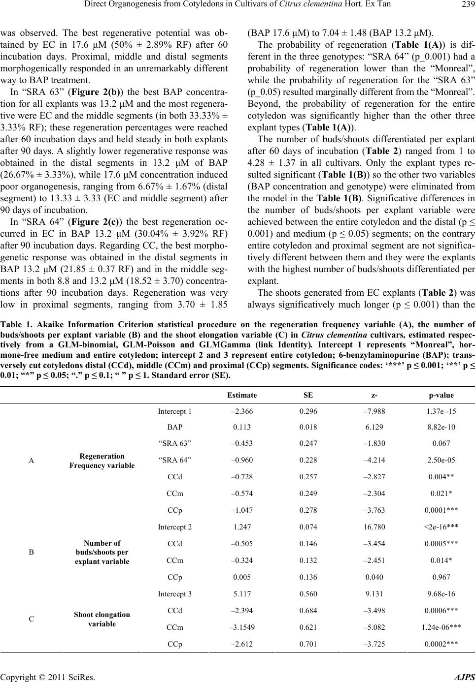 Direct Organogenesis from Cotyledons in Cultivars of Citrus clementina Hort. Ex Tan Copyright © 2011 SciRes. AJPS 239 was observed. The best regenerative potential was ob- tained by EC in 17.6 μM (50% ± 2.89% RF) after 60 incubation days. Proximal, middle and distal segments morphogenically responded in an unremarkably different way to BAP treatment. In “SRA 63” (Figure 2(b)) the best BAP concentra- tion for all explants was 13.2 μM and the most regenera- tive were EC and the middle segments (in both 33.33% ± 3.33% RF); these regeneration percentages were reached after 60 incubation days and held steady in both explants after 90 days. A slightly lower regenerative response was obtained in the distal segments in 13.2 μM of BAP (26.67% ± 3.33%), while 17.6 μM concentration induced poor organogenesis, ranging from 6.67% ± 1.67% (distal segment) to 13.33 ± 3.33 (EC and middle segment) after 90 days of incubation. In “SRA 64” (Figure 2(c)) the best regeneration oc- curred in EC in BAP 13.2 μM (30.04% ± 3.92% RF) after 90 incubation days. Regarding CC, the best morpho- genetic response was obtained in the distal segments in BAP 13.2 μM (21.85 ± 0.37 RF) and in the middle seg- ments in both 8.8 and 13.2 μM (18.52 ± 3.70) concentra- tions after 90 incubation days. Regeneration was very low in proximal segments, ranging from 3.70 ± 1.85 (BAP 17.6 μM) to 7.04 ± 1.48 (BAP 13.2 μM). The probability of regeneration (Table 1(A)) is dif- ferent in the three genotypes: “SRA 64” (p_0.001) had a probability of regeneration lower than the “Monreal”, while the probability of regeneration for the “SRA 63” (p_0.05) resulted marginally different from the “Monreal”. Beyond, the probability of regeneration for the entire cotyledon was significantly higher than the other three explant types (Table 1(A)). The number of buds/shoots differentiated per explant after 60 days of incubation (Table 2) ranged from 1 to 4.28 ± 1.37 in all cultivars. Only the explant types re- sulted significant (Table 1(B)) so the other two variables (BAP concentration and genotype) were eliminated from the model in the Table 1(B). Significative differences in the number of buds/shoots per explant variable were achieved between the entire cotyledon and the distal (p ≤ 0.001) and medium (p ≤ 0.05) segments; on the contrary entire cotyledon and proximal segment are not significa- tively different between them and they were the explants with the highest number of buds/shoots differentiated per explant. The shoots generated from EC explants (Table 2) was always significatively much longer (p ≤ 0.001) than the Table 1. Akaike Information Criterion statistical procedure on the regeneration frequency variable (A), the number of buds/shoots per explant variable (B) and the shoot elongation variable (C) in Citrus clementina cultivars, estimated respec- tively from a GLM-binomial, GLM-Poisson and GLMGamma (link Identity). Intercept 1 represents “Monreal”, hor- mone-free medium and entire cotyledon; intercept 2 and 3 represent entire cotyledon; 6-benzylaminopurine (BAP); trans- versely cut cotyledons distal (CCd), middle (CCm) and proximal (CCp) segments. Significance codes: ‘***’ p ≤ 0.001; ‘**’ p ≤ 0.01; “*” p ≤ 0.05; “.” p ≤ 0.1; “ ” p ≤ 1. Standard error (SE). Estimate SE z- p-value Intercept 1 –2.366 0.296 –7.988 1.37e -15 BAP 0.113 0.018 6.129 8.82e-10 “SRA 63” –0.453 0.247 –1.830 0.067 “SRA 64” –0.960 0.228 –4.214 2.50e-05 CCd –0.728 0.257 –2.827 0.004** CCm –0.574 0.249 –2.304 0.021* A Regeneration Frequency variable CCp –1.047 0.278 –3.763 0.0001*** Intercept 2 1.247 0.074 16.780 <2e-16*** CCd –0.505 0.146 –3.454 0.0005*** CCm –0.324 0.132 –2.451 0.014* B Number of buds/shoots per explant variable CCp 0.005 0.136 0.040 0.967 Intercept 3 5.117 0.560 9.131 9.68e-16 CCd –2.394 0.684 –3.498 0.0006*** CCm –3.1549 0.621 –5.082 1.24e-06*** C Shoot elongation variable CCp –2.612 0.701 –3.725 0.0002*** 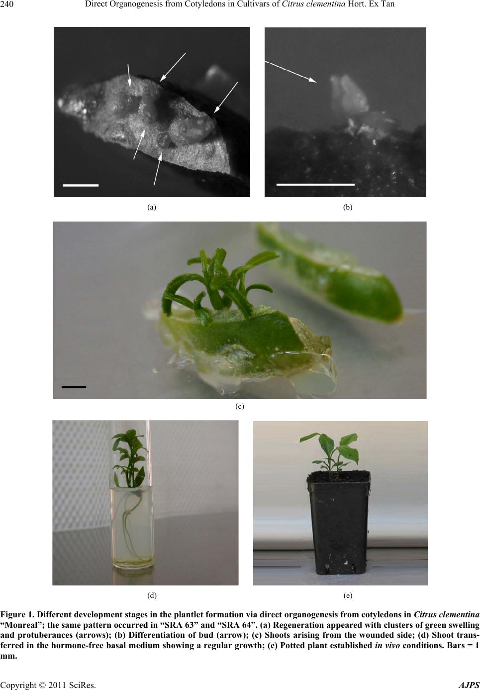 Direct Organogenesis from Cotyledons in Cultivars of Citrus clementina Hort. Ex Tan 240 (a) (b) (c) (d) (e) Figure 1. Different development stages in the plantlet formation via direct organogenesis from cotyledons in Citrus clementina “Monreal”; the same pattern occurred in “SRA 63” and “SRA 64”. (a) Regeneration appeared with clusters of green swelling and protuberances (arrows); (b) Differentiation of bud (arrow); (c) Shoots arising from the wounded side; (d) Shoot trans- ferred in the hormone-free basal medium showing a regular growth; (e) Potted plant established in vivo conditions. Bars = 1 mm. Copyright © 2011 SciRes. AJPS 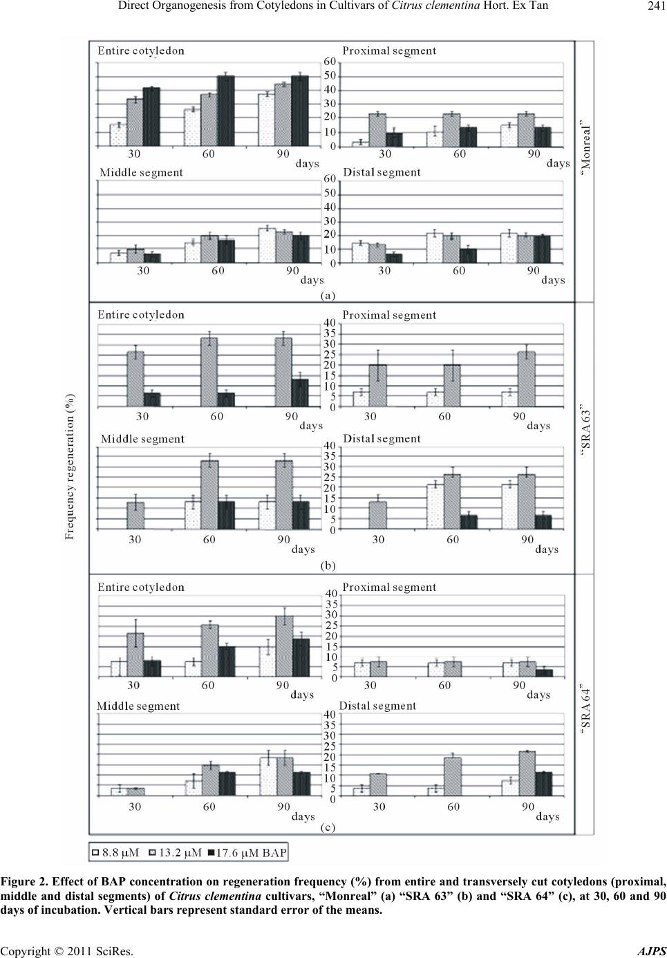 Direct Organogenesis from Cotyledons in Cultivars of Citrus clementina Hort. Ex Tan241 Figure 2. Effect of BAP concentration on regeneration frequency (%) from entire and transversely cut cotyledons (proximal, middle and distal segments) of Citrus clementina cultivars, “Monreal” (a) “SRA 63” (b) and “SRA 64” (c), at 30, 60 and 90 days of incubation. Vertical bars represent standard error of the means. Copyright © 2011 SciRes. AJPS 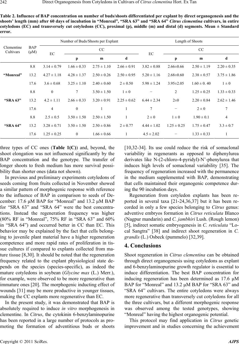 Direct Organogenesis from Cotyledons in Cultivars of Citrus clementina Hort. Ex Tan Copyright © 2011 SciRes. AJPS 242 Table 2. Influence of BAP concentration on number of buds/shoots differentiated per explant by direct organogenesis and the shoots’ length (mm) after 60 days of incubation in “Monreal”, “SRA 63” and “SRA 64” Citrus clementina cultivars, in entire cotyledons (EC) and transversely cut cotyledons (CC), proximal (p), middle (m) and distal (d) segments. Mean ± Standard error. Number of Buds/Shoots per Explant Length of Shoots CC CC Clementine Cultivars BAP (μM) EC p m d EC p m d 8.8 3.14 ± 0.791.66 ± 0.33 2.75 ± 1.102.66 ± 0.913.82 ± 0.882.66±0.66 2.50 ± 1.19 2.20 ± 0.35 13.2 4.27 ± 1.184.28 ± 1.37 2.50 ± 0.262.50 ± 0.955.20 ± 1.162.68±0.60 2.38 ± 0.57 3.75 ± 1.86 “Monreal” 17.6 3.6 ± 0.68 3.25 ± 1.10 2.40 ± 0.602 ± 0.50 5.98 ± 1.243.95±2.05 1.60 ± 0. 40 1 ± 0 8.8 0 7 3.50 ± 1.501 ± 0 − 2 1.25 ± 0.25 1.33 ± 0.33 13.2 4.2 ± 1.11 2.66 ± 0.33 3.20 ± 0.912.25 ± 0.626.44 ± 2.342±0 2.20 ± 0.84 2.62 ± 1.46 “SRA 63” 17.6 4 0 1 1 7 − 2 ± 0 7 8.8 2.5 ± 0.5 3.50 ± 1.50 2.50 ± 1.501 2 ± 0 1 ± 0 1.90 ± 0.1 4 13.2 3.28 ± 0.713.50 ± 1.50 2.50 ± 0.862 ± 0.77 4.44 ± 1.021.25 ± 0.25 1.75 ± 0.47 3.3 ± 0.7 “SRA 64” 17.6 1.25 ± 0.250 1.66 ± 0.661 4.5 ± 2.02 − 1.33 ± 0.33 1 three types of CC ones (Table 1(C)) and, beyond, the shoot elongation was not influenced significantly by the BAP concentration and the genotype. The transfer of longer shoots to fresh medium has more survival possi- bility than shorter ones (data not shown). In previous and preliminary experiments cotyledons of seeds coming from fruits collected in November showed a similar pattern of morphogenic response with reference to the influence of BAP in comparison to seeds of De- cember: 17.6 μM BAP for “Monreal” and 13.2 μM BAP for “SRA 63” and “SRA 64” were the best concentra- tions. Instead the regeneration frequency was higher (80% RF in “Monreal”, 75% RF in “SRA 63” and 60% in “SRA 64”) and occurred better in CC than EC. This behavior may be explained by the fact that cells belong- ing to juvenile plant material have a higher regeneration competence and more rapid rates of proliferation in tis- sue cultures if compared to explants collected from ma- ture tissue [8,30]. It should be noted that the regeneration frequency related to the explant physiological state de- pends on the species (species-specific), as indeed the mature cotyledons in soybean (Glycine max (L.) Merr.), for example, were observed to be more regenerative than immature ones [20]. The morphogenic inducting effect of wounds [31] may be more productive in younger tissues, making the CC explants more regenerative than EC. In the present study, it was demonstrated that BAP is absolutely required to induce in vitro morphogenesis in clementine. In Citrus, the cytokinin 6-benzylaminopurine has been reported in a large number of protocols as pro- moting the formation of adventitious buds or shoots [10,32-34]. Its use could reduce the risk of somaclonal variability in regenerants as opposed to diphenylurea derivates like N-(2-chloro-4-pyridyl)-N’-phenylurea that induces high levels of somaclonal variability [35]. The frequency of regeneration increased with the permanence in the medium supplemented with BAP, demonstrating that cells maintained their organogenic competence dur- ing the 90 incubation days. Regeneration from cotyledon explants has been re- ported in several taxa [21-24,36,37] but it has been re- corded in only a few species belonging to Citrus genus: adventive embryos formation in Citrus reticulata Blanco (Nagpur mandarin) and C. jambhiri Lush. (Rough lemon) [5], indirect somatic embryogenesis in C. reticulata “Lo- cal Sangtra” [38] and indirect shoot regeneration in C. grandis (L.) Osbeck (pummelo) [32,39]. 4. Conclusions Shoot regeneration in Citrus clementina can be obtained through direct organogenesis using cotyledons as explant and 6-benzylaminopurine growth regulator is essential to induce differentiation. The best BAP concentration for inducing regeneration has been determined as 17.6 μM BAP for “Monreal” and 13.2 μM BAP for “SRA 63” and “SRA 64” cultivars. The entire cotyledons were always more regenerative than transversely cut cotyledons for all the three cultivars, but a different morphogenic response was observed among the tested genotypes, showing “Monreal” having the highest organogenic potential. This protocol may find application in Citrus genetic improvement and in studies concerning the achievement 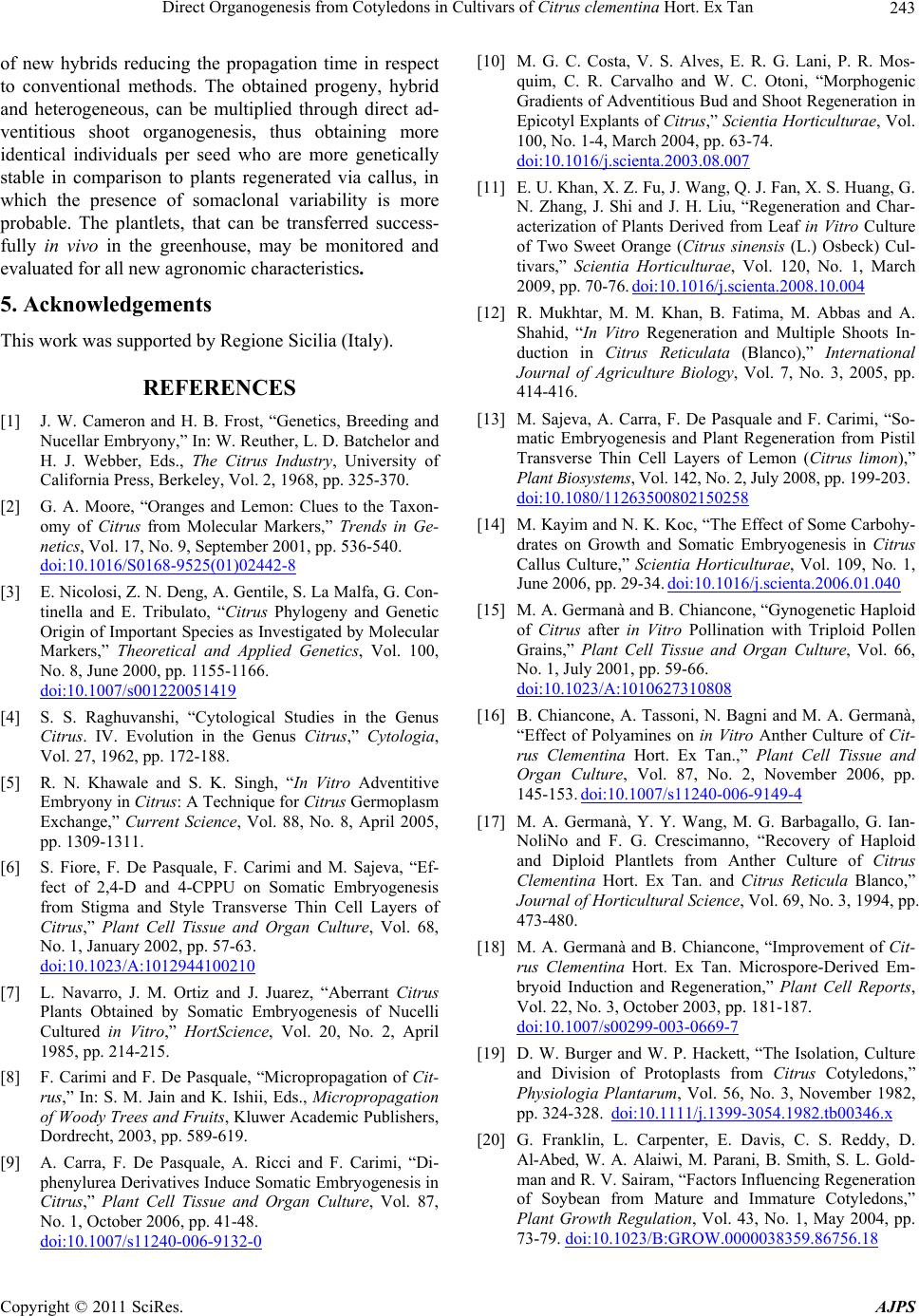 Direct Organogenesis from Cotyledons in Cultivars of Citrus clementina Hort. Ex Tan243 of new hybrids reducing the propagation time in respect to conventional methods. The obtained progeny, hybrid and heterogeneous, can be multiplied through direct ad- ventitious shoot organogenesis, thus obtaining more identical individuals per seed who are more genetically stable in comparison to plants regenerated via callus, in which the presence of somaclonal variability is more probable. The plantlets, that can be transferred success- fully in vivo in the greenhouse, may be monitored and evaluated for all new agronomic characteristics. 5. Acknowledgements This work was supported by Regione Sicilia (Italy). REFERENCES [1] J. W. Cameron and H. B. Frost, “Genetics, Breeding and Nucellar Embryony,” In: W. Reuther, L. D. Batchelor and H. J. Webber, Eds., The Citrus Industry, University of California Press, Berkeley, Vol. 2, 1968, pp. 325-370. [2] G. A. Moore, “Oranges and Lemon: Clues to the Taxon- omy of Citrus from Molecular Markers,” Trends in Ge- netics, Vol. 17, No. 9, September 2001, pp. 536-540. doi:10.1016/S0168-9525(01)02442-8 [3] E. Nicolosi, Z. N. Deng, A. Gentile, S. La Malfa, G. Con- tinella and E. Tribulato, “Citrus Phylogeny and Genetic Origin of Important Species as Investigated by Molecular Markers,” Theoretical and Applied Genetics, Vol. 100, No. 8, June 2000, pp. 1155-1166. doi:10.1007/s001220051419 [4] S. S. Raghuvanshi, “Cytological Studies in the Genus Citrus. IV. Evolution in the Genus Citrus,” Cytologia, Vol. 27, 1962, pp. 172-188. [5] R. N. Khawale and S. K. Singh, “In Vitro Adventitive Embryony in Citrus: A Technique for Citrus Germoplasm Exchange,” Current Science, Vol. 88, No. 8, April 2005, pp. 1309-1311. [6] S. Fiore, F. De Pasquale, F. Carimi and M. Sajeva, “Ef- fect of 2,4-D and 4-CPPU on Somatic Embryogenesis from Stigma and Style Transverse Thin Cell Layers of Citrus,” Plant Cell Tissue and Organ Culture, Vol. 68, No. 1, January 2002, pp. 57-63. doi:10.1023/A:1012944100210 [7] L. Navarro, J. M. Ortiz and J. Juarez, “Aberrant Citrus Plants Obtained by Somatic Embryogenesis of Nucelli Cultured in Vitro,” HortScience, Vol. 20, No. 2, April 1985, pp. 214-215. [8] F. Carimi and F. De Pasquale, “Micropropagation of Cit- rus,” In: S. M. Jain and K. Ishii, Eds., Micropropagation of Woody Trees and Fruits, Kluwer Academic Publishers, Dordrecht, 2003, pp. 589-619. [9] A. Carra, F. De Pasquale, A. Ricci and F. Carimi, “Di- phenylurea Derivatives Induce Somatic Embryogenesis in Citrus,” Plant Cell Tissue and Organ Culture, Vol. 87, No. 1, October 2006, pp. 41-48. doi:10.1007/s11240-006-9132-0 [10] M. G. C. Costa, V. S. Alves, E. R. G. Lani, P. R. Mos- quim, C. R. Carvalho and W. C. Otoni, “Morphogenic Gradients of Adventitious Bud and Shoot Regeneration in Epicotyl Explants of Citrus,” Scientia Horticulturae, Vol. 100, No. 1-4, March 2004, pp. 63-74. doi:10.1016/j.scienta.2003.08.007 [11] E. U. Khan, X. Z. Fu, J. Wang, Q. J. Fan, X. S. Huang, G. N. Zhang, J. Shi and J. H. Liu, “Regeneration and Char- acterization of Plants Derived from Leaf in Vitro Culture of Two Sweet Orange (Citrus sinensis (L.) Osbeck) Cul- tivars,” Scientia Horticulturae, Vol. 120, No. 1, March 2009, pp. 70-76. doi:10.1016/j.scienta.2008.10.004 [12] R. Mukhtar, M. M. Khan, B. Fatima, M. Abbas and A. Shahid, “In Vitro Regeneration and Multiple Shoots In- duction in Citrus Reticulata (Blanco),” International Journal of Agriculture Biology, Vol. 7, No. 3, 2005, pp. 414-416. [13] M. Sajeva, A. Carra, F. De Pasquale and F. Carimi, “So- matic Embryogenesis and Plant Regeneration from Pistil Transverse Thin Cell Layers of Lemon (Citrus limon),” Plant Biosystems, Vol. 142, No. 2, July 2008, pp. 199-203. doi:10.1080/11263500802150258 [14] M. Kayim and N. K. Koc, “The Effect of Some Carbohy- drates on Growth and Somatic Embryogenesis in Citrus Callus Culture,” Scientia Horticulturae, Vol. 109, No. 1, June 2006, pp. 29-34. doi:10.1016/j.scienta.2006.01.040 [15] M. A. Germanà and B. Chiancone, “Gynogenetic Haploid of Citrus after in Vitro Pollination with Triploid Pollen Grains,” Plant Cell Tissue and Organ Culture, Vol. 66, No. 1, July 2001, pp. 59-66. doi:10.1023/A:1010627310808 [16] B. Chiancone, A. Tassoni, N. Bagni and M. A. Germanà, “Effect of Polyamines on in Vitro Anther Culture of Cit- rus Clementina Hort. Ex Tan.,” Plant Cell Tissue and Organ Culture, Vol. 87, No. 2, November 2006, pp. 145-153. doi:10.1007/s11240-006-9149-4 [17] M. A. Germanà, Y. Y. Wang, M. G. Barbagallo, G. Ian- NoliNo and F. G. Crescimanno, “Recovery of Haploid and Diploid Plantlets from Anther Culture of Citrus Clementina Hort. Ex Tan. and Citrus Reticula Blanco,” Journal of Horticultural Science, Vol. 69, No. 3, 1994, pp. 473-480. [18] M. A. Germanà and B. Chiancone, “Improvement of Cit- rus Clementina Hort. Ex Tan. Microspore-Derived Em- bryoid Induction and Regeneration,” Plant Cell Reports, Vol. 22, No. 3, October 2003, pp. 181-187. doi:10.1007/s00299-003-0669-7 [19] D. W. Burger and W. P. Hackett, “The Isolation, Culture and Division of Protoplasts from Citrus Cotyledons,” Physiologia Plantarum, Vol. 56, No. 3, November 1982, pp. 324-328. doi:10.1111/j.1399-3054.1982.tb00346.x [20] G. Franklin, L. Carpenter, E. Davis, C. S. Reddy, D. Al-Abed, W. A. Alaiwi, M. Parani, B. Smith, S. L. Gold- man and R. V. Sairam, “Factors Influencing Regeneration of Soybean from Mature and Immature Cotyledons,” Plant Growth Regulation, Vol. 43, No. 1, May 2004, pp. 73-79. doi:10.1023/B:GROW.0000038359.86756.18 Copyright © 2011 SciRes. AJPS  Direct Organogenesis from Cotyledons in Cultivars of Citrus clementina Hort. Ex Tan Copyright © 2011 SciRes. AJPS 244 [21] K. Sujatha, B. M. Panda and S. Hazra, “De Novo Or- ganogenesis and Plant Regeneration in Pongamia Pinnata, Oil Producing Tree Legume,” Trees, Vol. 22, No. 5, Oc- tober 2008, pp. 711-716. [22] R. V. Sairam, G. Franklin, R. Hassel, B. Smith, K. Meeker, N. Kashikar, M. Parani, D. Al-Abed, S. Ismail, K. Berry and S. L. Goldman, “A Study on the Effect of Genotypes, Plant Growth Regulators and Sugars in Pro- moting Plant Regeneration via Organogenesis from Soy- bean Cotyledonary Nodal Callus,” Plant Cell Tissue and Organ Culture, Vol. 75, No. 1, October 2003, pp. 79-85. doi:10.1023/A:1024649122748 [23] A. K. Singh, S. Chand, S. Pattnaik and P. K. Chand, “Adventitious Shoot Organogenesis and Plant Regenera- tion from Cotyledons of Dalbergia Sissoo Roxb., a Tim- ber Yielding Tree Legume,” Plant Cell Tissue and Organ Culture, Vol. 68, No. 2, February 2002, pp. 203-209. doi:10.1023/A:1013870803937 [24] A. Joshi and S. L. Kothari, “High Copper Levels in the Medium Improves Shoot Bud Differentiation and Elonga- tion from the Cultured Cotyledons of Capsicum Annum L.,” Plant Cell Tissue and Organ Culture, Vol. 88, No. 2, February 2007, pp. 127-133. doi:10.1007/s11240-006-9171-6 [25] R. Chaturvedi and S. P. Bhatnagar, “High-Frequency Shoot Regeneration from Cotyledon Explants of Watermelon cv. Sugar Baby,” In Vitro Cellular & Developmental Biol- ogy-Plant, Vol. 37, No. 2, March 2001, pp. 255-258. oi:10.1007/s11627-001-0045-7 [26] T. Murashige and F. Skoog, “A Revised Medium for Rapid Growth and Bioasseys with Tobacco Tissue Cul- ture,” Physiologia Plantarum, Vol. 15, No. 3, July 1962, pp. 473-497. doi:10.1111/j.1399-3054.1962.tb08052.x [27] P. McCullagh and J. A. Nelder, “Generalized Linear Models,” Chapman and Hall, London, 1989. [28] H. Akaike, “A New Look at the Statistical Model Identi- fication,” IEEE Transactions on Automatic Control, Vol. 19, No. 6, December 1974, pp. 716-723. doi:10.1109/TAC.1974.1100705 [29] R Development Core Team, “R: A Language and Envi- ronment for Statistical Computing,” R Foundation for Statistical Computing, Vienna, 2005. http://www.R-project.org. [30] P. J. Ainsley, F. A. Hammerschlag, T. Bertozzi, G. G. Collins and M. Sedley, “Regeneration of Almond from Immature Seed Cotyledons,” Plant Cell Tissue and Organ Culture, Vol. 67, No. 3, December 2001, pp. 221-226. doi:10.1023/A:1012700714085 [31] S. Ogita, E. C. Yeung, H. Sasamoto, “Histological Analy- sis in Shoot Organogenesis from Hypocotyl Explants of Kandelia Candel (Rhizophoraceae),” Journal of Plant Research, Vol. 117, No. 6, December 2004, pp. 457-464. doi:10.1007/s10265-004-0180-4 [32] F. Begum, M. N. Amin, S. Islam, M. A. K. Azad and M. M. Rehman, “In Vitro Plant Regeneration from Cotyle- don-Derived Callus of Three Varieties Pummelo (Citrus Grandis (L.) Osb.),” Journal of Biological Science, Vol. 3, No. 8, November 2003, pp. 751-759. doi:10.3923/jbs.2003.751.759 [33] Y. Bordon, J. L. Guardiola and A. Garcia-Luis, “Geno- type Affects the Morphogenic Response in Vitro of Epi- cotyl Segments of Citrus Rootstocks,” Annals of Botany, Vol. 86, No. 1, July 2000, pp. 159-166. doi:10.1006/anbo.2000.1177 [34] B. Chakravarty and B. C. Goswami, “Plantlet Regenera- tion from Long-Term Callus Cultures of Citrus Acida Roxb. and Uniformity of Regenerated Plants,” Scientia Horticulturae, Vol. 82, No. 1-2, December 1999, pp. 159-169. doi:10.1016/S0304-4238(99)00047-3 [35] M. Siragusa, A. Carra, L. Salvia, A. M. Puglia, F. De Pasquale and F. Carimi, “Genetic Instability in Cala- mondin (Citrus Madurensis Lour.) Plants Derived from Somatic Embryogenesis Induced by Diphenylurea Deri- vates,” Plant Cell Reports, Vol. 26, No. 8, August 2007, pp. 1289-1296. doi:10.1007/s00299-007-0326-7 [36] N. Du and P. M. Pijut, “Regeneration of Plants from Fraxinus Pennsylvanica Hypocotyls and Cotyledons,” Scientia Horticulturae, Vol. 118, No. 1, September 2008, pp. 74-79. doi:10.1016/j.scienta.2008.05.014 [37] H. Saafi and D. Borthakur, “In Vitro Plantlet Regenera- tion from Cotyledons of the Tree-Legume Leucaena Leucocephala,” Plant Growth Regulation, Vol. 38, No. 3, November 2002, pp. 279-285. [38] M. I. S. Gill, Z. Singh, B. S. Dhillon and S. S. Gosal, “Somatic Embryogenesis and Plantlet Regeneration in Mandarin (Citrus Reticula Blanco),” Scientia Horticul- turae, Vol. 63, No. 3-4, September 1995, pp. 167-174. doi:10.1016/0304-4238(95)00812-8 [39] C. J. Goh, G. E. Sim, C. L. Morales and C. S. Loh, “Plantlet Regeneration through Different Morphogenic Pathways in Pommelo Tissue Culture,” Plant Cell Tissue and Organ Culture, Vol. 43, No. 3, December 1995, pp. 301-303. |