

Paper Menu >>
Journal Menu >>
 Journal of Cancer Therapy, 2011, 2, 161-166 doi:10.4236/jct.2011.22019 Published Online June 2011 (http://www.SciRP.org/journal/jct) Copyright © 2011 SciRes. JCT 161 Early Diagnosis of Cardiac Toxicity Related to Antineoplastic Treatment Maria Maiello1, Rakesh K. Sharma2, Marco Matteo Ciccone3, Humananth K. Reddy4, Pasquale Palmiero5* 1Cardiology Department, ASL BR, Brindisi, Italy; 2Cardiology Chair, University of Arkansas for Medical Sciences, El Dorado, USA; 3Cardiology Chair, University of Bari, Bari, Italy; 4Clinical Professor of Medicine, University of St. Louis, Associate Medical Director Medical Center of South Arkansas St. Louis, Arkansas, USA; 5Contract Professor of Cardiology, University of Heart Station, Brindisi, Italy. Email: [email protected] Received April 13th, 2011; revised May 23rd, 2011; accepted May 31st, 2011. ABSTRACT Background: breast cancer because of radiotherapy and/or chemotherapy causes cardiac disease, often it occurs on women just affected by hypertension and/or diabetes. All these conditions may affect left ventricular (LV) geometry, mass and diastolic function. The purpose of this study is to early detect these affections to improve heart failure preven- tion. Patients and methods: 134 women, affected by breast cancer, underwent to conventional transthoracic echocar- diography (TTE) and pulse wave tissue Doppler imaging (PW-TDI). A control group (CG) of 80 women unaffected by breast cancer, hypertension and diabetes was enrolled. Results: 54 of 134 women (40.2%), were affected by hyperten- sion or diabetes too (Prone Group = PG), 8 by both, 80 (59.8%) were free (FG). Mean age of all patients was 45.4 years. The rates of LV eccentric hypertrophy and LV distolic dysfunction were statistically significant higher on FG group than controls, and on PG group than FG group. Conclusions: an abnormal LV diastolic function is more com- mon among women affected by breast can cer after treatment than in general population , the same for LV eccentric hy- pertrophy but at a lower rate. 40% of women were affected by hypertension, diabetes or both, and as expected they have a higher rate of LV eccentric hypertrophy and diastolic dysfunction. This high prevalence of LV eccentric hyper- trophy and diastolic dysfunction, on asymptomatic women, affected by breast cancer, is a predictor of heart failure; Doppler-echo techniques may be helpful in early diagnosis. Keywords: Breast Cancer, Radiotherapy, Chemotherapy, Hypertension, Diabetes, Left Ventricular Geometry, Left Ventricular Mass, Diastolic Function, Eccentric Hypertrophy 1. Introduction Cancer treatment actually is a combination of surgery associated to chemotherapy [1] and radiotherapy [2] to prolong life duration and quality. Unfortunately this combination can cause cardiotoxicity, it can precociously on set but also arise after many years from therapy inter- ruption [3] Many different chemotherapeutic agents can determine cardiovascular complications such as heart failure for left ventricular function impairment, all kinds of coronary artery diseases and, at a lower rate, throm- boembolism, and arrhythmias [4]. The same happens for radiotherapy because of mediastinal irradiation [5]. It is important to early recognize cardiotoxicity onset because of its significant impact on the cardiac function and sur- vival of cancer patients. To assess LV function, a base- line evaluation of LV ejection fraction (EF) is always obtained, before chemo and/or radio therapy start, and it is recommended that the same methodology be used for serial assessment [6] It was first established in clinical practice since 1979 [7], based on their experiences, algo- rithms have been developed for serial monitoring of LVEF during chemiotherapy [8,9]. Measuring systolic function through evaluation of the LVEF with echocar- diography is one of the most commonly used measure- ments in monitoring and diagnosing chemotherapy- in- duced cardiomyopathy; however, it is not sensitive for early detection of pre-clinical cardiac disease (subclini- cal), and it is influenced by contractility and pre-load /afterload effects leading to transient changes. Therefore, other measurements of LV function (e.g., E/A ratio) have been used to detect early cardiotoxicity in addition to  Early Diagnosis of Cardiac Toxicity Related to Antineoplastic Treatment 162 LVEF [10]. Due to an increasing aging population of patients with cancer and the introduction of many new cancer therapies, breast cancer often occurs on women just affected by hypertension and/or diabetes [11] All these conditions may affect left ventricular (LV) geome- try, mass and diastolic function, but in what proportion it is unknown. Early detection of these conditions may contribute to improve heart failure prevention. The pur- pose of our study was to determine the prevalence of left ventricular changes of geometry, mass and diastolic dys- function on a population of women affected by breast cancer, submitted to radiotherapy and chemotherapy, who present preserved LVEF. 2. Patients and Methods We screened by our heart station 134 consecutive women, all submitted to radiotherapy and/or chemotherapy, be- cause of breast cancer, with a mean age of 52 ± 7 years, affected by breast cancer and a group of 80 consecutive women unaffected by breast cancer, hypertension and diabetes, mentioned as control group, population’s char- acteristics are reported on Table 1. During last three years they underwent to conventional transthoracic echocardiography (TTE), it was performed by GE Vivid 3 Expert echocardiography, an echo-Doppler system equipped with a multifrequency transducer. Exclusion criteria were diagnosis of: LV ejection fraction < 50%, wall motion abnormalities, severe valvular disease, atrial fibrillation and pulmonary hypertension, estimated from the tricuspid regurgitation velocity by modified Bernoulli equation. All patients were examined in a partially supine position, with the head of the examining table elevated by 30°. Recording followed a standardized protocol, the parasternal acoustic window was used to record at least 10 consecutive beats, in two-dimensional and M-mode recordings, to measure the LV internal diameter and wall thicknesses, at or just below the tips of the anterior mitral valve leaflets in both the long- and short-axis views. The apical acoustic window was used to record at least 10 Table 1. Characteristics of enrolled women. Women screened: 214 Women enrolled with breast cancer: 134 Women control group: 80 Left ventricular ejection fraction > 50%: 214 (100%) Mean age women with breast cancer: 52 ± 7 years Mean age women of control group: 52 ± 2 years Height: 160 ± 10 cm. Weight 68 ± 19 kg. Body surface area (BSA): 1.58 ± 0.5 m2 Diastolic dysfunction (DD): 52 (38.8%) Left ventricular hypertrophy (LVH): 38 (28.4%) Both DD and LVH: 4 (2.9%) Hypertensive women: 31 (23.1%) Diabetic women: 23 (17.2%) Diabetic and hypertensive women 8 (6%) cycles of two- and four-chamber images to assess LV wall motion. Pulsed Doppler sample volumes were placed at the center of the mitral annulus in the four- chamber view to record diastolic transmitral blood flow. The Doppler beam was aligned to produce the narrowest possible angle between the beam and the blood flow vector. Pulsed-wave (PW) Doppler was performed in the apical 4-chamber view to obtain mitral inflow velocities to assess LV filling [12]. Continuous Wave (CW) Dop- pler to assess peak E (early diastolic) and A (late dia- stolic) velocities. On mitral inflow we measured peak early filling (E-wave) and late diastolic filling (A-wave) velocities, E/A ratio, deceleration time (DT) of early fill- ing velocity, and isovolumic relaxation time (IVRT) [13]. PW DTI is performed in apical views to acquire mitral annular velocities, sample volume was positioned at or 1 cm within the septal and lateral insertion sites of the mi- tral leaflets. To measure systolic and early (E′) and late (A′) diastolic velocities. For the assessment of global LV diastolic function, we acquired and measured tissue Doppler signals at the septal and lateral sides of the mi- tral annulus and their average. E′ was used to correct for the effect of LV relaxation on mitral E velocity, and the E/E′ ratio was applied for the prediction of LV filling pressures. On all patients E/E′ ratio was calculated. An average ratio ≤ 8 identified patients with normal LV fill- ing pressures, whereas a ratio ≥ 13 indicates an increase in LV filling pressures. 86 When the ratio is between 9 and 13, a change in E/A ratio with the Valsalva maneu- ver of ≥ 0.5 and maximal LA volume ≥ 34 mL/m2 were indicative of increased LV filling pressures other meas- urements are essential. The grading scheme was mild or grade I (impaired relaxation pattern: mitral E/A ratio, < 0.8, DT > 200 ms, IVRT ≥ 100 ms, annular E′ < 8 cm/s, E/E′ ratio < 8), moderate or grade II (PNF: mitral E/A ratio 0.8 - 1.5, E/E′ ratio 9 to 12, and E < 8 cm/s), and severe (restrictive filling) or grade III (E/A ratio ≥ 2, DT < 160 ms, IVRT ≤ 60 ms, systolic filling fraction ≤ 40%, E/E′ ratio > 13 [14]. Adequate measurements of diastolic function were obtained in all patients. Hypertension and or diabetic status was assessed after at least 6 months of antihypertensive or antidiabetic therapy. LV mass was estimated by M-mode echocardiography. Patients with segmental LV dysfunction were excluded. LVMI was derived by dividing LV mass by body surface area. LVH was defined as LV mass index > 110 g/m2 for women and > 134 g/m2 for men [12,15-17]. LVEF was deter- mined on M-mode and checked on B-mode by Simp- son’s rule. Left ventricular geometry was assessed on the basis of relative wall thickness, classed as concentric when ≥ 0.45 and as eccentric when ≤ 0.34 [18]. To determine intra- and inter-observer variability, each of two investigators duplicated each measurement in 10 Copyright © 2011 SciRes. JCT 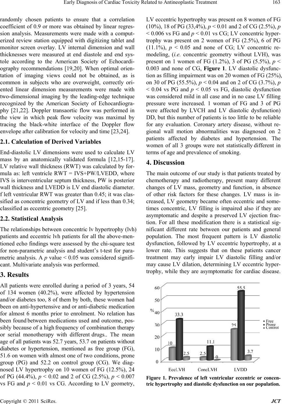 Early Diagnosis of Cardiac Toxicity Related to Antineoplastic Treatment163 randomly chosen patients to ensure that a correlation coefficient of 0.9 or more was obtained by linear regres- sion analysis. Measurements were made with a comput- erized review station equipped with digitizing tablet and monitor screen overlay. LV internal dimension and wall thicknesses were measured at end diastole and end sys- tole according to the American Society of Echocardi- ography recommendations [19,20]. When optimal orien- tation of imaging views could not be obtained, as is common in subjects who are overweight, correctly ori- ented linear dimension measurements were made with two-dimensional imaging by the leading-edge technique recognized by the American Society of Echocardiogra- phy [21,22]. Doppler transaortic flow was performed in the view in which peak flow velocity was maximal by tracing the black-white interface of the Doppler flow envelope after calibration for velocity and time [23,24]. 2.1. Calculation of Derived Variables End-diastolic LV dimensions were used to calculate LV mass by an anatomically validated formula [12,15-17]. LV relative wall thickness (RWT) was calculated by for- mula as: left ventricle RWT = IVS+PW/LVEDD, where IVS is interventricular septum thickness, PW is posterior wall thickness and LVEDD is LV end diastolic diameter. f left ventricular RWT was greater than 0.45; it was clas- sified as concentric geometry of LV and if less than 0.34; classified as eccentric geometry [25]. 2.2. Statistical Analysis The relationships between concentric lv hypertrophy (lvh) patients and eccentric lvh patients for all the above-men- tioned echo findings were assessed by the chi-square test for non-parametric analysis and student’s t-test for para- metric analysis. A p value < 0.05 was considered signifi- cant. Multivariate analysis was performed. 3. Results All patients were enrolled during a period of 3 years, 54 of 134 women (40.2%), were affected by hypertension and/or diabetes too, 8 of them by both, these women had been on anti-hypertensive and or anti-diabetic medication for almost 6 months prior to enrolment. No relation has been found between medications used and outcome, pos- sibly because of a high frequency of combination therapy or serial monotherapy with different drugs.. The mean age of all patients was 52.7 years, 53.7 on patients without diabetes or hypertension, mentioned as free group (FG), 51.6 on women with almost one of two conditions, prone group (PG) and 52.2 on control group (CG). We diag- nosed LV hypertrophy on 10 women of FG (12.5%), 24 of PG (44.4%), p < 0.02 and 2 of CG (2.5%), p < 0.007 vs FG and p < 0.01 vs CG. According to LV geometry, LV eccentric hypertrophy was present on 8 women of FG (10%), 18 of PG (33,4%), p < 0.01 and 2 of CG (2.5%), p < 0.006 vs FG and p < 0.01 vs CG; LV concentric hyper- trophy was present on 2 women of FG (2.5%), 6 of PG (11.1%), p < 0.05 and none of CG; LV concentric re- modeling, (i.e. concentric geometry without LVH), was present on 1 women of FG (1.2%), 3 of PG (5.5%), p < 0.003 and none of CG, Figure 1. LV diastolic dysfunc- tion as filling impairment was on 20 women of FG (25%), on 30 of PG (55.5%), p < 0.04 and on 2 of CG (3.7%), p < 0.04 vs PG and p < 0.05 vs FG, diastolic dysfunction was considered mild in all case and in no case LV filling pressure were increased. 1 woman of FG and 3 of PG were affected by LVCH and LV diastolic dysfunction) DD, but this number of patients is too little to be reliable for any evaluation. Coronary artery disease, without re- gional wall motion abnormalities was diagnosed on 2 patients affected by diabetes and hypertension. The women of all 3 groups were not statistically different in terms of age and prevalence of smoking. 4. Discussion The main outcome of our study is that patients treated by chemotherapy and radiotherapy, present many different changes of LV mass, geometry and function, in absence of other risk factors for these changes. LV mass is in- creased, LV geometry became often eccentric and some- times concentric, LV filling is impaired also if they are asymptomatic and despite a preserved LV ejection frac- tion. For all these modification there is a statistical sig- nificant different rate between our patients and general population. The most frequent pattern is LV diastolic dysfunction, followed by LV eccentric hypertrophy, at a lower rate. This suggests that on these patients cancer treatment may early impair LV diastolic filling and/or may cause LV dilation, determining LV eccentric hyper- trophy, while they are asymptomatic for cardiac disease. Figure 1. Prevalence of left ventricular eccentric or concen- tric hypertrophy and diastolic dysf unction on ou r popu lation. Copyright © 2011 SciRes. JCT  Early Diagnosis of Cardiac Toxicity Related to Antineoplastic Treatment 164 LV filling pressure was normal in all patients, but we have to consider that we are looking for early cardiac dysfunction. We know that a combined systo-diastolics dysfunction is relatively common during on women who underwent to cancer therapy [26-28], so it is reasonable to perform serial evaluation of LVEF [6], as prescribed by current guidelines, but when LVEF became impaired, it occurs a rapid development of severe heart failure. Early diagnosis of LV diastolic dysfunction and/or LV eccentric hypertrophy can be achieved by a simple tool such M-B mode echocardiogram, associated to PW and TDI Doppler, and it is more sensitive than LVEF in the early detection of LV unfavourable changes in geometry, as assessed by LV relative wall thickness and function. LV geometry eccentric pattern is common among our patients, not the same for concentric pattern, so we can suppose that cancer therapy doesn’t determine changes on peripheral resistances, but directly affect myocardial cells, the same explanation can be accounted for LV dia- stolic dysfunction not due to increased peripheral resis- tances but to direct myocardial cell damage. The bio- logical mechanisms underlying cancer therapy-associated cardiac dysfunction remain to be fully elucidated, but generation of reactive oxygen species (ROS) and induc- tion of cardiac myocyte apoptosis are hypothesized to play a central role. Concerning, chemotherapy ROS may continue to be produced by a drug retained within myo- cytes contributing to late-occurring cardiovascular injury [29]. Also about radiotherapy, the generation of ROS is thought to play a major role [30]. Another important out- come is that all above mentioned changes in LV mass, geometry and function are present at a statistical signifi- cant different higher rate among a population of women treated by chemotherapy and radiotherapy, but just af- fected by cardiovascular risk factors as diabetes and or hypertension [31], compared with our population of women treated for cancer but free from diabetes and or hypertension. So that the cardiac unfavorable changes, due to cancer therapy, add up to those due to diabetes and or hypertension, increasing their cardiovascular risk of events [31,32]. All these patients need of a careful monitoring of heart condition to prevent heart failure and other cardiac events. The main limitation of our study is the limited number of patients enrolled, so we cannot individuate subgroups according to different treatments, not even we can differentiate diabetic from hypertensive women, otherwise it is one of the first studies concerning breast cancer patient treated by both chemotherapy and radiotherapy, with preserved LV ejection fraction. 5. Conclusions An abnormal diastolic function is more common among women affected by breast cancer and treated for it than in general population, the same happens for LV eccentric hypertrophy but at a lower rate. 40% of women were affected by hypertension, diabetes or both, and they have a higher rate of LV eccentric hypertrophy and diastolic dysfunction than women treated for cancer but unaf- fected. We know that patients affected by these LV changes had worse event-free survival than patients with normal LV mass, geometry and function. LV eccentric hypertrophy and diastolic dysfunction are predictors of heart failure on asympomatic women. Screening with Doppler echo techniques may be helpful in identifying patients at high risk for subsequent cardiac events. 6. Disclosures All authors have no financial disclosures, they report no biomedical financial interests or potential conflicts of interest. 7. Acknowledgements The authors thank the nurses Grazia Quaranta, Emmia Donativo and Anna Paola Gennaro, for the support to the survey of the clinical data. REFERENCES [1] M. N. Levine and T. Whelan, “Adjuvant Chemotherapy for Breast Cancer—30 Years Later,” The New England Journal of Medicine, Vol. 355, No. 18, 2006, pp. 1920- 1922. doi:10.1056/NEJMe068204 [2] Early Breast Cancer Trialists’ Collaborative Group, “Ef- fects of Radiotherapy and Surgery in Early Breast Cancer. An Overview of the Randomized Trials,” The New Eng- land Journal of Medicine, Vol. 333, No. 22, 1995, pp. 1444-1455. doi:10.1056/NEJM199511303332202 [3] M. J. Hooning, et al., “Long-Term Risk of Cardiovascular Disease in 10-year Survivors of Breast Cancer,” Journal of the National Cancer Institute, Vol. 99, No. 5, 2007, pp. 365-375. doi:10.1093/jnci/djk064 [4] T. Hampton, “Cancer Therapy Can Be Hard on the Heart: Researchers Aim to Explain—and Avoid—Cardiotoxic- ity,” Journal of the American Medical Association, Vol. 303, No. 11, 2010, pp. 1019-1020. doi:10.1001/jama.2010.269 [5] P. A. Heidenreich, et al., “Diastolic Dysfunction after Mediastinal Irradiation,” American Heart Journal, Vol. 150, No. 5, 2005, pp. 977-982. doi:10.1016/j.ahj.2004.12.026 [6] M. T. Meinardi, et al., “Evaluation of Long Term Car- diotoxicity after Epirubicin Containing Adjuvant Che- motherapy and Locoregional Radiotherapy for Breast Cancer Using Various Detection Techniques,” Heart, Vol. 88, No. 1, 2002, pp. 81-82. doi:10.1136/heart.88.1.81 [7] J. Alexander, et al., “Serial Assessment of Doxorubicin Cardiotoxicity with Quantitative Radionuclide Angiocar- diography,” The New England Journal of Medicine, Vol. Copyright © 2011 SciRes. JCT  Early Diagnosis of Cardiac Toxicity Related to Antineoplastic Treatment165 300, No. 6, 1979, pp. 278-283. doi:10.1056/NEJM197902083000603 [8] R. G. Schwartz, et al., “Congestive Heart Failure and Left Ventricular Dysfunction Complicating Doxorubicin Therapy. Seven-Year Experience Using Serial Radionu- clide Angiocardiography,” American Journal of Medicine, Vol. 82, No. 6, 1987, pp. 1109-1118. doi:10.1016/0002-9343(87)90212-9 [9] L. Mosca, et al., “Evidence-Based Guidelines for Car- diovascular Disease Prevention in Women: 2007 Up- date,” Journal of the American College of Cardiology, Vol. 49, No. 11, 2007, pp. 1230-1250. doi:10.1016/j.jacc.2007.02.020 [10] W. I. Ganz, et al., “Review of Tests for Monitoring Doxorubicin-Induced Cardiomyopathy,” Oncology, Vol. 53, No. 6, 1996, pp. 461-470. doi:10.1159/000227621 [11] R. Yancik, et al., “Effect of Age and Comorbidity in Postmenopausal Breast Cancer Patients Aged 55 Years and Older,” Journal of the American Medical Association, Vol. 285, No. 7, 2001, pp. 885-892. doi:10.1001/jama.285.7.885 [12] C. P. Appleton, et al., “Doppler Evaluation of Left and Right Ventricular Diastolic Function: A Technical Guide for Obtaining Optimal Flow Velocity Recordings,” Jour- nal of the American Society of Echocardiography, Vol. 10, No. 3, 1997, pp. 271-292. doi:10.1016/S0894-7317(97)70063-4 [13] J. W. Ha, et al., “Triphasic Mitral Inflow Velocity with Middiastolic Filling: Clinical Implications and Associated Echocardiographic Findings,” Journal of the American Society of Echocardiography, Vol. 17, No. 5, 2004, pp. 428-431. doi:10.1016/j.echo.2004.02.007 [14] S. F. Nagueh, et al., “Recommendations for the Evalua- tion of Left Ventricular Diastolic Function by Echocardi- ography,” Journal of the American Society of Echocardi- ography, Vol. 22, No. 2, 2009, pp. 107-133. doi:10.1016/j.echo.2008.11.023 [15] R. B. Devereux and N. Reichek, “Echocardiographic Determination of Left Ventricular Mass in Man. Anat- omic Validation of the Method,” Circulation, Vol. 55, No. 4, 1977, pp. 613-618. [16] H. W. Hense, et al., “The Associations of Body Size and Body Composition with Left Ventricular Mass: Impacts for Indexation in Adults,” Journal of the American Col- lege of Cardiology, Vol. 32, No. 2, 1998, pp. 451-457. doi:10.1016/S0735-1097(98)00240-X [17] G. P. Aurigemma, M. R. Zile and W. H. Gaasch, “Con- tractile Behavior of the Left Ventricle in Diastolic Heart Failure: With Emphasis on Regional Systolic Function,” Circulation, Vol. 113, No. 2, 2006, pp. 296-304. doi:10.1161/CIRCULATIONAHA.104.481465 [18] G. de Simone, et al., “Evaluation of Concentric Left Ven- tricular Geometry in Humans: Evidence for Age-Related Systematic Underestimation,” Hypertension, Vol. 45, No. 1, 2005, pp. 64-68. [19] D. J. Sahn, et al., “Recommendations Regarding Quanti- tation in M-mode Echocardiography: Results of a Survey of Echocardiographic Measurements,” Circulation, Vol. 58, No. 6, 1978, pp. 1072-1083. [20] M. M. Redfield, et al., “Burden of Systolic and Diastolic Ventricular Dysfunction in the Community: Appreciating the Scope of the Heart Failure Epidemic,” Journal of the American Society of Echocardiography, Vol. 289, No. 2, 2003, pp. 194-202. doi:10.1001/jama.289.2.194 [21] N. B. Schiller, et al., “Recommendations for Quantitation of the Left Ventricle by Two-Dimensional Echocardi- ography. American Society of Echocardiography Com- mittee on Standards, Subcommittee on Quantitation of Two-Dimensional Echocardiograms,” Journal of the American Society of Echocardiography, Vol. 2, No. 5, 1989, pp. 358-367. [22] R. A. Nishimura, and A. J. Tajik, “Evaluation of Diastolic Filling of Left Ventricle in Health and Disease: Doppler Echocardiography is the Clinician’s Rosetta Stone,” Journal of the American College of Cardiology, Vol. 30, No. 1, 1997, pp. 8-18. [23] J. Dubin, et al., “Comparative Accuracy of Doppler Echo- cardiographic Methods for Clinical Stroke Volume De- termination,” American Heart Journal, Vol. 120, No. 1, 1990, pp. 116-123. doi:10.1016/0002-8703(90)90168-W [24] S. R. Ommen, et al., “Clinical Utility of Doppler Echo- cardiography and Tissue Doppler Imaging in the Estima- tion of Left Ventricular Filling Pressures: A Comparative Simultaneous Doppler-Catheterization Study,” Circula- tion, Vol. 102, No. 15, 2000, pp. 1788-1794. [25] A. Ganau, et al., “Patterns of Left Ventricular Hypertro- phy and Geometric Remodeling in Essential Hyperten- sion,” Journal of the American College of Cardiology, Vol. 19, No. 7, 1992, pp. 1550-1558. doi:10.1016/0735-1097(92)90617-V [26] J. J. Doyle, et al., “Chemotherapy and Cardiotoxicity in Older Breast Cancer Patients: A Population-Based Study,” Journal of Clinical Oncology, Vol. 23, No. 34, 2005, pp. 8597-8605. doi:10.1200/JCO.2005.02.5841 [27] E. A. Perez, et al., “Effect of Doxorubicin Plus Cyclo- phosphamide on Left Ventricular Ejection Fraction in Pa- tients with Breast Cancer in the North Central Cancer Treatment Group N9831 Intergroup Adjuvant Trial,” Journal of Clinical Oncology, Vol. 22, No. 18, 2004, pp. 3700-3704. doi:10.1200/JCO.2004.03.516 [28] E. T. Yeh, “Cardiotoxicity Induced by Chemotherapy and Antibody Therapy,” Annual Review of Medicine, Vol. 57, 2006, pp. 485-498. doi:10.1146/annurev.med.57.121304.131240 [29] D. Lebrecht, et al., “Time-Dependent and Tissue-Specific Accumulation of mtDNA and Respiratory Chain Defects in Chronic Doxorubicin Cardiomyopathy,” Circulation, Vol. 108, No. 19, 2003, pp. 2423-2429. doi:10.1161/01.CIR.0000093196.59829.DF [30] M. S. Anscher and Z. Vujaskovic, “Mechanisms and Po- tential Targets for Prevention and Treatment of Normal Tissue Injury after Radiation Therapy,” Seminars in On- cology, Vol. 32, No. 2 (Supplement), 2005, pp. S86-91. doi:10.1053/j.seminoncol.2005.03.015 Copyright © 2011 SciRes. JCT  Early Diagnosis of Cardiac Toxicity Related to Antineoplastic Treatment Copyright © 2011 SciRes. JCT 166 [31] D. M. Lloyd-Jones, et al., “Prediction of Lifetime Risk for Cardiovascular Disease by Risk Factor Burden at 50 Years of Age,” Circulation, Vol. 113, No. 6, 2006, pp. 791-798. doi:10.1161/CIRCULATIONAHA.105.548206 [32] P. M. Ridker, et al., “Development and Validation of Improved Algorithms for the Assessment of Global Car- diovascular Risk in Women: The Reynolds Risk Score,” Journal of the American Society of Echocardiography, Vol. 297, No. 6, 2007, pp. 611-619. doi:10.1001/jama.297.6.611 |