

Paper Menu >>
Journal Menu >>
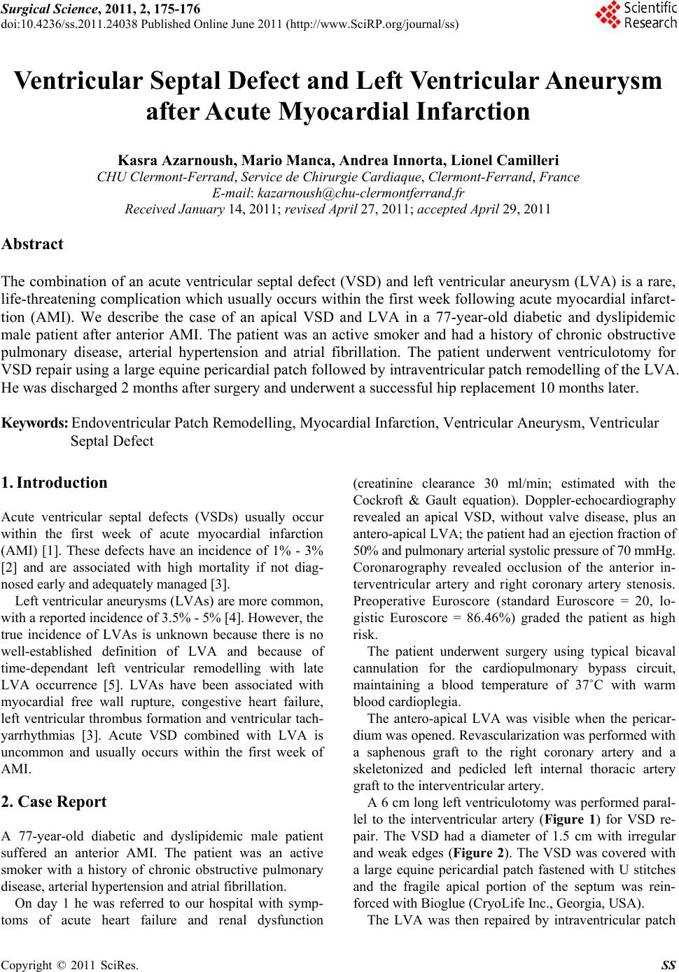 Surgical Science, 2011, 2, 175-176 doi:10.4236/ss.2011.24038 Published Online June 2011 (http://www.SciRP.org/journal/ss) Copyright © 2011 SciRes. SS Ventricular Septal Defect and Left Ventricular Aneurysm after Acute Myocardial Infarction Kasra Azarnoush, Mario Manca, Andrea Innorta, Lionel Camilleri CHU Clermont-Ferrand, Service de Chirurgie Cardiaque, Clermont-Ferrand, France E-mail: [email protected] Received January 14, 2011; revised April 27, 2011; accepted April 29, 2011 Abstract The combination of an acute ventricular septal defect (VSD) and left ventricular aneurysm (LVA) is a rare, life-threatening complication which usually occurs within the first week following acute myocardial infarct- tion (AMI). We describe the case of an apical VSD and LVA in a 77-year-old diabetic and dyslipidemic male patient after anterior AMI. The patient was an active smoker and had a history of chronic obstructive pulmonary disease, arterial hypertension and atrial fibrillation. The patient underwent ventriculotomy for VSD repair using a large equine pericardial patch followed by intraventricular patch remodelling of the LVA. He was discharged 2 months after surgery and underwent a successful hip replacement 10 months later. Keywords: Endoventricular Patch Remodelling, Myocardial Infarction, Ventricular Aneurysm, Ventricular Septal Defect 1. Introduction Acute ventricular septal defects (VSDs) usually occur within the first week of acute myocardial infarction (AMI) [1]. These defects have an incidence of 1% - 3% [2] and are associated with high mortality if not diag- nosed early and adequately managed [3]. Left ventricular aneurysms (LVAs) are more common, with a reported incidence of 3.5% - 5% [4]. However, the true incidence of LVAs is unknown because there is no well-established definition of LVA and because of time-dependant left ventricular remodelling with late LVA occurrence [5]. LVAs have been associated with myocardial free wall rupture, congestive heart failure, left ventricular thrombus formation and ventricular tach- yarrhythmias [3]. Acute VSD combined with LVA is uncommon and usually occurs within the first week of AMI. 2. Case Report A 77-year-old diabetic and dyslipidemic male patient suffered an anterior AMI. The patient was an active smoker with a history of chronic obstructive pulmonary disease, arterial hypertension and atrial fibrillation. On day 1 he was referred to our hospital with symp- toms of acute heart failure and renal dysfunction (creatinine clearance 30 ml/min; estimated with the Cockroft & Gault equation). Doppler-echocardiography revealed an apical VSD, without valve disease, plus an antero-apical LVA; the patient had an ejection fraction of 50% and pulmonary arterial systolic pressure of 70 mmHg. Coronarography revealed occlusion of the anterior in- terventricular artery and right coronary artery stenosis. Preoperative Euroscore (standard Euroscore = 20, lo- gistic Euroscore = 86.46%) graded the patient as high risk. The patient underwent surgery using typical bicaval cannulation for the cardiopulmonary bypass circuit, maintaining a blood temperature of 37˚C with warm blood cardioplegia. The antero-apical LVA was visible when the pericar- dium was opened. Revascularization was performed with a saphenous graft to the right coronary artery and a skeletonized and pedicled left internal thoracic artery graft to the interventricular artery. A 6 cm long left ventriculotomy was performed paral- lel to the interventricular artery (Figure 1) for VSD re- pair. The VSD had a diameter of 1.5 cm with irregular and weak edges (Figure 2). The VSD was covered with a large equine pericardial patch fastened with U stitches and the fragile apical portion of the septum was rein- forced with Bioglue (CryoLife Inc., Georgia, USA). The LVA was then repaired by intraventricular patch 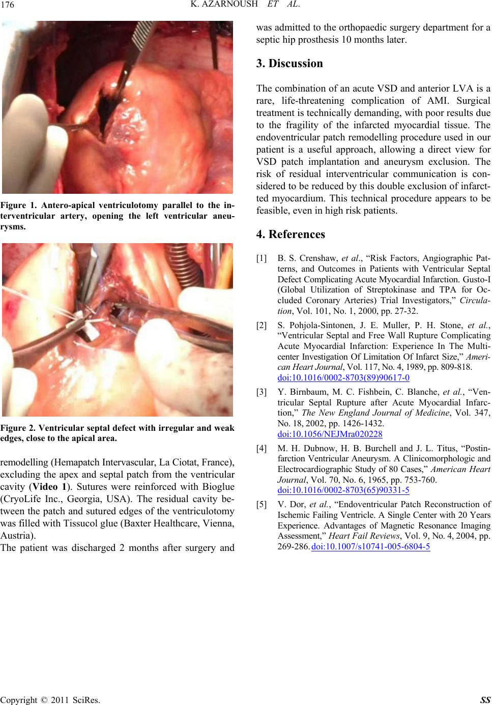 176 K. AZARNOUSH ET AL. Figure 1. Antero-apical ventriculotomy parallel to the in- terventricular artery, opening the left ventricular aneu- rysms. Figure 2. Ventricular septal defect with irregular and weak edges, close to the apical area. remodelling (Hemapatch Intervascular, La Ciotat, France), excluding the apex and septal patch from the ventricular cavity (Video 1). Sutures were reinforced with Bioglue (CryoLife Inc., Georgia, USA). The residual cavity be- tween the patch and sutured edges of the ventriculotomy was filled with Tissucol glue (Baxter Healthcare, Vienna, Austria). The patient was discharged 2 months after surgery and was admitted to the orthopaedic surgery department for a septic hip prosthesis 10 months later. 3. Discussion The combination of an acute VSD and anterior LVA is a rare, life-threatening complication of AMI. Surgical treatment is technically demanding, with poor results due to the fragility of the infarcted myocardial tissue. The endoventricular patch remodelling procedure used in our patient is a useful approach, allowing a direct view for VSD patch implantation and aneurysm exclusion. The risk of residual interventricular communication is con- sidered to be reduced by this double exclusion of infarct- ted myocardium. This technical procedure appears to be feasible, even in high risk patients. 4. References [1] B. S. Crenshaw, et al., “Risk Factors, Angiographic Pat- terns, and Outcomes in Patients with Ventricular Septal Defect Complicating Acute Myocardial Infarction. Gust o- I (Global Utilization of Streptokinase and TPA for Oc- cluded Coronary Arteries) Trial Investigators,” Circula- tion, Vol. 101, No. 1, 2000, pp. 27-32. [2] S. Pohjola-Sintonen, J. E. Muller, P. H. Stone, et al., “Ventricular Septal and Free Wall Rupture Complicating Acute Myocardial Infarction: Experience In The Multi- center Investigation Of Limitation Of Infarct Size,” Ameri- can Heart Journal, Vol. 117, No. 4, 1989, pp. 809-818. doi:10.1016/0002-8703(89)90617-0 [3] Y. Birnbaum, M. C. Fishbein, C. Blanche, et al., “Ven- tricular Septal Rupture after Acute Myocardial Infarc- tion,” The New England Journal of Medicine, Vol. 347, No. 18, 2002, pp. 1426-1432. doi:10.1056/NEJMra020228 [4] M. H. Dubnow, H. B. Burchell and J. L. Titus, “Postin- farction Ventricular Aneurysm. A Clinicomorp hologic and Electrocardiographic Study of 80 Cases,” American Heart Journal, Vol. 70, No. 6, 1965, pp. 753-760. doi:10.1016/0002-8703(65)90331-5 [5] V. Dor, et al., “Endoventricular Patch Reconstruction of Ischemic Failing Ventricle. A Single Center with 20 Years Experience. Advantages of Magnetic Resonance Imaging Assessment,” Heart Fail Reviews, Vol. 9, No. 4, 2004, pp. 269-286. doi:10.1007/s10741-005-6804-5 Copyright © 2011 SciRes. SS |