

Paper Menu >>
Journal Menu >>
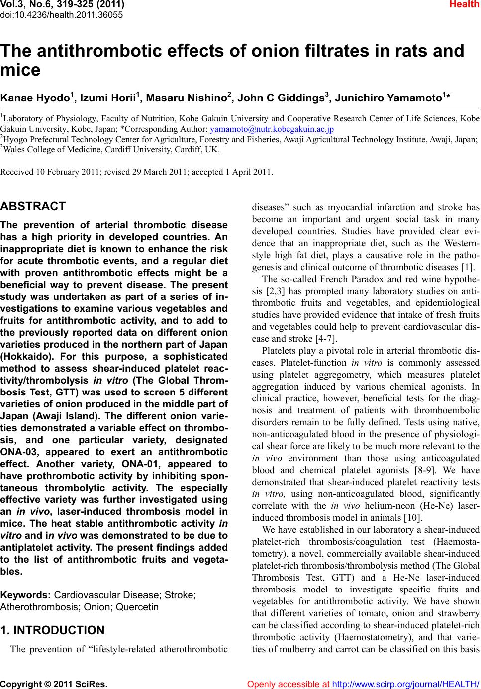 Vol.3, No.6, 319-325 (2011) doi:10.4236/health.2011.36055 C opyright © 2011 SciRes. Openly accessible at http://www.scirp.org/journal/HEALTH/ Health The antithrombotic effects of onion filtrates in rat s and mice Kanae Hyodo1, Izumi Horii1, Masaru Nishino2, John C Giddings3, Junichiro Yamamoto1* 1Laboratory of Physiology, Faculty of Nutrition, Kobe Gakuin University and Cooperative Research Center of Life Sciences, Kobe Gakuin University, Kobe, Japan; *Corresponding Author: [email protected] 2Hyogo Prefectural Technology Center for Agriculture, Forestry and Fisheries, Awaji Agricultural Technology Institute, Awaji, Japan; 3Wales College of Medicine, Cardiff University, Cardiff, UK. Received 10 February 2011; revised 29 March 2011; accepted 1 April 2011. ABSTRACT The prevention of arterial thrombotic disease has a high priority in developed countries. An inappropriate diet is known to enhance the risk for acute thrombotic events, and a regular diet with proven antithrombotic effects might be a beneficial way to prevent disease. The present study was undertaken as part of a series of in- vestigations to examine various vegetables and fruits for antithrombotic activity, and to add to the previously reported data on different onion varieties produced in the northern part of Japan (Hokkaido). For this purpose, a sophisticated method to assess shear-induced platelet reac- tivity/thrombolysis in vitro (The Global Throm- bosis Test, GTT) was used to screen 5 different varieties o f on ion p roduced in the middle part of Japan (Awaji Island). The different onion varie- ties demonstrated a variable effect on thrombo- sis, and one particular variety, designated ONA-03, appeared to exert an antithrombotic effect. Another variety, ONA-01, appeared to have prothrombotic activity by inhibiting spon- taneous thrombolytic activity. The especially effective variety was further investigated using an in vivo, laser-induced thrombosis model in mice. The heat stable antithrombotic activity in vitro and in vivo w as demonstrated to be due to antiplatelet activity. The present findings added to the list of antithrombotic fruits and vegeta- bles. Keywords: Cardiovascular Dise ase; Stroke; Atherothrombosis; Onion; Quercetin 1. INTRODUCTION The prevention of “lifestyle-related atherothrombotic diseases” such as myocardial infarction and stroke has become an important and urgent social task in many developed countries. Studies have provided clear evi- dence that an inappropriate diet, such as the Western- style high fat diet, plays a causative role in the patho- genesis and clinical outcome of thrombotic diseases [1]. The so-called French Paradox and red wine hypothe- sis [2,3] has prompted many laboratory studies on anti- thrombotic fruits and vegetables, and epidemiological studies have provided evidence that intake of fresh fruits and vegetables could help to prevent cardiovascular dis- ease and stroke [4-7]. Platelets play a pivotal role in arterial thrombotic dis- eases. Platelet-function in vitro is commonly assessed using platelet aggregometry, which measures platelet aggregation induced by various chemical agonists. In clinical practice, however, beneficial tests for the diag- nosis and treatment of patients with thromboembolic disorders remain to be fully defined. Tests using native, non-anticoagulated blood in the presence of physiologi- cal shear force are likely to be much more relevant to the in vivo environment than those using anticoagulated blood and chemical platelet agonists [8-9]. We have demonstrated that shear-induced platelet reactivity tests in vitro, using non-anticoagulated blood, significantly correlate with the in vivo helium-neon (He-Ne) laser- induced thrombosis model in animals [10]. We have established in our laboratory a shear-induced platelet-rich thrombosis/coagulation test (Haemosta- tometry), a novel, commercially available shear-induced platelet-rich thrombosis/thrombolysis method (The Global Thrombosis Test, GTT) and a He-Ne laser-induced thrombosis model to investigate specific fruits and vegetables for antithrombotic activity. We have shown that different varieties of tomato, onion and strawberry can be classified according to shear-induced platelet-rich thrombotic activity (Haemostatometry), and that varie- ties of mulberry and carrot can be classified on this basis 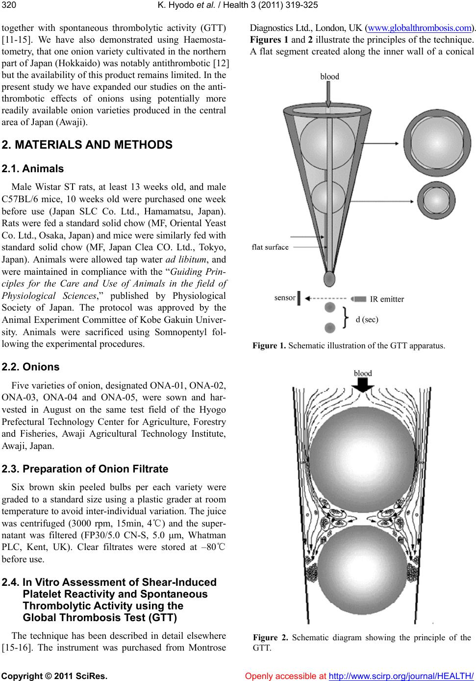 K. Hyodo et al. / Health 3 (2011) 319-325 Copyright © 2011 SciRes. Openly accessible at http://www.scirp.org/journal/HEALTH/ 320 together with spontaneous thrombolytic activity (GTT) [11-15]. We have also demonstrated using Haemosta- tometry, that one onion variety cultivated in the northern part of Japan (Hokkaido) was notably antithrombotic [12] but the availability of this product remains limited. In the present study we have expanded our studies on the anti- thrombotic effects of onions using potentially more readily available onion varieties produced in the central area of Japan (Awaji). 2. MATERIALS AND METHODS 2.1. Animals Male Wistar ST rats, at least 13 weeks old, and male C57BL/6 mice, 10 weeks old were purchased one week before use (Japan SLC Co. Ltd., Hamamatsu, Japan). Rats were fed a standard solid chow (MF, Oriental Yeast Co. Ltd., Osaka, Japan) and mice were similarly fed with standard solid chow (MF, Japan Clea CO. Ltd., Tokyo, Japan). Animals were allowed tap water ad libitum, and were maintained in compliance with the “Guiding Prin- ciples for the Care and Use of Animals in the field of Physiological Sciences,” published by Physiological Society of Japan. The protocol was approved by the Animal Experiment Committee of Kobe Gakuin Univer- sity. Animals were sacrificed using Somnopentyl fol- lowing the experimental procedures. 2.2. Onions Five varieties of onion, designated ONA-01, ONA-02, ONA-03, ONA-04 and ONA-05, were sown and har- vested in August on the same test field of the Hyogo Prefectural Technology Center for Agriculture, Forestry and Fisheries, Awaji Agricultural Technology Institute, Awaji, Japan. 2.3. Preparation of Onion Filtrate Six brown skin peeled bulbs per each variety were graded to a standard size using a plastic grader at room temperature to avoid inter-individual variation. The juice was centrifuged (3000 rpm, 15min, 4℃) and the super- natant was filtered (FP30/5.0 CN-S, 5.0 μm, Whatman PLC, Kent, UK). Clear filtrates were stored at –80℃ before use. 2.4. In Vitro Assessment of Shear-Induced Platelet Reactivity and Spontaneous Thrombolytic Activity using the Global Thrombosis Test (GTT) The technique has been described in detail elsewhere [15-16]. The instrument was purchased from Montrose Diagnostics Ltd., London, UK (www.globalthrombosis.com). Figures 1 and 2 illustrate the principles of the technique. A flat segment created along the inner wall of a conical Figure 1. Schematic illustration of the GTT apparatus. Figure 2. Schematic diagram showing the principle of the GTT.  K. Hyodo et al. / Health 3 (2011) 319-325 Copyright © 2011 SciRes. Openly accessible at http://www.scirp.org/journal/HEALTH/ 321321 plastic tube forms the basis of the technique. When per- fectly round steel ball-bearings are placed into the coni- cal tube, the flat segment prevents the spheres from oc- cluding the lumen. Blood is added, flows through the narrow gaps and exits in droplets into an adjacent col- lecting tube. The latter is trans-illuminated by a light emitter, and a sensor opposite the light source generates a signal whenever a drop of blood interrupts the light path. In essence, the instrument detects the time interval (d, sec) between consecutive blood drops. At the start, blood flow is rapid and hence (d) is small. Subsequently, the flow rate gradually decreases and hence (d) increases. When the actual (d) exceeds 15 seconds (occlusion-d), the instrument displays “Occlusion Time (OT)”, which is the time elapsed from the detection of the first drop of blood until (occlusion-d). Later, flow is completely ar- rested. Eventually, due to thrombolysis, flow is restored as indicated by the further detection of blood droplets. There is also an arbitrarily pre-set (d) (200 seconds) for lysis measurement (lysis-d). When (d) between the last drop before and the first drop after occlusion exceeds this (lysis-d), the instrument displays “Lysis Time (LT)”. Hence, lysis time is calculated as follows: LT = (time of first drop with d > (lysis-d)) – (time of last drop with d < (lysis-d)). Blood flows by gravity at 37oC through the narrow gaps formed between the larger ball bearing and the inner wall of the tube, and the resulting high shear stress (175 dyne/cm2) activates platelets. These activated platelets remain single, since the very short transit time and high shear prevent aggregation. In contrast, in the space distal to the large ball bearing, i.e. between the two ball bearings, the low shear and turbulent flow favor large platelet aggregate formation. Furthermore, in this space between the ball bearings, activated platelets gen- erate thrombin and initiate blood coagulation. Flow then carries these fibrin-stabilised platelet aggregates into the lower gaps where they are captured, resulting in occlu- sion and arrest of flow. Increased or decreased OT indi- cates inhibition or enhancement of platelet reactivity, respectively. Increased or decreased LT indicates inhibi- tion or enhancement of spontaneous thrombolysis, re- spectively. Measurements were made six times (n = 6) in each sample. Animals were fasted overnight but allowed water ad libitum. Blood was obtained from the abdominal aorta 30 minutes after anesthesia with sodium pentobarbital (Somnopentyl, 64.8 mg/ml, Kyoetsu Seiyaku Co. Ltd., Tokyo, Japan)diluted 5 times with saline (65 mg/kg, intramuscularly). Non-anticoagulated blood was mixed with saline (1:1). 3.6 ml of the diluted blood and 0.4 ml of onion filtrate or saline (control) (blood: filtrate = 9:1) were mixed in a syringe by inversion and the mixture was applied to GTT tube. 2.5. In Vivo Assessment of the Antithrombotic Effects Using the Laser-Induced Thrombosis Test in Mice Carotid Arteries The He-Ne laser-induced platelet-rich thrombosis method has been previously described in detail [17,18]. Mice were anaesthetised with Somnopentyl (65 mg/kg, intramuscularly). A polyethylene tube (PE10, Becton Dickinson and Company, New Jersey, USA) was placed into the left femoral artery to inject dye and the carotid artery (450 - 500 μm in diameter) was exposed by inci- sion. The mouse was placed on a specially adapted mi- croscope stage (Olympus Model BH-2, Olympus Co. Ltd., Tokyo, Japan) and Evans blue dye (30 mg/kg, Merck, Darnstadt, Germany) was injected intra-arterially. The centre of the exposed carotid artery was irradiated with a laser beam (Model Neo-50MS, 25 mW under an objective lens, Neoark Co. Ltd., Osaka, Japan). Throm- bus formation at the site of irradiation was monitored under epi-illumination and simultaneously recorded on videotape using a CCD camera (Model TMC-7, Take- naka System Co. Ltd., Kyoto, Japan). 2.6. Oral Ad ministration of Onion Filtrates to Mice The vegetable filtrate or saline only (control) was ad- ministered through a gastric tube at 15.4 ml/kg (un- heated filtrate) and 15.4 or 30.8 ml/kg (heated filtrate). Half volume of filtrate or saline (7.7 ml/kg (unheated filtrate) and 7.7 or 15.4 ml/kg (heated filtrate) was re- peatedly given at 30 minute interval as previously de- scribed [11]. The mouse was then anaesthetised and the thrombosis experiments commenced 90 min after the second oral administration. Antithrombotic or prothrom- botic effects were assessed by estimating total thrombus size. 2.7. Calculation of Thrombus Size Details of this technique have been described else- where [18]. Images of thrombus formation were com- puter-analysed at intervals of ten seconds. The area of thrombus was delineated and the mass of thrombus cal- culated by multiplying gray scale and area using Image J software (Image Processing and Analysis Java version 1.30, National Institutes of Health, Maryland, USA). Thrombotic status was expressed as the total sum of thrombus mass after the first 10 minutes of irradiation. 2.8. Statistical Analysis Data from GTT were analysed by repeated ANOVA (General Linear Model), followed by the multiple com- parison test (Dunnett), data from the laser-induced  K. Hyodo et al. / Health 3 (2011) 319-325 Copyright © 2011 SciRes. http://www.scirp.org/journal/HEALTH/Openly accessible at 322 thrombosis experiments by the unpaired t-test. Values were expressed as means ± SEM. P < 0.05 was consid- ered as the limit of significance. Analyses were per- formed using Statistical Package Unistat 5.6 Light (London, UK). 3. RESULTS 3.1. Effects of Raw Filtrates from Onion Varieties on Shear-Induced Platelet Thrombosis and Spontaneous Thrombolysis in Vitro The results are shown in Table 1. The filtrate from ONA-01 did not affect occlusion time (OT) but signifi- cantly inhibited spontaneous thrombolytic activity (LT), indicating that this variety had prothrombotic activity. ONA-02, ONA-04 and ONA-05 did not affect signifi- cantly either OT or LT. In contrast, the undiluted filtrate from ONA-03 significantly prolonged OT but did not affect LT, indicating that this particular variety had anti- thrombotic activity both in vitro and in vivo. 3.2. Heat Stability of Antithrombotic Effect of ONA-03 The antithrombotic effects of ONA-03 in vivo, were re-examined after the raw filtrate was heated in boiling water for 10 minutes. The filtrate was cooled to room temperature and the OT and LT were measured. The results are shown in Table 2. Similar results to those using unheated filtrate were obtained. The heated and undiluted (x1) filtrate significantly prolonged OT but had no significant effect on LT, again suggesting anti- thrombotic activity in vivo. Table 1. Effects of raw filtrates from five onion varieties on shear-induced platelet thrombosis (occlusion time, OT) and spontaneous thrombolysis (lysis time, LT). Variety Dilution Occlusion time Lysis time ONA-01 control 307.9 ± 9.9 989.7 ± 70.9 × 1 265.0 ± 6.3 1520.3 ± 110.9** × 3 327.7 ± 12.1 983.3 ± 103.4 × 10 311.5 ± 20.7 1024.3 ± 46.5 ONA-02 control 295.6 ± 4.7 855.3 ± 39.6 × 1 260.4 ± 16.2 1022.5 ± 90.2 × 3 289.7 ± 14.2 972.4 ± 76.9 × 10 289.1 ± 16.9 841.7 ± 57.9 ONA-03 control 329.1 ± 12.9 944.8 ± 24.8 × 1 457.4 ± 36.1** 977.3 ± 102.7 × 3 352.0 ± 22.5 935.3 ± 99.7 × 10 344.7 ± 16.5 900.6 ± 55.4 ONA-04 control 287.0 ± 20.7 985.0 ± 48.5 × 1 272.9 ± 25.1 1042.8 ± 80.7 × 3 294.0 ± 25.5 895.5 ± 52.2 × 10 299.5 ± 18.1 913.3 ± 78.5 ONA-05 control 304.0 ± 18.2 976.0 ± 32.4 × 1 367.1 ± 37.6 1032.4 ± 102.3 × 3 341.9 ± 19.4 891.8 ± 26.6 × 10 311.7 ± 14.9 889.3 ± 56.7 **: P < 0.01 Table 2. Effects of heat treatment on the inhibitory activity of ONA-03 in shear-induced platelet thrombosis. Variety Dilution factor of filtrate Occlusion time Lysis time ONA-03 saline (control) 302.7 ± 12.5 1066.7 ± 66.7 × 1 446.0 ± 19.4** 1249.6 ± 41.3 × 3 345.6 ± 9.3 981.5 ± 75.0 × 10 331.5 ± 12.7 959.2 ± 69.7 **: P < 0.01  K. Hyodo et al. / Health 3 (2011) 319-325 Copyright © 2011 SciRes. Openly accessible at http://www.scirp.org/journal/HEALTH/ 323323 3.3. Antithrombotic Activity of Orally Administered ONA-03 Unheated or heated onion filtrate was orally adminis- tered to mice and the antithrombotic effect was assessed using the He-Ne laser-induced thrombosis test on ex- posed carotid arteries. Results are shown in Figure 3. Unheated and undiluted (x1) filtrate from ONA-03 sig- nificantly inhibited He-Ne laser-induced thrombosis (A). The same volume of heated filtrate did not inhibit thrombosis (B) but administration of twice the volume significantly inhibited thrombus formation (C). This inconsistency between the in vitro GTT test and the in vivo laser-induced thrombosis test might have been be due to different sensitivity of the methods or the effects of intestinal absorption. Nevertheless, the results dem- onstrated that the filtrate from ONA-03 was relatively heat stable. 4. DISCUSSION Arterial platelet-rich thrombotic diseases are a social problem in many countries, and reliable tests to assess thrombotic tendency could be critically important in attempts to prevent these diseases. Various methods have been developed, including agonist-induced platelet ag- gregation tests using anticoagulated whole blood or platelet rich plasma. Biomarkers of coagulation and fi- brinolysis have also been devised [19-24]. It is widely appreciated, however, that thrombus formation in vivo is governed by interactions between blood flow, blood components and the blood vessel wall as proposed by Virchow, and the pathophysiological relevance of these tests to thrombotic status in vivo is not clear. Alterna- tively, innovative tests, in which non-anticoagulated blood is used and platelets are activated by shear force under flow, have been proposed [8,9,15]. Clinical and experimental studies have suggested that these tests may be more relevant to thrombotic status in vivo [8-10,16]. We have utilized in vivo tests of this nature to charac- terize various fruits and vegetables on the basis of anti- thrombotic activity. In particular, Haemostatometry, the GTT and the He-Ne laser-induced thrombosis test [17, 18] have enabled classification into different subgroups [11-14]. In an earlier study we used Haemostatometry and the He-Ne laser-induced thrombosis model to identify a specific antithrombotic onion variety produced in the northern part of Japan [12]. Distribution of this variety was limited, however, and further studies were devised to examine onion varieties from central Japan (Awaji Island). These varieties were potentially more available than others, and were classified using the GTT in place of Haemostatometry. The principle aim of our current series of investiga- tions is to broadly identify fruits and vegetables with antithrombotic activity. In addition, thrombus evolution is largely determined by a balance between pro-aggregatory and procoagulant mechanisms on the one hand, and fi- brinolytic processes on the other [25], and the relation- ship between the concentration of specific antithrom- botic substances and overall antithrombotic activity of fruits and vegetables remains to be defined. In this re- spect, quercetin is reported to be important in inhibiting collagen-stimulated platelet aggregation [26-28], but a high concentration of quercetin in onions does not nec- essarily mean that onions are antithrombotic, and we Figure 3. Antithrombotic activity of unheated and heated onion filtrates from ONA-03 n: number of measurements; *: P < 0.05, **: P < 0.01.  K. Hyodo et al. / Health 3 (2011) 319-325 Copyright © 2011 SciRes. Openly accessible at http://www.scirp.org/journal/HEALTH/ 324 have previously demonstrated that there was no signifi- cant correlation between quercetin concentration of on- ions and overall antiplatelet activity using shear-induced platelet function tests in vitro (Haemostatometry; 12). Furthermore, the fibrinolytic properties of onions have been the focus of some antithrombotic studies [29] but significant fibrinolytic activity was not evident in any of the five varieties of onions used in the present investiga- tions. Moreover, for clinical purposes it is important to note that the amounts of onion extracts given to animals in the present studies would be approximately equivalent to 40 kg onion given to a person with a body weight 70 kg. This is clearly impracticable, and further studies are re- quired to identify and isolate antithrombotic substances that could be scientifically important and lead to signifi- cant clinical benefit. Detailed analysis of this nature is outside the scope of the present studies, but nevertheless, our characterization of particular prothrombotic and an- tithrombotic fruits and vegetables could lead to the de- velopment of novel therapeutic products to help to pre- vent arterial thrombotic diseases. REFERENCES [1] Lichtenstein, A.H., Appel, L.J., Brands, M., Carnethon, M., Daniels, S., Franch, H.A., Franklin, B., Kris-Etherton, P., Harris, W.S., Howard, B., Karanja, N., Lefevre, M., Rudel, L., Sacks, F., Van Horn, L., Winston, M. and Wylie-Rosett, J. (2006) Summary of american heart as- sociation diet and lifestyle recommendations revision 2006. Arteriosclerosis Th rombos is and Vascular Biology, 26, 2186-2191. doi:10.1161/01.ATV.0000238352.25222.5e [2] Ulbricht, T.L. and Southgate, D.A. (1991) Coronary heart disease: seven dietary factors. Lancet, 338, 985- 992. doi:10.1016/0140-6736(91)91846-M [3] Renaud, S. and de Lorgeril, M. (1992) Wine, alcohol, platelets, and the French paradox for coronary heart dis- ease. Lancet, 339, 1523-1526. doi:10.1016/0140-6736(92)91277-F [4] Joshipura, K.J., Ascherio, A., Manson, J.E., Stampfer, M.J., Rimm, E.B., Speizer, F.E., Hennekens, C.H., Spie- gelman, D. and Willett, W.C. (1999) Fruit and vegetable intake in relation to risk of ischemic stroke. Journal of the American Medical Associat ion , 282, 1233-1239. doi:10.1001/jama.282.13.1233 [5] Liu, S., Manson, J.E., Lee, I.M., Cole, S.R., Hennekens, C.H., Willett, W.C. and Buring, J.E. (2000) Fruit and vegetable intake and risk of cardiovascular disease: the women’s health study. American Journal of Clinical Nu- trition, 72, 922-928. [6] Joshipura, K.J., Hu, F.B., Manson, J.E., Stampfer, M.J., Rimm, E.B., Speizer, F.E., Colditz, G., Ascherio, A., Rosner, B., Spiegelman, D. and Willett W.C. (2001) The effect of fruit and vegetable intake on risk for coronary heart disease. Annals of Internal Medicine, 134, 1106 -1114. [7] Bazzano L.A, He J, Ogden L.G, Loria C.M, Vupputuri S, Myers L. and Whelton P.K (2002) Fruit and vegetable intake and risk of cardiovascular disease in US adults: the first national health and nutrition examination survey epidemiologic follow-up study. American Journal of Clinical Nutrition, 76, 93-99. [8] Ratnatunga, C.P., Edomondson, S.F., Rees, G.M. and Kovacs, I.B. (1992) High-dose aspirin inhibits shear-in- duced platelet reaction involving thrombin generation. Circulation, 85, 1077-1082. [9] Gorog, D.A. and Kovacs, I.B. (1995) Thrombotic status analyser. Journal of Thrombosis and Haemostasis, 73, 514-520. [10] Yamamoto, J. (2007) Prediction of thrombotic status by a new test and prevention of thrombotic disorders by foods with antithrombotic activity. The 7th TTM Forum Report 2007, 203-220, (Japanese). [11] Yamamoto, J., Taka, T., Yamada, K., Ijiri, Y., Murakami, M., Hirata, Y., Naemura, A., Hashimoto, M., Yamashita, T., Oiwa, K., Seki, J., Suganuma, H., Inakuma, T. and Yoshida, T. (2003) Tomatoes have natural antithrombotic effects. British Journal of Nutrition, 90, 1031-1038. doi:10.1079/BJN2003994 [12] Yamada, K., Naemura, A., Sawashita, N., Noguchi, Y. and Yamamoto, J. (2004) An onion variety has natural anti- thrombotic effect as assessed by thrombosis/thrombolysis. Thrombosis Research, 114, 213-220. doi:10.1016/j.thromres.2004.06.007 [13] Naemura, A., Mitani, T., Ijiri, Y., Tamura, Y., Yamashita, T., Okimura, M. and Yamamoto, J. (2005) Anti-throm- botic effect of strawberries. Blood Coagulation & Fibri- nolysis, 16, 501-509. doi:10.1097/01.mbc.0000184737.50594.a8 [14] Yamamoto, J., Naemura, A., Ura, M., Ijiri, Y., Yamashita, T., Kurioka, A. and Koyama, A. (2006) Testing various fruits for anti-thrombotic effect: I. Mulberries. Platelets, 17, 555-564. doi:10.1080/09537100600759295 [15] Yamamoto, J., Yamashita, T., Ikarugi, H., Taka, T., Hashimoto, M., Ishii, H., Watanabe, S. and Kovacs, I.B. (2003) Gorog Thrombosis Test: a global in-vitro test of platelet function and thrombolysis. Blood Coagulation & Fibrinolysis, 14, 31-39. doi:10.1097/00001721-200301000-00007 [16] Saraf, S., Wellsted, D., Sharma, S. and Gorog, D.A. (2009) Shear-induced global thrombosis test of native blood: pivotal role of ADP allows monitoring of P2Y12 antagonist therapy. Thrombosis Research, 124, 447- 451. doi:10.1016/j.thromres.2009.04.013 [17] Kovacs, I.B., Tigyi-Sebes, A., Trombitas, K. and Gorog, P. (1975) Evans blue: an ideal energy-absorbing material to produce intravascular microinjury by He-Ne gas laser. Microvascular Research, 10, 107-124. doi:10.1016/0026-2862(75)90025-4 [18] Ijiri, Y., Miura, M., Hashimoto, M., Fukunaga, C., Wata- nabe, S., Kubota, A., Oiwa, K., Okuda, T. and Yamamoto, J. (2002) A new model to evaluate the diet-induced prothrombotic status, using He-Ne laser-induced throm- bogenesis in the carotid artery of apolipoprotein E-deficient and low-density lipoprotein receptor-deficient mice. Blood Coagulation & Fibrinolysis, 13, 497-504. doi:10.1097/00001721-200209000-00004  K. Hyodo et al. / Health 3 (2011) 319-325 Copyright © 2011 SciRes. Openly accessible at http://www.scirp.org/journal/HEALTH/ 325325 [19] Nakajima, S., Noguchi, T., Taka, T., Ueda, T., Kaizu, K., Fukamizu, M., Fujita, S., Tabuchi, M. and Yamamoto, J. (2000) A global platelet test of thrombosis and throm- bolysis detects a prothrombotic state in some patients with non-insulin dependent diabetes and in some patients with stroke. Platelets, 11, 459-466. doi:10.1080/09537100020027833 [20] Harrison, P. (2005) Platelet function analysis. Blood Re- views, 19, 111-123. doi:10.1016/j.blre.2004.05.002 [21] Ferreiro, J.L., Sibbing, D. and Angiolillo, D.J. (2010) Platelet function testing and risk of bleeding complica- tions. Thrombosis and Haemostasis, 103, 1128-1135. doi:10.1160/TH09-11-0799 [22] Gorog, D.A. (2010) Prognostic value of plasma fibrinoly- sis activation markers in cardiovascular disease. Journal of the American College of Cardiology, 55, 2701-2709. doi:10.1016/j.jacc.2009.11.095 [23] Lippi, G., Cervellin, G., Franchini, M. and Favaloro, E.J. (2010) Biochemical markers for the diagnosis of venous thromboembolism: the past, present and future. Journal of Thr ombosis and Thr ombolysis, 30, 459-471. doi:10.1007/s11239-010-0460-x [24] Saraf, S., Christopoulos, C., Salha, I.B., Stott, D.J. and Gorog, D.A. (2010) Impaired endogenous thrombolysis in acute coronary syndrome patients predicts cardiovas- cular death and nonfatal myocardial infarction. Journal of the American College of Cardiology, 55, 2107- 2115. doi:10.1016/j.jacc.2010.01.033 [25] Bodary, P.F., Wickenheiser, K.J. and Eitzman, D.T. (2002) Recent advances in understanding endogenous fibrinoly- sis: implications for molecular-based treatment of vascu- lar disorders. Expert Reviews in Molecular Medicine, 4, 1-10. doi:10.1017/S1462399402004362 [26] Hubbard, G.P., Stevens, J.M., Cicmil, M., Sage, T., Jordan, P.A., Williams, C.M., Lovegrove, J.A. and Gibbins, J.M. (2003) Quercetin inhibits collagen-stimulated platelet ac- tivation through inhibition of multiple components of the glycoprotein VI signaling pathway. Journal of Thrombo- sis and Haemostasis, 1, 1079-1088. doi:10.1046/j.1538-7836.2003.00212.x [27] Hubbard, G.P., Wolffram, S., Lovegrove, J.A. and Gibbins, J.M. (2004) Ingestion of quercetin inhibits platelet ag- gregation and essential components of the collagen- stimulated platelet activation pathway in humans. Jour- nal of Thrombosis and Haemostasis, 2, 2138-2145. doi:10.1111/j.1538-7836.2004.01067.x [28] Hubbard, G.P., Wolffram, S., de Vos, R., Bovy, A., Gib- bins, J.M. and Lovegrove, J.A. (2006) Ingestion of onion soup high in quercetin inhibits platelet aggregation and essential components of the collagen-stimulated platelet activation pathway in man: a pilot study. British Journal of Nutrition, 96, 482-488. [29] Nagda, K.K., Ganeriwal, S.K., Nagda, K.C. and Diwan, A.M. (1983) Effect of onion and garlic on blood coagula- tion and fibrinolysis in vitro. Indian Journal of Physiol- ogy & Pharmacology, 27, 141-145. |