

Paper Menu >>
Journal Menu >>
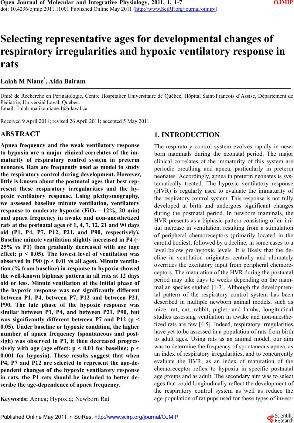 Open Journal of Molecular and Integrative Physiology, 2011, 1, 1-7 doi: 10.4236/ojmip.2011.11001 Published Online May 2011 (http://www.SciRP.org/journal/ojmip/ OJMIP ). Published Online May 2011 in SciRes. http://www.scirp.org/journal/OJMIP Selecting representative ages for developmental changes of respiratory irregularities and hypoxic ventilatory response in rats Lalah M Niane*, Aida Bairam Unité de Recherche en Périnatologie, Centre Hospitalier Universitaire de Québec, Hôpital Saint-François d’Assise, Département de Pédiatrie, Université Laval, Québec. Email: *[email protected] Received 9 April 2011; revised 26 April 2011; accepted 5 May 2011. ABSTRACT Apnea frequency and the weak ventilatory response to hypoxia are a major clinical correlates of the im- maturity of respiratory control system in preterm neonates. Rats are frequently used as model to study the respiratory control during development. However, little is known about the postnatal ages that best rep- resent these respiratory irregularities and the hy- poxic ventilatory response. Using plethysmography, we assessed baseline minute ventilation, ventilatory response to moderate hypoxia (FiO2 = 12%, 20 min) and apnea frequency in awake and non-anesthetized rats at the postnatal ages of 1, 4, 7, 12, 21 and 90 days old (P1, P4, P7, P12, P21, and P90, respectively). Baseline minute ventilation slightly increased in P4 (~ 25% vs P1) then gradually decreased with age (age effect: p < 0.05). The lowest level of ventilation was observed in P90 (p < 0.01 vs all ages). Minute ventila- tion (% from baseline) in response to hypoxia showed the well-known biphasic pattern in all rats at 12 days old or less. Minute ventilation at the initial phase of the hypoxic response was not significantly different between P1, P4, between P7, P12 and between P21, P90. The late phase of the hypoxic response was similar between P1, P4, and between P21, P90, but was significantly different between P7 and P12 (p < 0.05). Under baseline or hypoxic condition, the higher number of apnea frequency (spontaneous and post- sigh) was observed in P1, it then decreased progres- sively with age (age effect: p < 0.01 for baseline; p < 0.001 for hypoxia). These results suggest that when P4, P7 and P12 are selected to represent the age-de- pendent changes of the hypoxic ventilatory response in rats, the P1 rats should be included to better de- scribe the age-dependence of apnea frequency. Keywords: Apnea; Hypoxia; Newborn Rat 1. INTRODUCTION The respiratory control system evolves rapidly in new- born mammals during the neonatal period. The major clinical correlates of the immaturity of this system are periodic breathing and apnea, particularly in preterm neonates. Accordingly, apnea in preterm neonates is sys- tematically treated. The hypoxic ventilatory response (HVR) is regularly used to evaluate the immaturity of the respiratory control system. This response is not fully developed at birth and undergoes significant changes during the postnatal period. In newborn mammals, the HVR presents as a biphasic pattern consisting of an ini- tial increase in ventilation, resulting from a stimulation of peripheral chemoreceptors (primarily located in the carotid bodies), followed by a decline, in some cases to a level below pre-hypoxic levels. It is likely that the de- cline in ventilation originates centrally and ultimately overrides the excitatory input from peripheral chemore- ceptors. The maturation of the HVR during the postnatal period may take days to weeks depending on the mam- malian species studied [1-3]. Although the developmen- tal pattern of the respiratory control system has been described in multiple newborn animal models, such as mice, rat, cat, rabbit, piglet, and lambs, longitudinal studies assessing ventilation in awake and non-anesthe- tized rats are few [4,5]. Indeed, respiratory irregularities have yet to be assessed in a population of rats from birth to adult ages. Using rats as an animal model, our aim was to determine the frequency of spontaneous apnea, as an index of respiratory irregularities, and to concurrently evaluate the HVR, as an index of maturation of the chemoreceptor reflex to hypoxia in specific postnatal age groups and as adult. The secondary aim was to select ages that could longitudinally reflect the development of the respiratory control system as well as reduce the age-population of rat pups used for these types of invest- 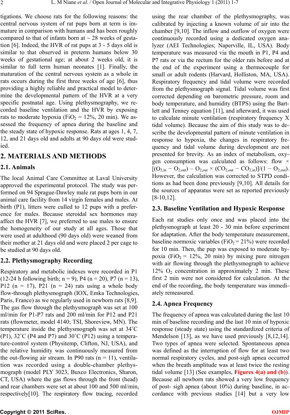 L. M Niane et al. / Open Journal of Molecular and Integrative Physiology 1 (2011) 1-7 2 tigations. We choose rats for the following reasons: the central nervous system of rat pups born at term is im- mature in comparison with humans and has been roughly compared to that of infants born at ~ 28 weeks of gesta- tion [6]. Indeed, the HVR of rat pups at 3 - 5 days old is similar to that observed in preterm humans below 30 weeks of gestational age; at about 2 weeks old, it is similar to full term human neonates [1]. Finally, the maturation of the central nervous system as a whole in rats occurs during the first three weeks of age [6], thus providing a highly reliable and practical model to deter- mine the developmental pattern of the HVR at a very specific postnatal age. Using plethysmography, we re- corded baseline ventilation and the HVR by exposing rats to moderate hypoxia (FiO2 = 12%, 20 min). We as- sessed the frequency of apnea during the baseline and the steady state of hypoxic response. Rats at ages 1, 4, 7, 12, and 21 days old and adults at 90 days old were stud- ied. 2. MATERIALS AND METHODS 2.1. Animals The local Animal Care Committee at Laval University approved the experimental protocol. The study was per- formed on 94 Sprague-Dawley male rat pups born in our animal care facility from 14 virgin females and males. At birth (P1), litters were culled to 12 pups with a prefer- ence for males. Because steroidal sex hormones may affect the HVR [7], we preferred to use males to ensure the homogeneity of our study at all ages. Those that were used at adulthood (90 days old) were weaned from their mother at 21 days old and were placed 2 per cage to be studied at 90 days old. 2.2. Plethysmography Recording Respiratory and metabolic indexes were recorded in P1 (12-24 h following birth; n = 9), P4 (n = 20), P7 (n = 13), P12 (n = 17), P21 (n = 24) rats using a whole body flow-through plethysmograph (IOX, Emka Technologies, Paris, France) as we regularly used in newborn rats [8,9]. The gas flow through the plethysmograph was set at 100 ml/min for P1-P7 rats and 200 ml/min for P12 and P21 rats (flowmeter, model 4140; TSI, Shoreview, MN). The temperature inside the plethysmograph was set at 34˚C (P1), 32˚C (P4 and P7) and 30˚C (P12) using a tempera- ture-control system (Physitemp, Clifton, NJ, USA), and the relative humidity was continuously measured from the out-flowing air stream. In P90 rats (n = 11), ventila- tion was recorded using a double-chamber plethys- mograph (model PLY 3023, Buxco Electronics, Sharon, CT, USA) where the gas flows through the front (head) and rear chambers were set at about 100 and 500 ml/min, respectively[10]. The respiratory flow tracing, recorded using the rear chamber of the plethysmography, was calibrated by injecting a known volume of air into the chamber [9,10]. The inflow and outflow of oxygen were continuously recorded using a dedicated oxygen ana- lyzer (AEI Technologies; Naperville, IL, USA). Body temperature was measured via the mouth in P1, P4 and P7 rats or via the rectum for the older rats before and at the end of the experiment using a thermocouple for small or adult rodents (Harvard, Holliston, MA, USA). Respiratory frequency and tidal volume were recorded from the plethysmograph signal. Tidal volume was first corrected depending on barometric pressure, room and body temperature, and humidity (BTPS) using the Bart- lett and Tenney equation [11], and afterward, it was used to calculate minute ventilation (respiratory frequency X tidal volume). Because the aim of this study was to de- scribe the developmental pattern of minute ventilation in response to hypoxia, the changes in respiratory fre- quency and tidal volume during development are not presented for brevity. As an index of metabolism, oxy- gen consumption was calculated as follows: flow × [(O2,in − O2,out) – O2,out × (CO2,out − CO2,in)]/(1 − O2,out). However, the calculation was corrected to STPD condi- tions as had been done previously [9,10]. All details for the sources of apparatus were set as reported previously [8-10,12]. 2.3. Baseline Ventilation and Hypoxic Response Each rat studies only once and was placed into the plethysmograph at least 20 - 30 min before experiment for adaptation. After the body temperature measurement, baseline normoxic variables (FiO2 = 21%) were recorded for 10 min. Then, the pup was exposed to moderate hy- poxia (FiO2 = 12%, 20 min) by mixing pure nitrogen with air flowing through the plethysmograph to achieve 12% O2 concentration in approximately 2 min. These first 2 min were not considered for calculation. At the end of the recording, the body temperature was immedi- ately remeasured. 2.4. Apnea Frequency The frequency of apnea was calculated during the last 10 min of baseline recording and the last 10 min of hypoxic response (steady state) using the standardized criteria of Mendelson [13], as we have used previously [8,12,14]. Two types of apnea were selected. Spontaneous apnea was defined as the interruption of flow for at least two normal respiratory cycles, and post-sigh apnea occurred when the breath amplitude was at least twice the resting tidal volume [13] (See examples, Figures 4(a) and (b)). Because all newborn rats showed a very low frequency of post- sigh apnea (about 10%) during baseline, in ac- cordance with previous studies [14] but a very low C opyright © 2011 SciRes. OJMIP 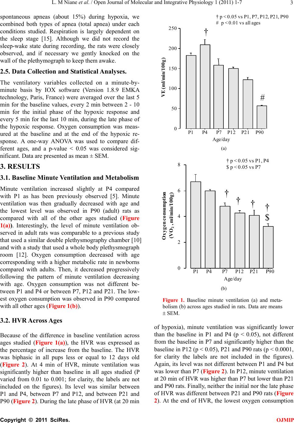 L. M Niane et al. / Open Journal of Molecular and Integrative Physiology 1 (2011) 1-7 Copyright © 2011 SciRes. 3 spontaneous apneas (about 15%) during hypoxia, we combined both types of apnea (total apnea) under each conditions studied. Respiration is largely dependent on the sleep stage [15]. Although we did not record the sleep-wake state during recording, the rats were closely observed, and if necessary we gently knocked on the wall of the plethymograph to keep them awake. 0 50 100 150 200 250 P1P4P7 P12P21P90 † # V E (m l /m i n /100g) † p < 0.05 vs P1, P7, P12, P21, P90 # p < 0.01 vs all ages Age/day 2.5. Data Collection and Statistical Analyses. The ventilatory variables collected on a minute-by- minute basis by IOX software (Version 1.8.9 EMKA technology, Paris, France) were averaged over the last 5 min for the baseline values, every 2 min between 2 - 10 min for the initial phase of the hypoxic response and every 5 min for the last 10 min, during the late phase of the hypoxic response. Oxygen consumption was meas- ured at the baseline and at the end of the hypoxic re- sponse. A one-way ANOVA was used to compare dif- ferent ages, and a p-value < 0.05 was considered sig- nificant. Data are presented as mean ± SEM. (a) Oxygen consumption (VO 2 , ml/min/100g) 0 2 4 6 8 P1P4P7P12 P21P90 †††† $ † p < 0.05 vs P1, P4 $ p < 0.05 vs P7 Age/day 3. RESULTS 3.1. Baseline Minute Ventilation and Metabolism Minute ventilation increased slightly at P4 compared with P1 as has been previously observed [5]. Minute ventilation was then gradually decreased with age and the lowest level was observed in P90 (adult) rats as compared with all of the other ages studied (Figure 1(a)). Interestingly, the level of minute ventilation ob- served in adult rats was comparable to a previous study that used a similar double plethysmography chamber [10] and with a study that used a whole body plethysmograph room [12]. Oxygen consumption decreased with age corresponding with a higher metabolic rate in newborns compared with adults. Then, it decreased progressively following the pattern of minute ventilation decreasing with age. Oxygen consumption was not different be- tween P1 and P4 or between P7, P12 and P21. The low- est oxygen consumption was observed in P90 compared with all other ages (Figure 1(b)). (b) Figure 1. Baseline minute ventilation (a) and meta- bolism (b) across ages studied in rats. Data are means SEM. 3.2. HVR Across Ages of hypoxia), minute ventilation was significantly lower than the baseline in P1 and P4 (p < 0.05), not different from the baseline in P7 and significantly higher than the baseline in P12 (p < 0.05), P21 and P90 rats (p < 0.0001, for clarity the labels are not included in the figures). Again, its level was not different between P1 and P4 but was lower than P7 (Figure 2). In P12, minute ventilation at 20 min of HVR was higher than P7 but lower than P21 and P90 rats. Finally, neither the initial nor the late phase of HVR was different between P21 and P90 rats (Figure 2). At the end of HVR, the lowest oxygen consumption Because of the difference in baseline ventilation across ages studied (Figure 1(a)), the HVR was expressed as the percentage of increase from the baseline. The HVR was biphasic in all pups less or equal to 12 days old (Figure 2). At 4 min of HVR, minute ventilation was significantly higher than baseline in all ages studied (P varied from 0.01 to 0.001; for clarity, the labels are not included on the figures). Its level was similar between P1 and P4, between P7 and P12, and between P21 and P90 (Figure 2). During the late phase of HVR (at 20 min OJMIP 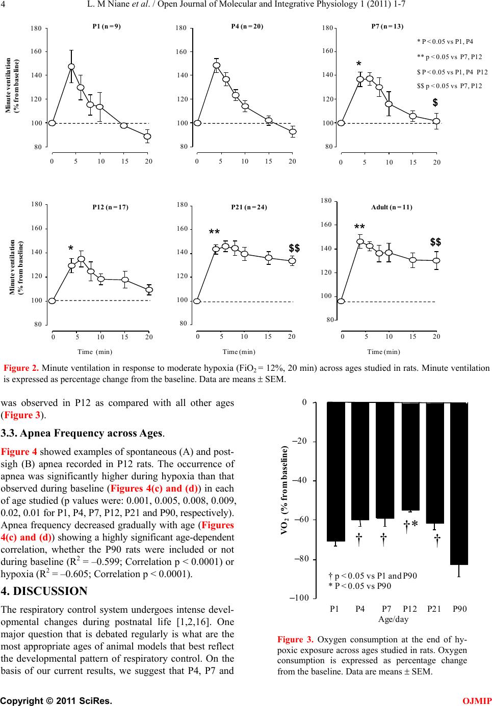 L. M Niane et al. / Open Journal of Molecular and Integrative Physiology 1 (2011) 1-7 4 Minute ventilation (% from bas el in e ) 0510 15 20 80 100 120 140 160 180 0510 15 20 80 100 120 140 160 180 05101520 80 100 120 140 160 180 P1 (n = 9)P4 (n = 20)P7 (n = 13) Time (min) 0510 15 20 80 100 120 140 160 180 P1 2 (n = 17) Time (min) 0510 15 20 100 120 140 160 180 Time (min) 0510 15 20 80 100 120 140 160 180 P21 (n = 24)Ad u l t (n = 11) Minute ventilation (% from baseline) * $ ** $$ $$ * P < 0.05 vs P1, P4 ** p < 0.05 vs P7, P12 ** $ P < 0.05 vs P1, P4 P12 $$ p < 0.05 vs P7, P12 * 80 Figure 2. Minute ventilation in response to moderate hypoxia (FiO2 = 12%, 20 min) across ages studied in rats. Minute ventilation is expressed as percentage change from the baseline. Data are means SEM. was observed in P12 as compared with all other ages (Figure 3). 3.3. Apnea Frequency across Ages. Figure 4 showed examples of spontaneous (A) and post- sigh (B) apnea recorded in P12 rats. The occurrence of apnea was significantly higher during hypoxia than that observed during baseline (Figures 4(c) and (d)) in each of age studied (p values were: 0.001, 0.005, 0.008, 0.009, 0.02, 0.01 for P1, P4, P7, P12, P21 and P90, respectively). Apnea frequency decreased gradually with age (Figures 4(c) and (d)) showing a highly significant age-dependent correlation, whether the P90 rats were included or not during baseline (R2 = –0.599; Correlation p < 0.0001) or hypoxia (R2 = –0.605; Correlation p < 0.0001). 4. DISCUSSION The respiratory control system undergoes intense devel- opmental changes during postnatal life [1,2,16]. One major question that is debated regularly is what are the most appropriate ages of animal models that best reflect the developmental pattern of respiratory control. On the basis of our current results, we suggest that P4, P7 and VO 2 (% from bas el in e) –100 –80 –60 –40 –20 0 P1P4P7P12 P21P90 †* †† † † p < 0.05 vs P1 and P90 * P < 0.05 vs P90 Age/day Figure 3. Oxygen consumption at the end of hy- poxic exposure across ages studied in rats. Oxygen consumption is expressed as percentage change from the baseline. Data are means SEM. C opyright © 2011 SciRes. OJMIP 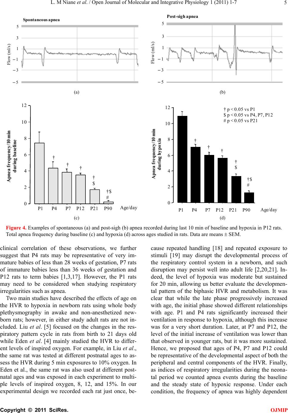 L. M Niane et al. / Open Journal of Molecular and Integrative Physiology 1 (2011) 1-7 5 S p on tan e ou s ap n ea Flow (ml/s) 5 3 1 –1 –3 –5 Pos t-s igh apn ea 5 3 1 –1 –3 –5 Flow (ml/s) (a) (b) 0 2 4 6 8 10 12 Apnea frequency/10 min during baseline P1P4P7 P12P21P90 † † † $ †$ # † Age/day Apnea frequency/10 min during hypoxia P1P4P7 P12P21P90 0 2 4 6 8 10 12 † p < 0.05 vs P1 $ p < 0.05 vs P4, P7, P12 # p < 0.05 vs P21 † † † $ †$ # † Age/day (c) (d) Figure 4. Examples of spontaneous (a) and post-sigh (b) apnea recorded during last 10 min of baseline and hypoxia in P12 rats. Total apnea frequency during baseline (c) and hypoxia (d) across ages studied in rats. Data are means SEM. clinical correlation of these observations, we further suggest that P4 rats may be representative of very im- mature babies of less than 28 weeks of gestation, P7 rats of immature babies less than 36 weeks of gestation and P12 rats to term babies [1,3,17]. However, the P1 rats may need to be considered when studying respiratory irregularities such as apnea. Two main studies have described the effects of age on the HVR to hypoxia in newborn rats using whole body plethysmography in awake and non-anesthetized new- born rats; however, in either study adult rats are not in- cluded. Liu et al. [5] focused on the changes in the res- piratory pattern cycle in rats from birth to 21 days old while Eden et al. [4] mainly studied the HVR to differ- ent levels of inspired oxygen. For example, in Liu et al., the same rat was tested at different postnatal ages to as- sess the HVR during 5 min exposures to 10% oxygen. In Eden et al., the same rat was also used at different post- natal ages and was exposed in each experiment to multi- ple levels of inspired oxygen, 8, 12, and 15%. In our experimental design we recorded each rat just once, be- cause repeated handling [18] and repeated exposure to stimuli [19] may disrupt the developmental process of the respiratory control system in a newborn, and such disruption may persist well into adult life [2,20,21]. In- deed, the level of hypoxia was moderate but sustained for 20 min, allowing us better evaluate the developmen- tal pattern of the biphasic HVR and metabolism. It was clear that while the late phase progressively increased with age, the initial phase showed different relationships with age. P1 and P4 rats significantly increased their ventilation in response to hypoxia, although this increase was for a very short duration. Later, at P7 and P12, the level of the initial increase of ventilation was lower than that observed in younger rats, but it was more sustained. Hence, we proposed that ages of P4, P7 and P12 could be representative of the developmental aspect of both the peripheral and central components of the HVR. Finally, as indices of respiratory irregularities during the neona- tal period we counted apnea events during the baseline and the steady state of hypoxic response. Under each condition, the frequency of apnea was highly dependent C opyright © 2011 SciRes. OJMIP 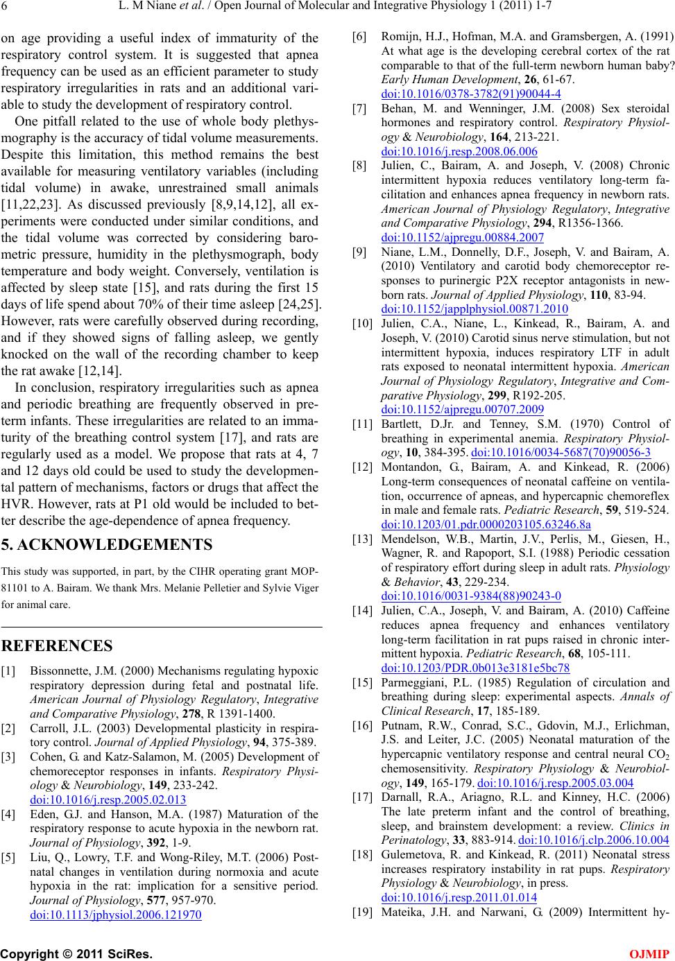 L. M Niane et al. / Open Journal of Molecular and Integrative Physiology 1 (2011) 1-7 6 on age providing a useful index of immaturity of the respiratory control system. It is suggested that apnea frequency can be used as an efficient parameter to study respiratory irregularities in rats and an additional vari- able to study the development of respiratory control. One pitfall related to the use of whole body plethys- mography is the accuracy of tidal volume measurements. Despite this limitation, this method remains the best available for measuring ventilatory variables (including tidal volume) in awake, unrestrained small animals [11,22,23]. As discussed previously [8,9,14,12], all ex- periments were conducted under similar conditions, and the tidal volume was corrected by considering baro- metric pressure, humidity in the plethysmograph, body temperature and body weight. Conversely, ventilation is affected by sleep state [15], and rats during the first 15 days of life spend about 70% of their time asleep [24,25]. However, rats were carefully observed during recording, and if they showed signs of falling asleep, we gently knocked on the wall of the recording chamber to keep the rat awake [12,14]. In conclusion, respiratory irregularities such as apnea and periodic breathing are frequently observed in pre- term infants. These irregularities are related to an imma- turity of the breathing control system [17], and rats are regularly used as a model. We propose that rats at 4, 7 and 12 days old could be used to study the developmen- tal pattern of mechanisms, factors or drugs that affect the HVR. However, rats at P1 old would be included to bet- ter describe the age-dependence of apnea frequency. 5. ACKNOWLEDGEMENTS This study was supported, in part, by the CIHR operating grant MOP- 81101 to A. Bairam. We thank Mrs. Melanie Pelletier and Sylvie Viger for animal care. REFERENCES [1] Bissonnette, J.M. (2000) Mechanisms regulating hypoxic respiratory depression during fetal and postnatal life. American Journal of Physiology Regulatory, Integrative and Comparative Physiology, 278, R 1391-1400. [2] Carroll, J.L. (2003) Developmental plasticity in respira- tory control. Journal of Applied Physiology, 94, 375-389. [3] Cohen, G. and Katz-Salamon, M. (2005) Development of chemoreceptor responses in infants. Respiratory Physi- ology & Neurobiology, 149, 233-242. doi:10.1016/j.resp.2005.02.013 [4] Eden, G.J. and Hanson, M.A. (1987) Maturation of the respiratory response to acute hypoxia in the newborn rat. Journal of Physiology, 392, 1-9. [5] Liu, Q., Lowry, T.F. and Wong-Riley, M.T. (2006) Post- natal changes in ventilation during normoxia and acute hypoxia in the rat: implication for a sensitive period. Journal of Physiology, 577, 957-970. doi:10.1113/jphysiol.2006.121970 [6] Romijn, H.J., Hofman, M.A. and Gramsbergen, A. (1991) At what age is the developing cerebral cortex of the rat comparable to that of the full-term newborn human baby? Early Human Development, 26, 61-67. doi:10.1016/0378-3782(91)90044-4 [7] Behan, M. and Wenninger, J.M. (2008) Sex steroidal hormones and respiratory control. Respiratory Physiol- ogy & Neurobiology, 164, 213-221. doi:10.1016/j.resp.2008.06.006 [8] Julien, C., Bairam, A. and Joseph, V. (2008) Chronic intermittent hypoxia reduces ventilatory long-term fa- cilitation and enhances apnea frequency in newborn rats. American Journal of Physiology Regulatory, Integrative and Comparative Physiology, 294, R1356-1366. doi:10.1152/ajpregu.00884.2007 [9] Niane, L.M., Donnelly, D.F., Joseph, V. and Bairam, A. (2010) Ventilatory and carotid body chemoreceptor re- sponses to purinergic P2X receptor antagonists in new- born rats. Journal of Applied Physiology, 110, 83-94. doi:10.1152/japplphysiol.00871.2010 [10] Julien, C.A., Niane, L., Kinkead, R., Bairam, A. and Joseph, V. (2010) Carotid sinus nerve stimulation, but not intermittent hypoxia, induces respiratory LTF in adult rats exposed to neonatal intermittent hypoxia. American Journal of Physiology Regulatory, Integrative and Com- parative Physiology, 299, R192-205. doi:10.1152/ajpregu.00707.2009 [11] Bartlett, D.Jr. and Tenney, S.M. (1970) Control of breathing in experimental anemia. Respiratory Physiol- ogy, 10, 384-395. doi:10.1016/0034-5687(70)90056-3 [12] Montandon, G., Bairam, A. and Kinkead, R. (2006) Long-term consequences of neonatal caffeine on ventila- tion, occurrence of apneas, and hypercapnic chemoreflex in male and female rats. Pediatric Research, 59, 519-524. doi:10.1203/01.pdr.0000203105.63246.8a [13] Mendelson, W.B., Martin, J.V., Perlis, M., Giesen, H., Wagner, R. and Rapoport, S.I. (1988) Periodic cessation of respiratory effort during sleep in adult rats. Physiology & Behavior, 43, 229-234. doi:10.1016/0031-9384(88)90243-0 [14] Julien, C.A., Joseph, V. and Bairam, A. (2010) Caffeine reduces apnea frequency and enhances ventilatory long-term facilitation in rat pups raised in chronic inter- mittent hypoxia. Pediatric Research, 68, 105-111. doi:10.1203/PDR.0b013e3181e5bc78 [15] Parmeggiani, P.L. (1985) Regulation of circulation and breathing during sleep: experimental aspects. Annals of Clinical Research, 17, 185-189. [16] Putnam, R.W., Conrad, S.C., Gdovin, M.J., Erlichman, J.S. and Leiter, J.C. (2005) Neonatal maturation of the hypercapnic ventilatory response and central neural CO2 chemosensitivity. Respiratory Physiology & Neurobiol- ogy, 149, 165-179. doi:10.1016/j.resp.2005.03.004 [17] Darnall, R.A., Ariagno, R.L. and Kinney, H.C. (2006) The late preterm infant and the control of breathing, sleep, and brainstem development: a review. Clinics in Perinatology, 33, 883-914. doi:10.1016/j.clp.2006.10.004 [18] Gulemetova, R. and Kinkead, R. (2011) Neonatal stress increases respiratory instability in rat pups. Respiratory Physiology & Neurobiology, in press. doi:10.1016/j.resp.2011.01.014 [19] Mateika, J.H. and Narwani, G. (2009) Intermittent hy- C opyright © 2011 SciRes. OJMIP  L. M Niane et al. / Open Journal of Molecular and Integrative Physiology 1 (2011) 1-7 Copyright © 2011 SciRes. 7 OJMIP poxia and respiratory plasticity in humans and other animals: does exposure to intermittent hypoxia promote or mitigate sleep apnoea? Experimental Physiology, 94, 279-296. doi:10.1113/expphysiol.2008.045153 [20] Bavis, R.W. and Mitchell, G.S. (2008) Long-term effects of the perinatal environment on respiratory control. Journal of Applied Physiology, 104, 1220-1229. doi:10.1152/japplphysiol.01086.2007 [21] Montandon, G., Bairam, A. and Kinkead, R. (2008) Neo- natal caffeine induces sex-specific developmental plas- ticity of the hypoxic respiratory chemoreflex in adult rats, American Journal of Physiology Regulatory, Integrative and Comparative Physiology, 295, R922-934. doi:10.1152/ajpregu.00059.2008 [22] Enhorning, G., Schaik, van S., Lundgren, C. and Vargas, I. (1998) Whole-body plethysmography, does it measure tidal volume of small animals? Canadian Journal of Physiology and Pharmacology, 76, 945-951. doi:10.1139/y99-002 [23] Mortola, J.P. and Frappell, P.B. (1998) On the barometric method for measurements of ventilation, and its use in small animals. Canadian Journal of Physiology and Pharmacology, 76, 937-944. doi:10.1139/y99-001 [24] Blumberg, M.S., Seelke, A.M., Lowen, S.B. and Karls- son, K.A. (2005) Dynamics of sleep-wake cyclicity in developing rats. Proceedings of the National Academy of Sciences, USA, 102, 14860-14864. doi:10.1073/pnas.0506340102 [25] Frank, M.G. and Heller, H.C. (1997) Development of REM and slow wave sleep in the rat. American Journal of Physiology, 272, R1792-1799. |