

Paper Menu >>
Journal Menu >>
 Vol.3, No.5, 304-311 (2011) doi:10.4236/health.2011.35053 C opyright © 2011 SciRes. Openly accessible at http://www.scirp.org/journal/HEALTH/ Health Endogenous prostaglandin D2 synthesis inhibits e-selectin generation in human umbilical vein endothelial cells Hideyuki Negoro1*, Hiroyuki Kobayashi2, Yoshio Uehara3 1Harvard Medical School, Internal Medicine, Boston, USA; *Corresponding Author: [email protected] 2 Department of Hospital Administration, Graduate School of Medicine, Juntendo University, Tokyo, Japan; 3University of Tokyo Internal Medicine, Tokyo, Japan. Received 14 March 2011; revised 25 April 2011; accepted 29 April 2011. ABSTRACT We examined the role of prostaglandin D2 (PGD2) in the formation of E-selectin following inter- leukin-1 (IL-1) stimulation in human umbilical vein endothelial cells (HUVEC) transfected with lipocaline-type PGD2 synthase (L-PGDS) genes. HUVEC were isolated from human umbilical vein and incubated with 20 U/mL IL-1 and vari- ous concentrations of authentic PGD2. The iso- lated HUVEC were also transfected with L-PGDS genes by electroporation. The L-PGDS-trans- fected HUVEC were used to investigate the role of endogenou s PGD2 in IL-1-stimulated E-selectin biosynthesis. We also used an anti-PGD2 anti- body to examine whether an intracrine mecha- nism was involved in E-selectin production. PGD2 and E-selectin levels were determined by radio-immunoassay and enzyme-immunoassay, respectively. E-selectin mRNA was assessed by real-time RT-PCR. IL-1-stimulated E-selectin production by HUVEC was dose-dependently inhibited by authentic PGD2 at concentrations greater than 10−6 mol/L. L-PGDS gene-trans- fected HUVEC produced more PGD2 than HU- VEC transfected with the reporter gene alone. IL-1 induced increases in E-selectin production in HUVEC transfected with the reporter genes alone. However, this effect was significantly attenuated in the case of IL-1 stimulation of HUVEC transfected with L-PGDS genes, and accompanied by an apparent suppression of E-selectin mRNA expression. Neutralization of extracellular PGD2 by anti-PGD 2- sp ecifi c antibody influenced neither E-selectin mRNA expression nor E-selectin biosynthesis. HUVEC transfected with L-PGDS genes showed increased PGD2 synthesis. This increase was associated with attenuation of both E-selectin generation and E-selectin mRNA expression. The results sug- gest that endogenous PGD2 decreases E-se- lectin synthesis and E-selectin mRNA expres- sion, probably through an intracrine mecha- nism. Keywords: Prostaglandin; E-Selectin; P GDS; Endothelial Cell 1. INTRODUCTION Adhesion molecules play an important role in the de- velopment and progression of atherosclerosis. The hy- pothesis proposed by Ross and its modifications have been generally accepted as a mechanism of atherosclero- sis where adhesion of circulating monocytes and lym- phocytes to vascular endothelium presumably initiates a series of events toward atherosclerosis [1]. Cellular ad- hesion molecules mediate the adhesion, margination, and transendothelial migration of circulating mononuclear cells from the blood stream to the extravascular com- partment to have an important part in the progression of atherosclerotic plaque [2]. Recent studies have eluci- dated further that to anchor leukocytes onto the endothe- lial cells, the adhesion molecules expressed on the sur- face of endothelial cells necessitated to bind to their ligands expressed on leukocytes. Endothelial cells are stimulated by inflammatory agents to express selectins, such as endothelial-leukocyte adhesion molecule-1 (E-selectin), which interact with carbohydrate ligands on leukocytes, and to express immunoglobulin superfamily proteins, such as vascular cell adhesion molecule-1 (VCAM-1). The adhesion molecules expressed on the endothelial cells include VCAM-1, intracellular adhe- sion molecule (ICAM)-1, P-selectin and E-selectin [3]. Selectins, including E-selectin and P-selectin, are in-  H. Negoro et al. / Health 3 (2011) 304-311 Copyright © 2011 SciRes. Openly accessible at http://www.scirp.org/journal/HEALTH/ 305305 volved in the first step of leukocyte adhesion at sites of inflammation or injury. Selectins are characterized by rolling and tethering of leukocytes to the endothelial surface, to platelets or to other leukocytes [4]. In fact, E-selectin is demonstrated to occur in atherosclerotic lesions in the coronary artery of humans [5]. Therefore, E-selectin is believed to be a key factor for the develop- ment of immune-mediated cardiovascular injury. Intrestngly, we have recently reported that PGD2 at- tenuates inducible nitric oxide generation in vascular smooth muscle cells [6]. Endogenous prostaglandin D2 synthesis reduces plasminogen activator inhibitor-1 gen- eration following cytokines stimulation in bovine endo- thelial cells [7]. Especially, PGD2 is synthesized in vas- cular components of atheromatous lesions including endothelial cells, macrophages, platelets, and mast cells [8] and lipocalin-type PGD2 synthase (L-PGDS) is demonstrated to occur in atheromatous lesions in the cardiovascular system [9]. These data strongly suggest that L-PGDS/PGD2 is upregulated in response to im- mune-related vascular lesions and in turn, the increase of L-PGDS/PGD2 is exerted to attenuate the progression of the arterial remodeling. Taken together, we proposed the hypothesis that PGD2 regulates E-selectin expression in endothelial cells, thereby contributing to leukocyte adhesion, an integral component of the development of vascular injury. How- ever, there had been few data investigating the crosstalk between endogenous L-PGDS/PGD2 system and the adhesion molecule expression by cytokines. In the pre- sent study, in order to test our hypothesis that L-PGDS/ PGD2 protects the vascular wall against immune-related vascular injury, we examined the relationship between endogenous PGD2 and E-selectin expression by endo- thelial cells and attempted to reveal its intracellular mechanism mediated by PGD2 using L-PGDS gene- transfected endothelial cells in culture. We also exam- ined whether the increases in intracellular PGD2 synthesis influenced E-selectin mRNA expression and E-selectin biosynthesis observed following interleukin-1b (IL-1) stimulation. 2. MATERIALS AND METHODS 2.1. Materials Eicosanoids and related compounds were purchased from Funakoshi chemicals (Tokyo, Japan). Arachidonic acid was purchased from Sigma (St. Louis, MO, USA). Recombinant murine IL-1 was purchased from R&D Systems (Minneapolis, MN, USA). Radioactively la- beled materials were purchased from Amersham (Tokyo, Japan). A 3-kb gene for rat brain PGDS [(5Z, 13E)- (15S)-9a, 11a -epidoxy-15-hydroxyprosta-5,13-dienoate D-isomerase, EC 5.3.99.2] was isolated from a rat ge- nomic DNA library by plaque hybridization with cDNA for the PGDS enzyme, as described in our previous studies [10]. A 3-kb BamHI fragment of rat PGDS, which belongs to the lipocalin family, was inserted into a pcD2 plasmid containing the SV40 promoter, along with a polyA signal at the XhoI site (Invitrogen, Carlsbad, CA, USA) [11]. The b-galactosidase gene with a cytomega- lovirus (CMV) promoter at an XbaI site was inserted into the pBluescript 2 KS+ plasmid (Stratagene, La Jolla, CA, USA). 2.2. Cell Culture Human umbilical vein endothelial cells (HUVEC) were harvested enzymatically as described previously [12]. They were maintained in medium 199 (GIBCO BRL, Gaithersburg, MD), containing Hepes, heparin (1%), endothelial cell growth factor (50 mg/ml), L-glutamin (1%), antibiotics, and 5% fetal bovine serum (FBS). When the cells reached confluence, they were replanted onto low pyrogen fibronectin at 20,000 cells/ cm2. HUVEC which were isolated from a confluent monolayer of polygonal cells. The cells expressed von Willbrand factor as determined by their content of spe- cific mRNA and immunoreactive protein. Cellular vi- ability was assessed by Trypan blue exclusion. 2.3. Effect of Exogenous PGD2 on E-Selectin Expression in Endothelial Cells Cultures of HUVEC were treated with 20 U/ml IL-1, according to the previous study, in the presence of vari- ous concentrations of PGD2 to be incubated for 18 h. Thereafter, the HUVEC were washed three times with FBS-free Dulbecco’s phosphate-buffered saline (D-PBS; Gibco) and the cells were re-incubated in 1 ml of fresh D-PBS for 2 h. Subsequently, the cells and culture su- pernatants were used in various assays. 2.4. Transfection of L-PGDS Genes into HUVEC HUVEC were transfected with L-PGDS genes using the Shimadzu GTE-10 electroporation device (Gene Transfer Equipment-10, Shimadzu Co., Ltd., Kyoto, Japan). This equipment transiently increases the perme- ability of plasma membranes of HUVEC, thereby facili- tating translocation of genes into the cytoplasm. Briefly, the cells were washed three times with FBS-free D-PBS and 10 mg of pcD2-rat PGDS in 0.5 ml of fresh D-PBS were added to each well [13]. A transient electrical cur- rent was applied onto HUVEC growing in the culture dishes, using a 35-mm round electrode (Model FTC-  H. Negoro et al. / Health 3 (2011) 304-311 Copyright © 2011 SciRes. Openly accessible at http://www.scirp.org/journal/HEALTH/ 306 33D3, Shimadzu Co., Ltd., Kyoto, Japan), after which the cells were incubated for 30 minutes. Following transfection, 2 ml of DMEM containing 10% FBS was added to the HUVEC and the dishes were incubated for an additional 24 hours. Endogenous PGD2 production was stimulated by adding 10–6 mol/l arachi- donic acid for 24 hours. Fresh medium containing 10–6 mol/l arachidonic acid was then added and E-selectin mRNA expression and E-selectin expression were stimulated for 18 hours by the addition of 20 U/ml IL-1. At the end of this incubation period, the HUVEC were washed three times with FBS-free D-PBS and incubated in 1 ml of fresh D-PBS for 2 hours. Finally, the cells and culture supernatants were collected for analysis. For comparison, HUVEC were transfected with b- galactosidase genes (b-gal) by electroporation to deter- mine the efficacy of gene transfection. Three days after the transfection, HUVEC were stained with X-gal [14], and b-galactasidase expression was measured. Transfec- tion efficacy was estimated as the ratio of the X-gal stained area to the sectional area of the HUVEC. 2.5. Neutralizing Extrinsic PGD2 Released from HUVEC We attempted to neutralize PGD2 using an anti-PGD2- specific antibody in order to investigate the effects of PGD2 released from L-PGDS-transfected HUVEC. We estimated the amount of anti-PGD2 antibody required to completely neutralize the secreted PGD2 using the Scatchard analysis. The binding affinity was 0.0051 ml/pg, and the Bmax (maximal binding capacity) was 25 pg/l [15]. Therefore, 1 liter of antibody had the capacity to bind 25 pg PGD2. Taking into account of these results, we used 200 ml of the antibody to inhibit the receptor- mediated actions of PGD2 in HUVEC under our culture conditions. The anti-PGD2 antibody was raised in our laboratories using PGD2-conjugated thyroglobulin and Freund’s complete adjuvant. The antibody cross-reacted 0.003% with thromboxane B2, 0.01% with prostaglandin E2 (PGE2), 0.009% with prostaglandin F2a (PGF2a), 0.008% with 6-keto-PGF1a and 0.01% with arachidonate [16]. Antibody activity was confirmed by suppression of intracellular cyclic AMP (cAMP) following PGD2 stimulation via PGD2 receptor, which acts as a second messenger for PGD2 signal transduction [17]. 2.6. Eicosanoid Radioimmunoassay Eicosanoids were determined in culture media using the direct radioimmunoassay method described previ- ously [16]. Briefly, 0.1 ml of sample, 0.1 ml of [3H] ei- cosanoid (5000 dpm) and 0.1 ml of the diluted antibody were mixed and incubated at 4˚C for 24 hours. To sepa- rate bound from free [3H] eicosanoid, 0.1 ml of dex- tran-coated charcoal in a 50 mmol/l phosphate buffer at pH 7.4 containing 0.1% gelatin and 100 mmol/l NaCl was added to the ice-chilled assay mixture. The mixture was vortexed and centrifuged at 3000 rpm for 5 min at 4˚C. The supernatant was assayed for [3H] eicosanoids bound to the antibody. Radioactivity was determined using an automatic liquid scintillation counter. The properties of the anti-6keto-PGF1a antibody was de- scribed previously [15,16]. The cross-reactivity and its properties of the anti-PGD2 antibody was detailed above. The low cross-reactivity of each antibody made it feasi- ble to measure directly the eicosanoid in media. 2.7. E-Secletin and E-Secletin mRNA Measurements We measured E-selectin expression by commercially available cell surface enzyme immunoassay as described previously (R&D Systems, Inc., USA). Cultured E-selectin transcripts were detected using the real-time reverse transcription-polymerase chain reaction (real-time RT-PCR) method using real-time RT-PCR machine as described previously [18]. 2.8. Statistical Analysis All values are expressed as the mean ± SE. The dif- ferences between values were assessed by an one-way ANOVA and Duncan’s multiple range test using the STATISTICA program (StatSoft, Tulsa, OK, USA) on a Gateway G6-400 computer system (Gateway Inc., N Sioux City, SD, USA) running the Windows 98 operat- ing system. P values less than 0.05 were considered sta- tistically significant. 3. RESULTS 3.1. Effect of Exogenous PGD2 on E-Selectin Expression in Endothelial Cells Stimulation of endothelial cells with IL-1 significantly increased E-selectin expression. The increase was re- duced in a dose-dependent manner by the addition of PGD2 at concentrations ranging from 10–7 to 10–4 mol/l (Figure 1). 3.2. Gene Transfection and Eicosanoid Generation in HUVEC We transfected HUVEC with L-PGDS genes in order to increase endogenous PGD2 formation. PGD2 was as- sayed using radioimmunoassay. The basal levels of PGD2 in reporter-gene-transfected HUVEC maintained in  H. Negoro et al. / Health 3 (2011) 304-311 Copyright © 2011 SciRes. Openly accessible at http://www.scirp.org/journal/HEALTH/ 307307 Figure 1. Effect of exogenous PGD2 on E-selectin generation in endothelial cells. Stimulation of endothelial cells with IL-1 sig- nificantly increased E-selectin generation (left two columns). The increase was reduced in a dose-dependent manner by the addition of PGD2 at concentrations ranging from 10–7 to 10–4 mol/l. All experiments were performed three different times with at least six replicates. Statistical differences were analyzed by one-way ANOVA and Duncan’s multiple range tests. *P < 0.01 vs the value at 0 mol/L PGD2. arachidonate-free media were as low as 51.5 ± 2.2 pg/106 cells/2 hours, and the addition of 10–6 mol/l ara- chidonate had no effect on PGD2 synthesis. In contrast, L-PGDS gene-transfected HUVEC showed an increase (178.0%) in PGD2 generation even in arachidonate-free media, as compared with control HUVEC carrying re- porter genes alone. Furthermore, 10–6 mol/l arachidonate markedly stimulated PGD2 biosynthesis by 640.7%, as compared with control HUVEC carrying vector genes alone (Figure 2(a)). Thereafter, PGI2 was assayed as 6-keto-PGF1a using radioimmunoassay. Basal levels of prostacyclin (PGI2), the major eicosanoid synthesized in HUVEC, were 1488 ± 66 pg/106 cells/2 hours in cells having reporter genes alone under arachidonate-free conditions. The PGI2 generation was a markedly in- creased by 327% when the cells were stimulated with 10–6 mol/l arachidonate (Figure 2(b)). L-PGDS gene transfection did not influence PGI2 synthesis in HUVEC maintained under either arachidonate-free or arachido- nate-stimulated conditions, as compared to HUVEC transfected with reporter genes. These data clearly sug- gest that the L-PGDS genes alter the phenotype of HU- VEC so that the recombinant cells acquire the capacity to produce PGD2 in response to arachidonate stimula- tion. 3.3. Effect of L-PGDS Gene Transfection on E-Selectin Expression in HUVEC Using the HUVEC having L-PGDS genes, we invest- (a) (b) Figure 2. PGD2 and PGI2 generation in endothelial cells. PGD2 was assayed using radioimmunoassay. 10–6 mol/l arachidonate did not stimulate PGD2 generation in cells having reporter genes alone (two columns to the left in Graph a). However, cells carrying the L-PGDS genes acquired the ability to produce PGD2 with or without arachidonate stimulation (two columns to the right in Graph a). (Graph a). PGI2 was assayed as 6-keto-PGF1a using radioimmunoassay. The addition of 10–6 mol/L arachidonate to the cultures increased PGI2 generation; however, there were no differences in PGI2 production between PGDS(–), AA(+) and PGDS(+), AA(+) lines. (Graph b). Statis- tical differences were assessed by Student’s t-test (n = 6). *P < 0.01. N.S. represents not statistically significant. tigated the effects of endogenous PGD2 on the expres- sion of E-selectin with or without IL-1 stimulation. The E-selectin expression was significantly increased upon IL-1 stimulation in endothelial cells transfected with transporter genes. This increase in E-selectin with or without IL-1 stimulation was significantly blunted in HUVEC transfected with L-PGDS genes that produced indeed endogenous PGD2 in response to arachidonate stimulation (Figure 3). 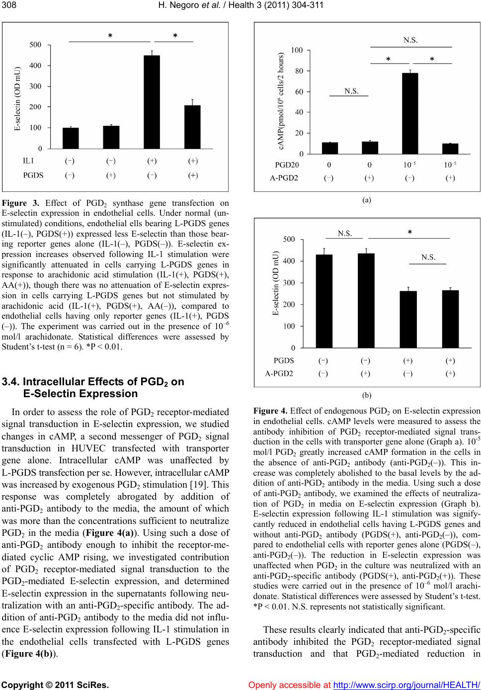 H. Negoro et al. / Health 3 (2011) 304-311 Copyright © 2011 SciRes. Openly accessible at http://www.scirp.org/journal/HEALTH/ 308 Figure 3. Effect of PGD2 synthase gene transfection on E-selectin expression in endothelial cells. Under normal (un- stimulated) conditions, endothelial ells bearing L-PGDS genes (IL-1(–), PGDS(+)) expressed less E-selectin than those bear- ing reporter genes alone (IL-1(–), PGDS(–)). E-selectin ex- pression increases observed following IL-1 stimulation were significantly attenuated in cells carrying L-PGDS genes in response to arachidonic acid stimulation (IL-1(+), PGDS(+), AA(+)), though there was no attenuation of E-selectin expres- sion in cells carrying L-PGDS genes but not stimulated by arachidonic acid (IL-1(+), PGDS(+), AA(–)), compared to endothelial cells having only reporter genes (IL-1(+), PGDS (–)). The experiment was carried out in the presence of 10–6 mol/l arachidonate. Statistical differences were assessed by Student’s t-test (n = 6). *P < 0.01. 3.4. Intracellular Effects of PGD2 on E-Selectin Expression In order to assess the role of PGD2 receptor-mediated signal transduction in E-selectin expression, we studied changes in cAMP, a second messenger of PGD2 signal transduction in HUVEC transfected with transporter gene alone. Intracellular cAMP was unaffected by L-PGDS transfection per se. However, intracellular cAMP was increased by exogenous PGD2 stimulation [19]. This response was completely abrogated by addition of anti-PGD2 antibody to the media, the amount of which was more than the concentrations sufficient to neutralize PGD2 in the media (Figure 4(a)). Using such a dose of anti-PGD2 antibody enough to inhibit the receptor-me- diated cyclic AMP rising, we investigated contribution of PGD2 receptor-mediated signal transduction to the PGD2-mediated E-selectin expression, and determined E-selectin expression in the supernatants following neu- tralization with an anti-PGD2-specific antibody. The ad- dition of anti-PGD2 antibody to the media did not influ- ence E-selectin expression following IL-1 stimulation in the endothelial cells transfected with L-PGDS genes (Figure 4(b)). (a) (b) Figure 4. Effect of endogenous PGD2 on E-selectin expression in endothelial cells. cAMP levels were measured to assess the antibody inhibition of PGD2 receptor-mediated signal trans- duction in the cells with transporter gene alone (Graph a). 10-5 mol/l PGD2 greatly increased cAMP formation in the cells in the absence of anti-PGD2 antibody (anti-PGD2(–)). This in- crease was completely abolished to the basal levels by the ad- dition of anti-PGD2 antibody in the media. Using such a dose of anti-PGD2 antibody, we examined the effects of neutraliza- tion of PGD2 in media on E-selectin expression (Graph b). E-selectin expression following IL-1 stimulation was signify- cantly reduced in endothelial cells having L-PGDS genes and without anti-PGD2 antibody (PGDS(+), anti-PGD2(–)), com- pared to endothelial cells with reporter genes alone (PGDS(–), anti-PGD2(–)). The reduction in E-selectin expression was unaffected when PGD2 in the culture was neutralized with an anti-PGD2-specific antibody (PGDS(+), anti-PGD2(+)). These studies were carried out in the presence of 10–6 mol/l arachi- donate. Statistical differences were assessed by Student’s t-test. *P < 0.01. N.S. represents not statistically significant. These results clearly indicated that anti-PGD2-specific antibody inhibited the PGD2 receptor-mediated signal transduction and that PGD2-mediated reduction in 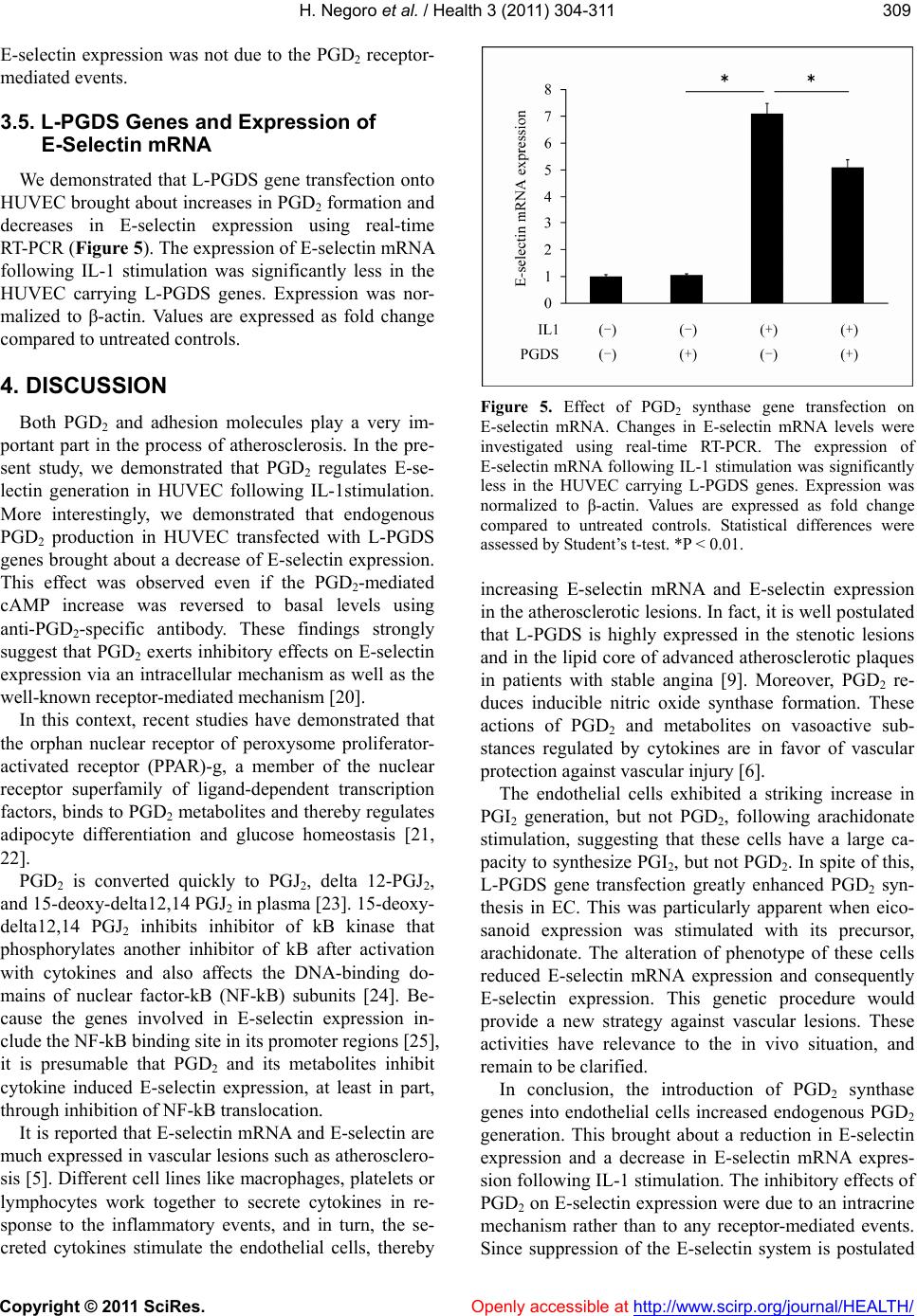 H. Negoro et al. / Health 3 (2011) 304-311 Copyright © 2011 SciRes. Openly accessible at http://www.scirp.org/journal/HEALTH/ 309309 E-selectin expression was not due to the PGD2 receptor- mediated events. 3.5. L-PGDS Genes and Expression of E-Selectin mRNA We demonstrated that L-PGDS gene transfection onto HUVEC brought about increases in PGD2 formation and decreases in E-selectin expression using real-time RT-PCR (Figure 5). The expression of E-selectin mRNA following IL-1 stimulation was significantly less in the HUVEC carrying L-PGDS genes. Expression was nor- malized to β-actin. Values are expressed as fold change compared to untreated controls. 4. DISCUSSION Both PGD2 and adhesion molecules play a very im- portant part in the process of atherosclerosis. In the pre- sent study, we demonstrated that PGD2 regulates E-se- lectin generation in HUVEC following IL-1stimulation. More interestingly, we demonstrated that endogenous PGD2 production in HUVEC transfected with L-PGDS genes brought about a decrease of E-selectin expression. This effect was observed even if the PGD2-mediated cAMP increase was reversed to basal levels using anti-PGD2-specific antibody. These findings strongly suggest that PGD2 exerts inhibitory effects on E-selectin expression via an intracellular mechanism as well as the well-known receptor-mediated mechanism [20]. In this context, recent studies have demonstrated that the orphan nuclear receptor of peroxysome proliferator- activated receptor (PPAR)-g, a member of the nuclear receptor superfamily of ligand-dependent transcription factors, binds to PGD2 metabolites and thereby regulates adipocyte differentiation and glucose homeostasis [21, 22]. PGD2 is converted quickly to PGJ2, delta 12-PGJ2, and 15-deoxy-delta12,14 PGJ2 in plasma [23]. 15-deoxy- delta12,14 PGJ2 inhibits inhibitor of kB kinase that phosphorylates another inhibitor of kB after activation with cytokines and also affects the DNA-binding do- mains of nuclear factor-kB (NF-kB) subunits [24]. Be- cause the genes involved in E-selectin expression in- clude the NF-kB binding site in its promoter regions [25], it is presumable that PGD2 and its metabolites inhibit cytokine induced E-selectin expression, at least in part, through inhibition of NF-kB translocation. It is reported that E-selectin mRNA and E-selectin are much expressed in vascular lesions such as atherosclero- sis [5]. Different cell lines like macrophages, platelets or lymphocytes work together to secrete cytokines in re- sponse to the inflammatory events, and in turn, the se- creted cytokines stimulate the endothelial cells, thereby Figure 5. Effect of PGD2 synthase gene transfection on E-selectin mRNA. Changes in E-selectin mRNA levels were investigated using real-time RT-PCR. The expression of E-selectin mRNA following IL-1 stimulation was significantly less in the HUVEC carrying L-PGDS genes. Expression was normalized to β-actin. Values are expressed as fold change compared to untreated controls. Statistical differences were assessed by Student’s t-test. *P < 0.01. increasing E-selectin mRNA and E-selectin expression in the atherosclerotic lesions. In fact, it is well postulated that L-PGDS is highly expressed in the stenotic lesions and in the lipid core of advanced atherosclerotic plaques in patients with stable angina [9]. Moreover, PGD2 re- duces inducible nitric oxide synthase formation. These actions of PGD2 and metabolites on vasoactive sub- stances regulated by cytokines are in favor of vascular protection against vascular injury [6]. The endothelial cells exhibited a striking increase in PGI2 generation, but not PGD2, following arachidonate stimulation, suggesting that these cells have a large ca- pacity to synthesize PGI2, but not PGD2. In spite of this, L-PGDS gene transfection greatly enhanced PGD2 syn- thesis in EC. This was particularly apparent when eico- sanoid expression was stimulated with its precursor, arachidonate. The alteration of phenotype of these cells reduced E-selectin mRNA expression and consequently E-selectin expression. This genetic procedure would provide a new strategy against vascular lesions. These activities have relevance to the in vivo situation, and remain to be clarified. In conclusion, the introduction of PGD2 synthase genes into endothelial cells increased endogenous PGD2 generation. This brought about a reduction in E-selectin expression and a decrease in E-selectin mRNA expres- sion following IL-1 stimulation. The inhibitory effects of PGD2 on E-selectin expression were due to an intracrine mechanism rather than to any receptor-mediated events. Since suppression of the E-selectin system is postulated 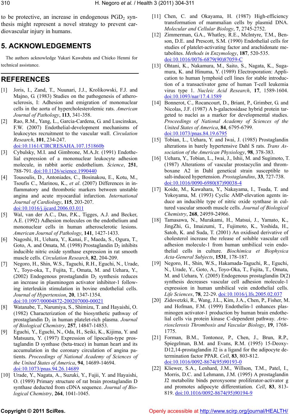 H. Negoro et al. / Health 3 (2011) 304-311 Copyright © 2011 SciRes. Openly accessible at http://www.scirp.org/journal/HEALTH/ 310 to be protective, an increase in endogenous PGD2 syn- thesis might represent a novel strategy to prevent car- diovascular injury in humans. 5. ACKNOWLEDGEMENTS The authors acknowledge Yukari Kawabata and Chieko Henmi for technical assistance. REFERENCES [1] Joris, I., Zand, T., Nuunari, J.J., Krolikowski, F.J. and Majno, G. (1983) Studies on the pathogenesis of athero- sclerosis, I: Adhesion and emigration of mononuclear cells in the aorta of hypercholesterolemic rats. American Journal of Pathology, 113, 341-358. [2] Rao, R.M., Yang, L., Garcia-Cardena, G. and Luscinskas, F.W. (2007) Endothelial-development mechanisms of leukocytes recruitment to the vascular wall. Circulation Research, 101, 234-247. doi:10.1161/CIRCRESAHA.107.151860b [3] Cybulsky, M.I. and Gimbrone, M.A.Jr. (1991) Endothe- lial expression of a mononuclear leukocyte adhesion molecule, in rabbit aortic endothelium. Science, 251, 788-791. doi:10.1126/science.1990440 [4] Tousoulis, D., Antoniades, C., Bosinakou, E., Kotu, M., Tsoufis C., Marinou, K., et al. (2007) Differences in in- flammatory and thrombotic markers between unstable angina and acute myocardial infarction. International Journal of Cardiology, 115, 203-207. doi:10.1016/j.ijcard.2006.03.011 [5] Wal, van der A.C., Das, P.K., Tigges, A.J. and Becker, A.E. (1992) Adhesion molecules on the endothelium and mononuclear cells in human atherosclerotic lesions. American Journal of Pathology, 141, 1427-1433. [6] Nagoshi, H., Uehara, Y., Kanai, F., Maeda, S., Ogura, T., Goto, A. and Omata, M. (1998) Prostaglandin D2 inhibits inducible nitric oxide synthase expression in rat smooth muscle cells. Circulation Research, 82, 204-209. [7] Negoro, H., Shin, W.S., Taguchi, R.H., Eguchi, N., Urade, Y., Toyo-oka, T., Fujita, T., Omata, M. and Uehara, Y., (2002) Endogenous prostaglandin D2 synthesis reduces an increase in plasminogen activator inhibitor-1 follow- ing interleukin stimulation in bovine endothelial cells. Journal of Hypertension, 20, 1347-1354. doi:10.1097/00004872-200207000-00021 [8] Watanabe, T., Narumiya, S., Shimizu, T. and Hayaishi, O. (1982) Characterization of the biosynthetic pathway of prostaglandin D2 in human platelet-rich plasma. Journal of Biological Chemistry, 257, 14847-14853. [9] Eguchi, Y., Eguchi, N., Oda, H., Seiki, K., Kijima, Y. and Matsuura, Y. (1997) Expression of lipocalin-type pros- taglandin D synthase (beta-trace) in human heart and its accumulation in the coronary circulation of angina pa- tients. Proceedings of National Academy of Sciences of the United States of America, 94, 14689-14694. doi:10.1073/pnas.94.26.14689 [10] Urade, Y., Nagata, A., Suzuki, Y., Fujii, Y. and Hayaishi, O. (1989) Primary structure of rat brain prostaglandin D synthase deducted from cDNA sequence. Journal of Bio- logical Chemistry, 264, 1041-1045. [11] Chen, C. and Okayama, H. (1987) High-efficiency transformation of mammalian cells by plasmid DNA. Molecular and Cellul ar B iology, 7, 2745-2752. [12] Zimmerman, G.A., Whatley, R.E., McIntyre, T.M., Ben- son, D.E. and Prescott, S.M. (1990) Endothelial cells for studies of platelet-activating factor and arachidonate me- tabolites. Methods in Enzymology, 187, 520-535. doi:10.1016/0076-6879(90)87059-C [13] Ohtani, K., Nakamura, M., Saito, S., Nagata, K., Suga- mura, K. and Hinuma, Y. (1989) Electroporation: Appli- cation to human lymphoid cell lines for stable introduc- tion of a transactivator gene of human T-cell leukemia virus type 1. Nucleic Acid Research, 17, 1589-1604. doi:10.1093/nar/17.4.1589 [14] Bonnerot, C., Rocancourt, D., Briant, P., Grimber, G. and Nicolas, J.F. (1987) A b-galactosidase hybrid protein tar- geted to nuclei as a marker for developmental studies. Proceedings of National Academy of Sciences of the United States of America, 84, 6795-6799. doi:10.1073/pnas.84.19.6795 [15] Tobian, L., Uehara, Y. and Iwai, J. (1985) Prostaglandin alterations in barely hypertensive Dahl S rats. Trans As- sociation of the American Physiology, 98, 378-383. [16] Uehara, Y., Tobian, L., Iwai, J., Ishii, M. and Sugimoto, T. (1987) Alterations of vascular prostacyclin and throm- boxane A2 in Dahl genetical strain susceptible to salt-induced hypertension. Prostaglandins, 33, 727-738. doi:10.1016/0090-6980(87)90038-4 [17] Koide, M., Kawahara, Y., Nakayama, I., Tsuda, T. and Yokoyama, M. (1993) Cyclic AMP-elevation agents in- duce an inducible type of nitric oxide synthase in cul- tured vascular smooth muscle cells. Journal of Biological Chemistry, 268, 24959-24966. [18] Tamasawa, N., Murakami, H., Matsui, J., Yamato, K., JingZhi, G., Imaizumi, T., Fujimoto, K., Yoshida, H., Satoh, K. and Suda, T. (2001) An oxidised derivative of cholesterol increase the release of soluble vascular cell adhesion molecule-1 from human umbilical vein endo- thelial cells in culture. Biochimica et Biophysica Acta-General Subjects, 1531, 178-187. [19] Negoro, H., Shin, W.S., Hakamada-Taguchi, R., Eguchi, N., Urade, Y., Goto, A., Toyo-Oka, T., Fujita, T., Omata, M. and Uehara, Y. (2005) Endogenous prostaglandin D(2) synthesis decreases vascular cell adhesion molecule-1 expression in human umbilical vein endothelial cells. Life Sciences, 78, 22-29. doi:10.1016/j.lfs.2005.02.037 [20] Zidovetzki, R., Wang, J.L., Kim, J.A., Chen, P., Fisher, M. and Hofman, F.M. (1999) Endothelin-1 enhances plas- minogen activator-1 production by human brain endothe- lial cells via protein kinase C-dependent pathway. Arte- riosclerosis Thrombosis and Vascular Biology, 19, 1768- 1775. [21] Forman, B.M., Tontonoz, P., Chen, J., Brun, R.P., Spiegelman, B.M. and Evans, R.M. (1995) 15-Deoxy- D12,14-prostaglandin J2 is a ligand for the adipocyte de- termination factor PPAR. Cell, 83, 803-812. doi:10.1016/0092-8674(95)90193-0 [22] Kliewer, S.A., Lenhard, J.M., Willson, T.M., Patel, I., Morris, D.C. and Lehmann, J.M. (1995) A prostaglandin J2 metabolite binds peroxysome proliferator-activator g and promotes adipocyte differentiation. Cell, 83, 813- 819. doi:10.1016/0092-8674(95)90194-9  H. Negoro et al. / Health 3 (2011) 304-311 Copyright © 2011 SciRes. http://www.scirp.org/journal/HEALTH/Openly accessible at 311311 [23] Kikwa, Y., Narumiya, S., Fukushima, M., Wakatsuka, H. and Hayaish, O. (1984) 9-deoxy-d9, d12-13,14-dihydro- prostaglandin D2, a metabolite of prostaglandin D2 formed in human plasma. Proceedings of National Academy of Sciences of the United States of America, 81, 1317-1321. doi:10.1073/pnas.81.5.1317 [24] Ricote, M., Li, A.C., Willson, T.M., Kelly, C.J. and Glass, C.K. (1998) The peroxysome proliferator-activated re- ceptor-g is a negative regulator of macrophage activation. Nature, 391, 79-82. doi:10.1038/34178 [25] Yu, G., Rux, A.H., Ma, P., Bdeir, K. and Sachais, B.S. (2005) Endothelial expression of E-selectin is induced by the platelet-specific chemokine platelet factor 4 through LRP in an NF-kappaB-dependent manner. Blood, 105, 3545-3551. doi:10.1182/blood-2004-07-2617 |