

Paper Menu >>
Journal Menu >>
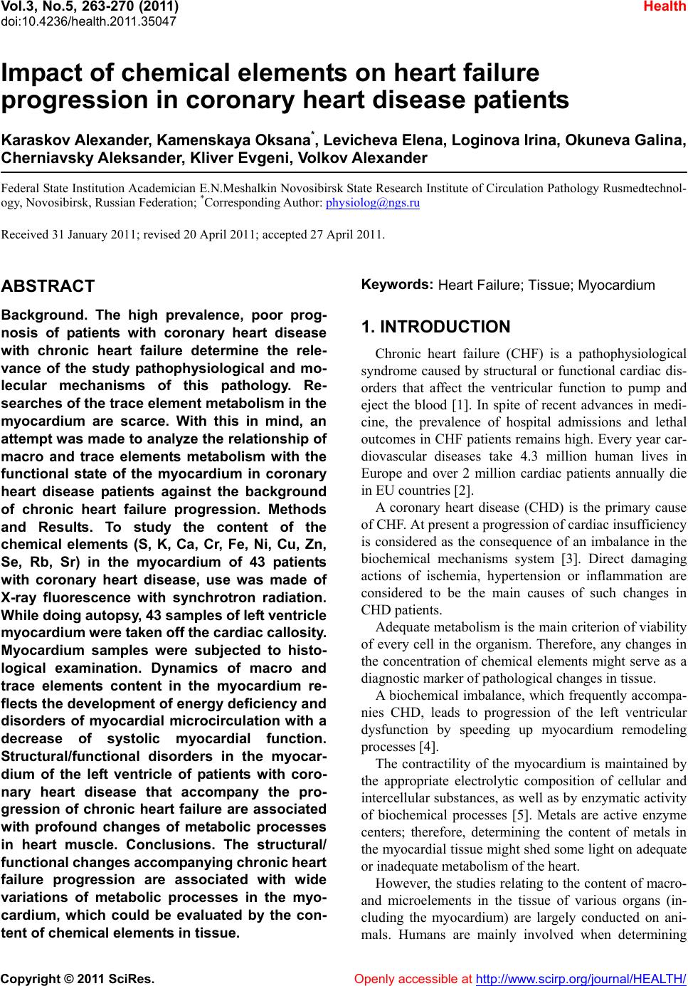 Vol.3, No.5, 263-270 (2011) doi:10.4236/health.2011.35047 C opyright © 2011 SciRes. Openly accessible at http://www.scirp.org/journal/HEALTH/ Health Impact of chemical elements on heart failure progression in coronary heart disease patients Karaskov Alexander, Kamenskaya Oksana*, Levicheva Elena, Loginova Irina, Okuneva Galina, Cherniavsky Aleksander, Kliver Evgeni, Volkov Alexander Federal State Institution Academician E.N.Meshalkin Novosibirsk State Research Institute of Circulation Pathology Rusmedtechnol- ogy, Novosibirsk, Russian Federation; *Corresponding Author: [email protected] Received 31 January 2011; revised 20 April 2011; accepted 27 April 2011. ABSTRACT Background. The high prevalence, poor prog- nosis of patients with coronary heart disease with chronic heart failure determine the rele- vance of the study pathophysiological and mo- lecular mechanisms of this pathology. Re- searches of the trace element metabolism in the myocardium are scarce. With this in mind, an attempt w as made to analyze the relationship of macro and trace elements metabolism with the functional state of the myocardium in coronary heart disease patients against the background of chronic heart failure progression. Methods and Results. To study the content of the chemical elements (S, K, Ca, Cr, Fe, Ni, Cu, Zn, Se, Rb, Sr) in the myocardium of 43 patients with coronary heart disease, use was made of X-ray fluorescence with synchrotron radiation. While doing autop sy, 43 samples of left ventricle myocardiu m were taken off the car diac callosit y. Myocardium samples were subjected to histo- logical examination. Dynamics of macro and trace elements content in the myocardium re- flects the deve lopment of energy deficiency and disorders of myocardial microcirculation with a decrease of systolic myocardial function. Structural/functional disorders in the myocar- dium of the left ventricle of patients with coro- nary heart disease that accompany the pro- gression of chronic heart failure are associated with profound changes of metabolic processes in heart muscle. Conclusions. The structural/ functional changes accomp anyi ng chronic heart failure progression are associated with wide variations of metabolic processes in the myo- cardium, which could be evaluated by the con- tent of chemical elements in tissue. Keywords: Heart Failure; Tissue; Myocardium 1. INTRODUCTION Chronic heart failure (CHF) is a pathophysiological syndrome caused by structural or functional cardiac dis- orders that affect the ventricular function to pump and eject the blood [1]. In spite of recent advances in medi- cine, the prevalence of hospital admissions and lethal outcomes in CHF patients remains high. Every year car- diovascular diseases take 4.3 million human lives in Europe and over 2 million cardiac patients annually die in EU countries [2]. A coronary heart disease (CHD) is the primary cause of CHF. At present a progression of cardiac insufficiency is considered as the consequence of an imbalance in the biochemical mechanisms system [3]. Direct damaging actions of ischemia, hypertension or inflammation are considered to be the main causes of such changes in CHD patients. Adequate metabolism is the main criterion of viability of every cell in the organism. Therefore, any changes in the concentration of chemical elements might serve as a diagnostic marker of pathological changes in tissue. A biochemical imbalance, which frequently accompa- nies CHD, leads to progression of the left ventricular dysfunction by speeding up myocardium remodeling processes [4]. The contractility of the myocardium is maintained by the appropriate electrolytic composition of cellular and intercellular substances, as well as by enzymatic activity of biochemical processes [5]. Metals are active enzyme centers; therefore, determining the content of metals in the myocardial tissue might shed some light on adequate or inadequate metabolism of the heart. However, the studies relating to the content of macro- and microelements in the tissue of various organs (in- cluding the myocardium) are largely conducted on ani- mals. Humans are mainly involved when determining 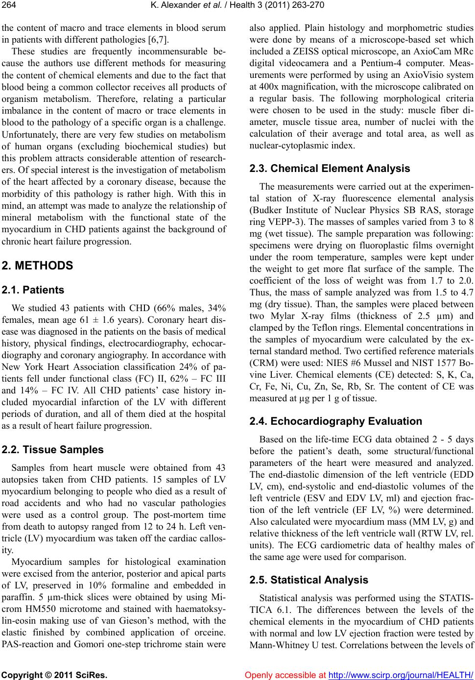 K. Alexander et al. / Health 3 (2011) 263-270 Copyright © 2011 SciRes. Openly accessible at http://www.scirp.org/journal/HEALTH/ 264 the content of macro and trace elements in blood serum in patients with different pathologies [6,7]. These studies are frequently incommensurable be- cause the authors use different methods for measuring the content of chemical elements and due to the fact that blood being a common collector receives all products of organism metabolism. Therefore, relating a particular imbalance in the content of macro or trace elements in blood to the pathology of a specific organ is a challenge. Unfortunately, there are very few studies on metabolism of human organs (excluding biochemical studies) but this problem attracts considerable attention of research- ers. Of special interest is the investigation of metabolism of the heart affected by a coronary disease, because the morbidity of this pathology is rather high. With this in mind, an attempt was made to analyze the relationship of mineral metabolism with the functional state of the myocardium in CHD patients against the background of chronic heart failure progression. 2. METHODS 2.1. Patients We studied 43 patients with CHD (66% males, 34% females, mean age 61 ± 1.6 years). Coronary heart dis- ease was diagnosed in the patients on the basis of medical history, physical findings, electrocardiography, echocar- diography and coronary angiography. In accordance with New York Heart Association classification 24% of pa- tients fell under functional class (FC) II, 62% – FC III and 14% – FC IV. All CHD patients’ case history in- cluded myocardial infarction of the LV with different periods of duration, and all of them died at the hospital as a result of heart failure progression. 2.2. Tissue Samples Samples from heart muscle were obtained from 43 autopsies taken from CHD patients. 15 samples of LV myocardium belonging to people who died as a result of road accidents and who had no vascular pathologies were used as a control group. The post-mortem time from death to autopsy ranged from 12 to 24 h. Left ven- tricle (LV) myocardium was taken off the cardiac callos- ity. Myocardium samples for histological examination were excised from the anterior, posterior and apical parts of LV, preserved in 10% formaline and embedded in paraffin. 5 µm-thick slices were obtained by using Mi- crom HM550 microtome and stained with haematoksy- lin-eosin making use of van Gieson’s method, with the elastic finished by combined application of orceine. PAS-reaction and Gomori one-step trichrome stain were also applied. Plain histology and morphometric studies were done by means of a microscope-based set which included a ZEISS optical microscope, an AxioCam MRc digital videocamera and a Pentium-4 computer. Meas- urements were performed by using an AxioVisio system at 400x magnification, with the microscope calibrated on a regular basis. The following morphological criteria were chosen to be used in the study: muscle fiber di- ameter, muscle tissue area, number of nuclei with the calculation of their average and total area, as well as nuclear-cytoplasmic index. 2.3. Chemical Element Analysis The measurements were carried out at the experimen- tal station of X-ray fluorescence elemental analysis (Budker Institute of Nuclear Physics SB RAS, storage ring VEPP-3). The masses of samples varied from 3 to 8 mg (wet tissue). The sample preparation was following: specimens were drying on fluoroplastic films overnight under the room temperature, samples were kept under the weight to get more flat surface of the sample. The coefficient of the loss of weight was from 1.7 to 2.0. Thus, the mass of sample analyzed was from 1.5 to 4.7 mg (dry tissue). Than, the samples were placed between two Mylar X-ray films (thickness of 2.5 µm) and clamped by the Teflon rings. Elemental concentrations in the samples of myocardium were calculated by the ex- ternal standard method. Two certified reference materials (CRM) were used: NIES #6 Mussel and NIST 1577 Bo- vine Liver. Chemical elements (CE) detected: S, K, Ca, Cr, Fe, Ni, Cu, Zn, Se, Rb, Sr. The content of CE was measured at µg per 1 g of tissue. 2.4. Echocardiography Evaluation Based on the life-time ECG data obtained 2 - 5 days before the patient’s death, some structural/functional parameters of the heart were measured and analyzed. The end-diastolic dimension of the left ventricle (EDD LV, cm), end-systolic and end-diastolic volumes of the left ventricle (ESV and EDV LV, ml) and ejection frac- tion of the left ventricle (EF LV, %) were determined. Also calculated were myocardium mass (MM LV, g) and relative thickness of the left ventricle wall (RTW LV, rel. units). The ECG cardiometric data of healthy males of the same age were used for comparison. 2.5. Statistical Analysis Statistical analysis was performed using the STATIS- TICA 6.1. The differences between the levels of the chemical elements in the myocardium of CHD patients with normal and low LV ejection fraction were tested by Mann-Whitney U test. Correlations between the levels of 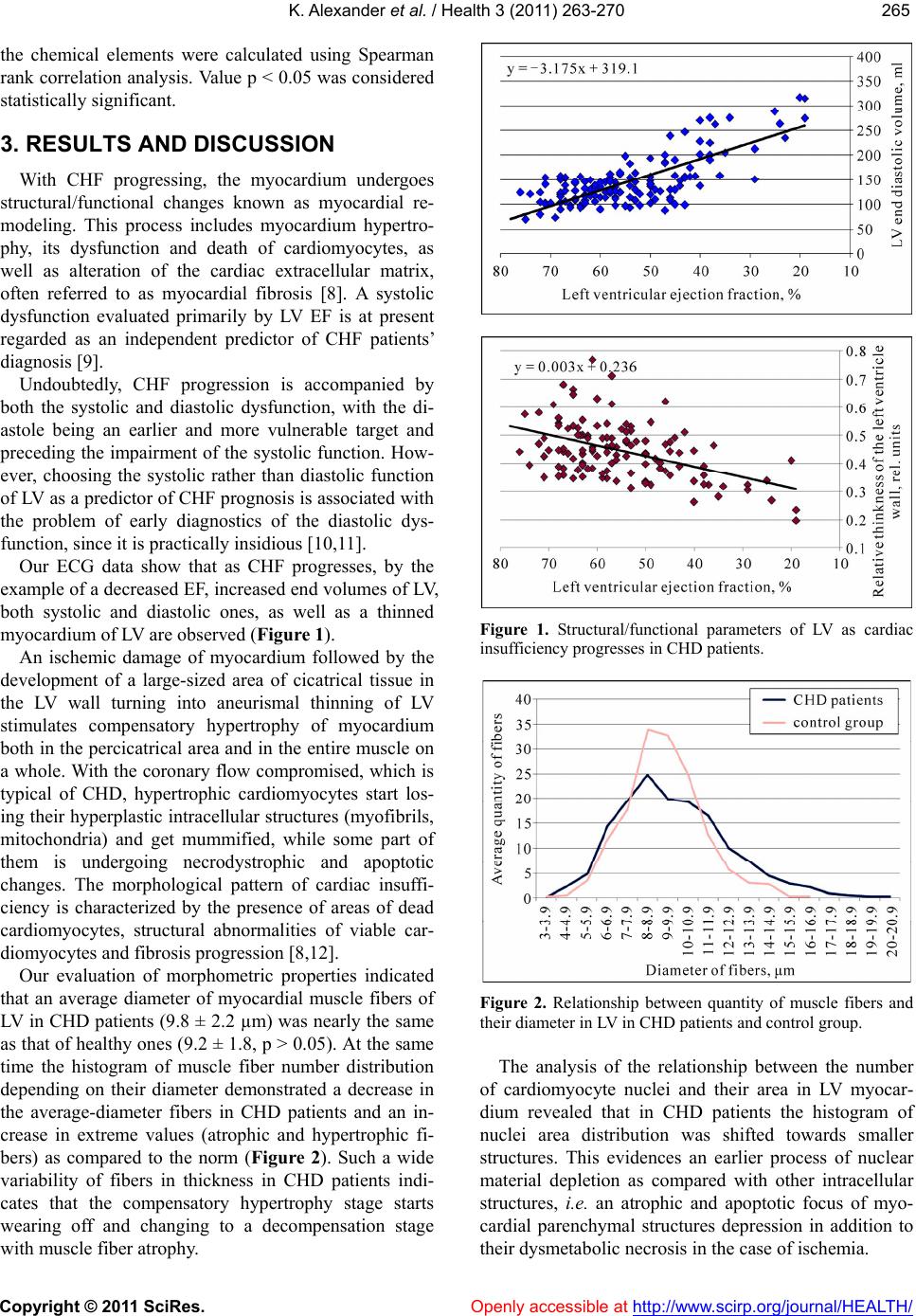 K. Alexander et al. / Health 3 (2011) 263-270 Copyright © 2011 SciRes. Openly accessible at http://www.scirp.org/journal/HEALTH/ 265265 the chemical elements were calculated using Spearman rank correlation analysis. Value p < 0.05 was considered statistically significant. 3. RESULTS AND DISCUSSION With CHF progressing, the myocardium undergoes structural/functional changes known as myocardial re- modeling. This process includes myocardium hypertro- phy, its dysfunction and death of cardiomyocytes, as well as alteration of the cardiac extracellular matrix, often referred to as myocardial fibrosis [8]. A systolic dysfunction evaluated primarily by LV EF is at present regarded as an independent predictor of CHF patients’ diagnosis [9]. Undoubtedly, CHF progression is accompanied by both the systolic and diastolic dysfunction, with the di- astole being an earlier and more vulnerable target and preceding the impairment of the systolic function. How- ever, choosing the systolic rather than diastolic function of LV as a predictor of CHF prognosis is associated with the problem of early diagnostics of the diastolic dys- function, since it is practically insidious [10,11]. Our ECG data show that as CHF progresses, by the example of a decreased EF, increased end volumes of LV, both systolic and diastolic ones, as well as a thinned myocardium of LV are observed (Figure 1). An ischemic damage of myocardium followed by the development of a large-sized area of cicatrical tissue in the LV wall turning into aneurismal thinning of LV stimulates compensatory hypertrophy of myocardium both in the percicatrical area and in the entire muscle on a whole. With the coronary flow compromised, which is typical of CHD, hypertrophic cardiomyocytes start los- ing their hyperplastic intracellular structures (myofibrils, mitochondria) and get mummified, while some part of them is undergoing necrodystrophic and apoptotic changes. The morphological pattern of cardiac insuffi- ciency is characterized by the presence of areas of dead cardiomyocytes, structural abnormalities of viable car- diomyocytes and fibrosis progression [8,12]. Our evaluation of morphometric properties indicated that an average diameter of myocardial muscle fibers of LV in CHD patients (9.8 ± 2.2 µm) was nearly the same as that of healthy ones (9.2 ± 1.8, p > 0.05). At the same time the histogram of muscle fiber number distribution depending on their diameter demonstrated a decrease in the average-diameter fibers in CHD patients and an in- crease in extreme values (atrophic and hypertrophic fi- bers) as compared to the norm (Figure 2). Such a wide variability of fibers in thickness in CHD patients indi- cates that the compensatory hypertrophy stage starts wearing off and changing to a decompensation stage with muscle fiber atrophy. Figure 1. Structural/functional parameters of LV as cardiac insufficiency progresses in CHD patients. Figure 2. Relationship between quantity of muscle fibers and their diameter in LV in CHD patients and control group. The analysis of the relationship between the number of cardiomyocyte nuclei and their area in LV myocar- dium revealed that in CHD patients the histogram of nuclei area distribution was shifted towards smaller structures. This evidences an earlier process of nuclear material depletion as compared with other intracellular structures, i.e. an atrophic and apoptotic focus of myo- cardial parenchymal structures depression in addition to their dysmetabolic necrosis in the case of ischemia. 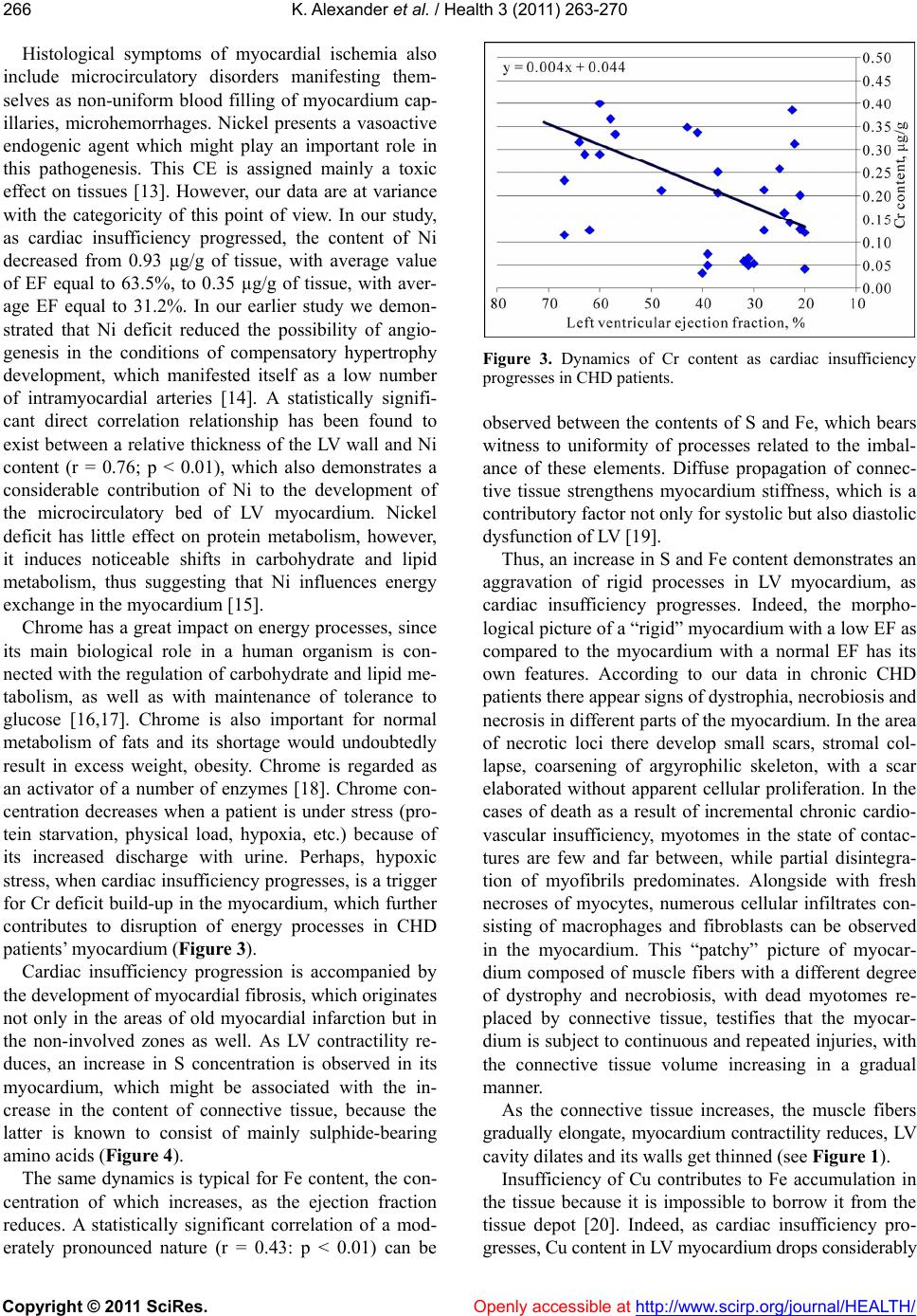 K. Alexander et al. / Health 3 (2011) 263-270 Copyright © 2011 SciRes. Openly accessible at http://www.scirp.org/journal/HEALTH/ 266 Histological symptoms of myocardial ischemia also include microcirculatory disorders manifesting them- selves as non-uniform blood filling of myocardium cap- illaries, microhemorrhages. Nickel presents a vasoactive endogenic agent which might play an important role in this pathogenesis. This CE is assigned mainly a toxic effect on tissues [13]. However, our data are at variance with the categoricity of this point of view. In our study, as cardiac insufficiency progressed, the content of Ni decreased from 0.93 µg/g of tissue, with average value of EF equal to 63.5%, to 0.35 µg/g of tissue, with aver- age EF equal to 31.2%. In our earlier study we demon- strated that Ni deficit reduced the possibility of angio- genesis in the conditions of compensatory hypertrophy development, which manifested itself as a low number of intramyocardial arteries [14]. A statistically signifi- cant direct correlation relationship has been found to exist between a relative thickness of the LV wall and Ni content (r = 0.76; p < 0.01), which also demonstrates a considerable contribution of Ni to the development of the microcirculatory bed of LV myocardium. Nickel deficit has little effect on protein metabolism, however, it induces noticeable shifts in carbohydrate and lipid metabolism, thus suggesting that Ni influences energy exchange in the myocardium [15]. Chrome has a great impact on energy processes, since its main biological role in a human organism is con- nected with the regulation of carbohydrate and lipid me- tabolism, as well as with maintenance of tolerance to glucose [16,17]. Chrome is also important for normal metabolism of fats and its shortage would undoubtedly result in excess weight, obesity. Chrome is regarded as an activator of a number of enzymes [18]. Chrome con- centration decreases when a patient is under stress (pro- tein starvation, physical load, hypoxia, etc.) because of its increased discharge with urine. Perhaps, hypoxic stress, when cardiac insufficiency progresses, is a trigger for Cr deficit build-up in the myocardium, which further contributes to disruption of energy processes in CHD patients’ myocardium (Figure 3). Cardiac insufficiency progression is accompanied by the development of myocardial fibrosis, which originates not only in the areas of old myocardial infarction but in the non-involved zones as well. As LV contractility re- duces, an increase in S concentration is observed in its myocardium, which might be associated with the in- crease in the content of connective tissue, because the latter is known to consist of mainly sulphide-bearing amino acids (Figure 4). The same dynamics is typical for Fe content, the con- centration of which increases, as the ejection fraction reduces. A statistically significant correlation of a mod- erately pronounced nature (r = 0.43: p < 0.01) can be Figure 3. Dynamics of Cr content as cardiac insufficiency progresses in CHD patients. observed between the contents of S and Fe, which bears witness to uniformity of processes related to the imbal- ance of these elements. Diffuse propagation of connec- tive tissue strengthens myocardium stiffness, which is a contributory factor not only for systolic but also diastolic dysfunction of LV [19]. Thus, an increase in S and Fe content demonstrates an aggravation of rigid processes in LV myocardium, as cardiac insufficiency progresses. Indeed, the morpho- logical picture of a “rigid” myocardium with a low EF as compared to the myocardium with a normal EF has its own features. According to our data in chronic CHD patients there appear signs of dystrophia, necrobiosis and necrosis in different parts of the myocardium. In the area of necrotic loci there develop small scars, stromal col- lapse, coarsening of argyrophilic skeleton, with a scar elaborated without apparent cellular proliferation. In the cases of death as a result of incremental chronic cardio- vascular insufficiency, myotomes in the state of contac- tures are few and far between, while partial disintegra- tion of myofibrils predominates. Alongside with fresh necroses of myocytes, numerous cellular infiltrates con- sisting of macrophages and fibroblasts can be observed in the myocardium. This “patchy” picture of myocar- dium composed of muscle fibers with a different degree of dystrophy and necrobiosis, with dead myotomes re- placed by connective tissue, testifies that the myocar- dium is subject to continuous and repeated injuries, with the connective tissue volume increasing in a gradual manner. As the connective tissue increases, the muscle fibers gradually elongate, myocardium contractility reduces, LV cavity dilates and its walls get thinned (see Figure 1). Insufficiency of Cu contributes to Fe accumulation in the tissue because it is impossible to borrow it from the tissue depot [20]. Indeed, as cardiac insufficiency pro- gresses, Cu content in LV myocardium drops considerably 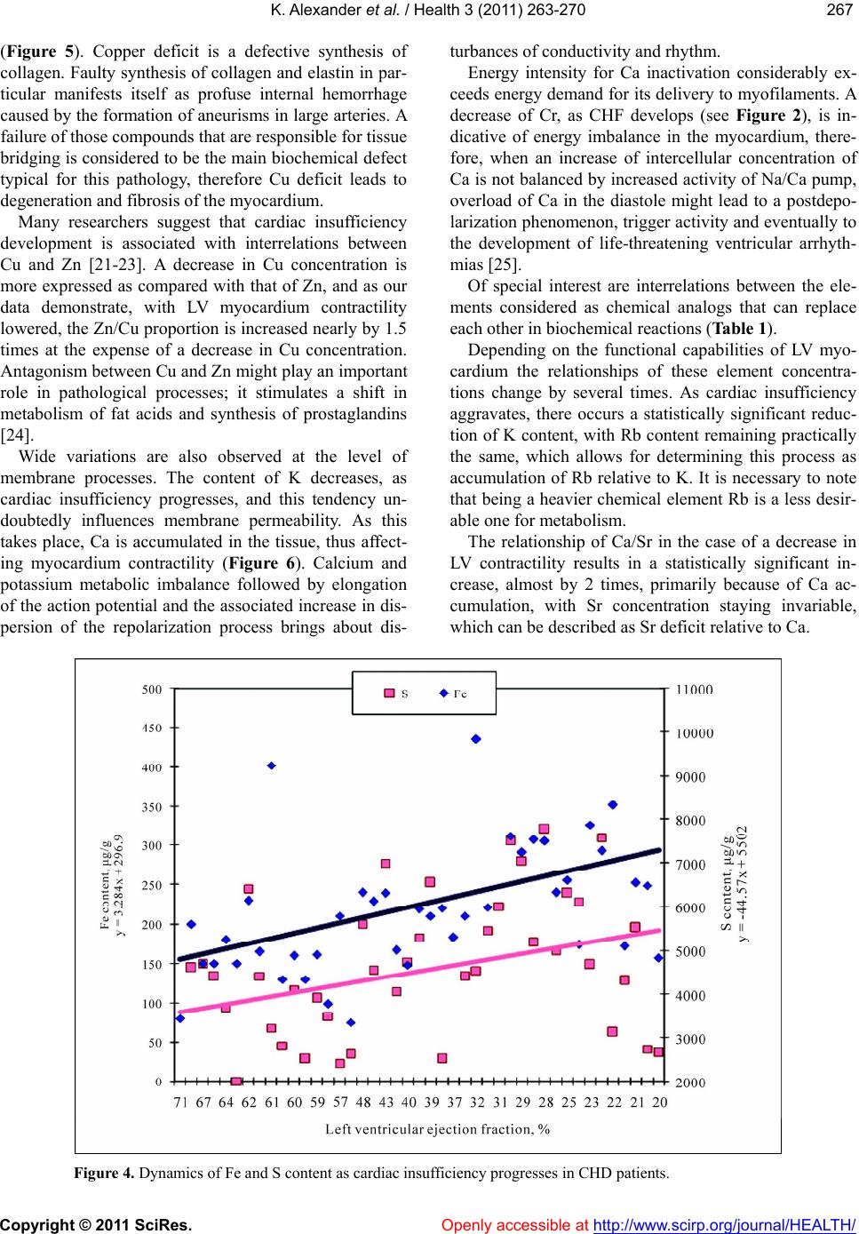 K. Alexander et al. / Health 3 (2011) 263-270 Copyright © 2011 SciRes. http://www.scirp.org/journal/HEALTH/ 267267 (Figure 5). Copper deficit is a defective synthesis of collagen. Faulty synthesis of collagen and elastin in par- ticular manifests itself as profuse internal hemorrhage caused by the formation of aneurisms in large arteries. A failure of those compounds that are responsible for tissue bridging is considered to be the main biochemical defect typical for this pathology, therefore Cu deficit leads to degeneration and fibrosis of the myocardium. Openly accessible at Many researchers suggest that cardiac insufficiency development is associated with interrelations between Cu and Zn [21-23]. A decrease in Cu concentration is more expressed as compared with that of Zn, and as our data demonstrate, with LV myocardium contractility lowered, the Zn/Cu proportion is increased nearly by 1.5 times at the expense of a decrease in Cu concentration. Antagonism between Cu and Zn might play an important role in pathological processes; it stimulates a shift in metabolism of fat acids and synthesis of prostaglandins [24]. Wide variations are also observed at the level of membrane processes. The content of K decreases, as cardiac insufficiency progresses, and this tendency un- doubtedly influences membrane permeability. As this takes place, Ca is accumulated in the tissue, thus affect- ing myocardium contractility (Figure 6). Calcium and potassium metabolic imbalance followed by elongation of the action potential and the associated increase in dis- persion of the repolarization process brings about dis- turbances of conductivity and rhythm. Energy intensity for Ca inactivation considerably ex- ceeds energy demand for its delivery to myofilaments. A decrease of Cr, as CHF develops (see Figure 2), is in- dicative of energy imbalance in the myocardium, there- fore, when an increase of intercellular concentration of Ca is not balanced by increased activity of Na/Ca pump, overload of Ca in the diastole might lead to a postdepo- larization phenomenon, trigger activity and eventually to the development of life-threatening ventricular arrhyth- mias [25]. Of special interest are interrelations between the ele- ments considered as chemical analogs that can replace each other in biochemical reactions (Table 1). Depending on the functional capabilities of LV myo- cardium the relationships of these element concentra- tions change by several times. As cardiac insufficiency aggravates, there occurs a statistically significant reduc- tion of K content, with Rb content remaining practically the same, which allows for determining this process as accumulation of Rb relative to K. It is necessary to note that being a heavier chemical element Rb is a less desir- able one for metabolism. The relationship of Сa/Sr in the case of a decrease in LV contractility results in a statistically significant in- crease, almost by 2 times, primarily because of Ca ac- cumulation, with Sr concentration staying invariable, which can be described as Sr deficit relative to Ca. Figure 4. Dynamics of Fe and S content as cardiac insufficiency progresses in CHD patients. 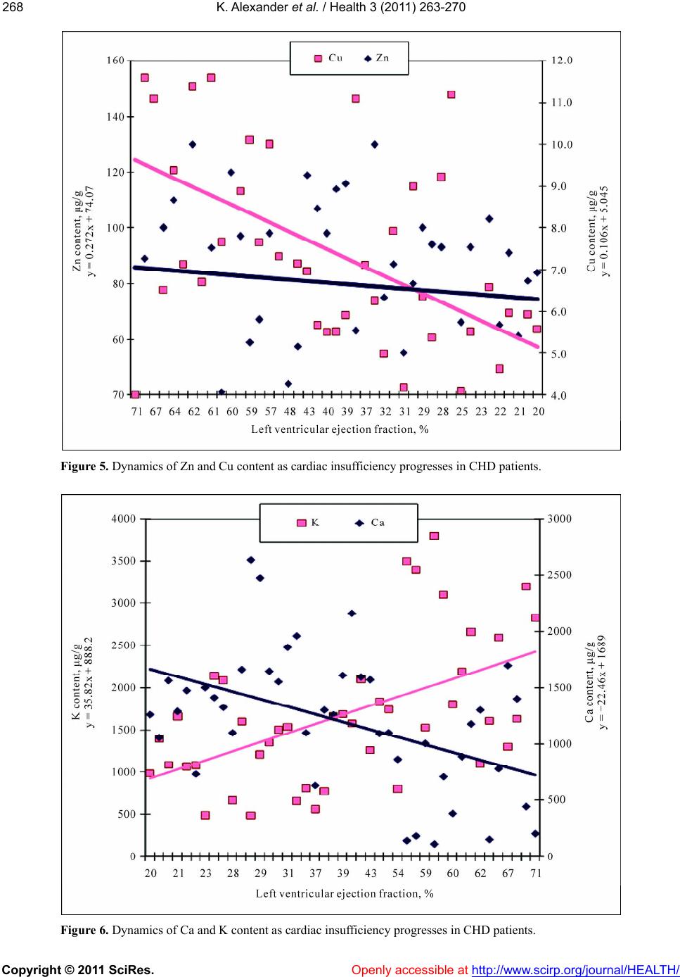 K. Alexander et al. / Health 3 (2011) 263-270 Copyright © 2011 SciRes. Openly accessible at http://www.scirp.org/journal/HEALTH/ 268 Figure 5. Dynamics of Zn and Cu content as cardiac insufficiency progresses in CHD patients. Figure 6. Dynamics of Ca and K content as cardiac insufficiency progresses in CHD patients. 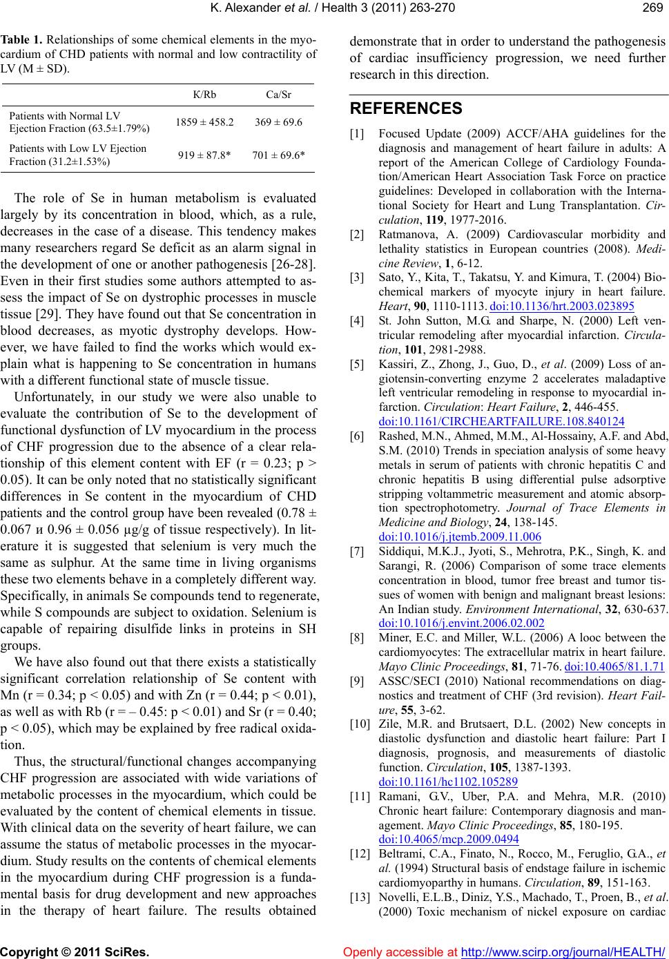 K. Alexander et al. / Health 3 (2011) 263-270 Copyright © 2011 SciRes. http://www.scirp.org/journal/HEALTH/ 269269 Table 1. Relationships of some chemical elements in the myo- cardium of CHD patients with normal and low contractility of LV (M ± SD). К/Rb Ca/Sr Patients with Normal LV Ejection Fraction (63.5±1.79%) 1859 ± 458.2 369 ± 69.6 Patients with Low LV Ejection Fraction (31.2±1.53%) 919 ± 87.8* 701 ± 69.6* Openly accessible at The role of Se in human metabolism is evaluated largely by its concentration in blood, which, as a rule, decreases in the case of a disease. This tendency makes many researchers regard Se deficit as an alarm signal in the development of one or another pathogenesis [26-28]. Even in their first studies some authors attempted to as- sess the impact of Se on dystrophic processes in muscle tissue [29]. They have found out that Se concentration in blood decreases, as myotic dystrophy develops. How- ever, we have failed to find the works which would ex- plain what is happening to Se concentration in humans with a different functional state of muscle tissue. Unfortunately, in our study we were also unable to evaluate the contribution of Se to the development of functional dysfunction of LV myocardium in the process of CHF progression due to the absence of a clear rela- tionship of this element content with EF (r = 0.23; p > 0.05). It can be only noted that no statistically significant differences in Se content in the myocardium of CHD patients and the control group have been revealed (0.78 ± 0.067 и 0.96 ± 0.056 µg/g of tissue respectively). In lit- erature it is suggested that selenium is very much the same as sulphur. At the same time in living organisms these two elements behave in a completely different way. Specifically, in animals Se compounds tend to regenerate, while S compounds are subject to oxidation. Selenium is capable of repairing disulfide links in proteins in SH groups. We have also found out that there exists a statistically significant correlation relationship of Se content with Mn (r = 0.34; p < 0.05) and with Zn (r = 0.44; p < 0.01), as well as with Rb (r = – 0.45: p < 0.01) and Sr (r = 0.40; p < 0.05), which may be explained by free radical oxida- tion. Thus, the structural/functional changes accompanying CHF progression are associated with wide variations of metabolic processes in the myocardium, which could be evaluated by the content of chemical elements in tissue. With clinical data on the severity of heart failure, we can assume the status of metabolic processes in the myocar- dium. Study results on the contents of chemical elements in the myocardium during CHF progression is a funda- mental basis for drug development and new approaches in the therapy of heart failure. The results obtained demonstrate that in order to understand the pathogenesis of cardiac insufficiency progression, we need further research in this direction. REFERENCES [1] Focused Update (2009) ACCF/AHA guidelines for the diagnosis and management of heart failure in adults: A report of the American College of Cardiology Founda- tion/American Heart Association Task Force on practice guidelines: Developed in collaboration with the Interna- tional Society for Heart and Lung Transplantation. Cir- culation, 11 9, 1977-2016. [2] Ratmanova, A. (2009) Cardiovascular morbidity and lethality statistics in European countries (2008). Medi- cine Review, 1, 6-12. [3] Sato, Y., Kita, T., Takatsu, Y. and Kimura, T. (2004) Bio- chemical markers of myocyte injury in heart failure. Heart, 90, 1110-1113. doi:10.1136/hrt.2003.023895 [4] St. John Sutton, M.G. and Sharpe, N. (2000) Left ven- tricular remodeling after myocardial infarction. Circula- tion, 101, 2981-2988. [5] Kassiri, Z., Zhong, J., Guo, D., et al. (2009) Loss of an- giotensin-converting enzyme 2 accelerates maladaptive left ventricular remodeling in response to myocardial in- farction. Circulation: Heart Failure, 2, 446-455. doi:10.1161/CIRCHEARTFAILURE.108.840124 [6] Rashed, M.N., Ahmed, M.M., Al-Hossainy, A.F. and Abd, S.M. (2010) Trends in speciation analysis of some heavy metals in serum of patients with chronic hepatitis C and chronic hepatitis B using differential pulse adsorptive stripping voltammetric measurement and atomic absorp- tion spectrophotometry. Journal of Trace Elements in Medicine and Biology, 24, 138-145. doi:10.1016/j.jtemb.2009.11.006 [7] Siddiqui, M.K.J., Jyoti, S., Mehrotra, P.K., Singh, K. and Sarangi, R. (2006) Comparison of some trace elements concentration in blood, tumor free breast and tumor tis- sues of women with benign and malignant breast lesions: An Indian study. Environment International, 32, 630-637. doi:10.1016/j.envint.2006.02.002 [8] Miner, E.C. and Miller, W.L. (2006) A looc between the cardiomyocytes: The extracellular matrix in heart failure. Mayo Clinic Proceedings, 81, 71-76. doi:10.4065/81.1.71 [9] ASSC/SECI (2010) National recommendations on diag- nostics and treatment of CHF (3rd revision). Heart Fail- ure, 55, 3-62. [10] Zile, M.R. and Brutsaert, D.L. (2002) New concepts in diastolic dysfunction and diastolic heart failure: Part I diagnosis, prognosis, and measurements of diastolic function. Circulation, 105, 1387-1393. doi:10.1161/hc1102.105289 [11] Ramani, G.V., Uber, P.A. and Mehra, M.R. (2010) Chronic heart failure: Contemporary diagnosis and man- agement. Mayo Clinic Proceedings, 85, 180-195. doi:10.4065/mcp.2009.0494 [12] Beltrami, C.A., Finato, N., Rocco, M., Feruglio, G.A., et al. (1994) Structural basis of endstage failure in ischemic cardiomyoparthy in humans. Circulation, 89, 151-163. [13] Novelli, E.L.B., Diniz, Y.S., Machado, T., Proen, B., et al. (2000) Toxic mechanism of nickel exposure on cardiac 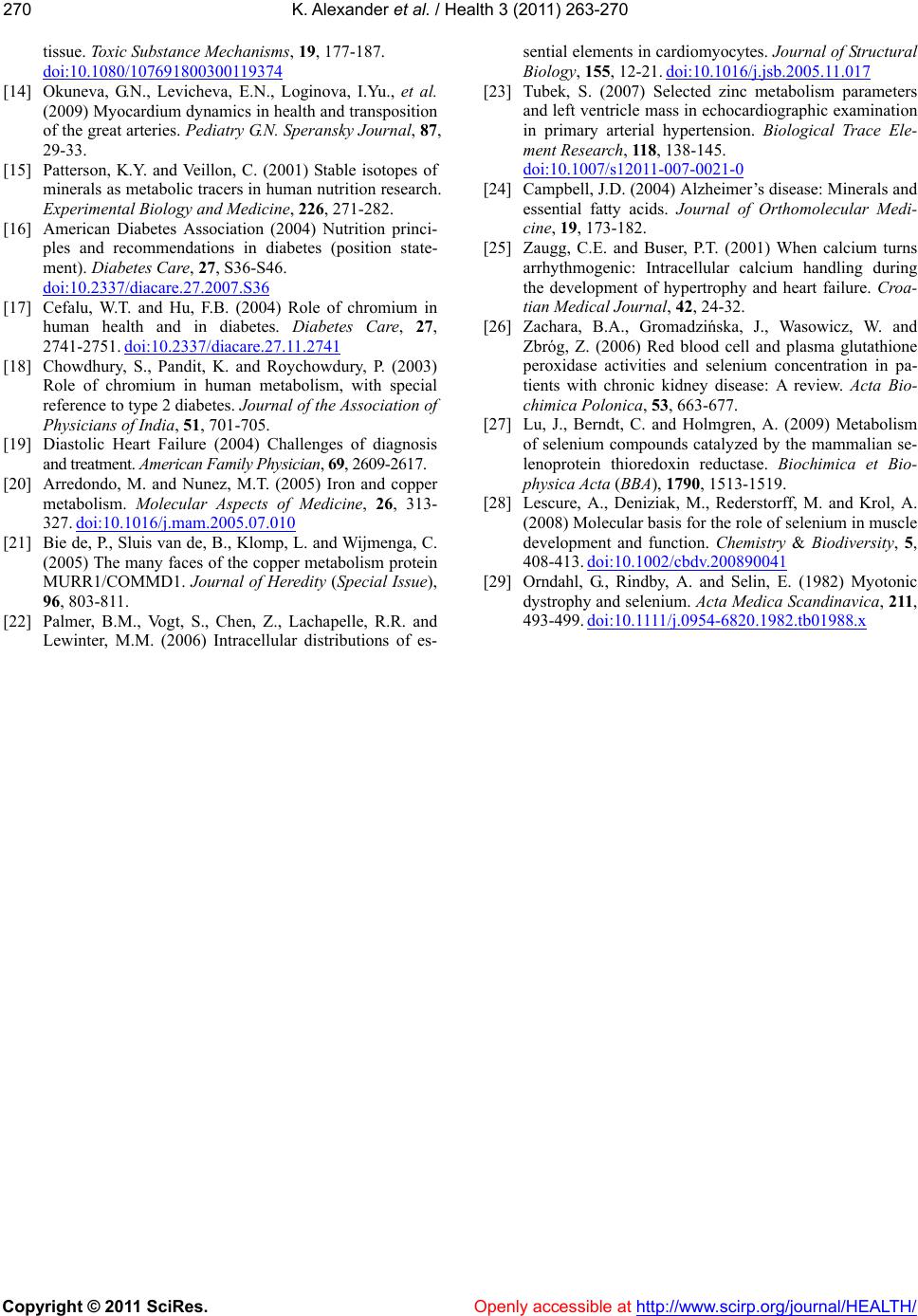 K. Alexander et al. / Health 3 (2011) 263-270 Copyright © 2011 SciRes. Openly accessible at http://www.scirp.org/journal/HEALTH/ 270 tissue. Toxic Substance Mechanisms, 19, 177-187. doi:10.1080/107691800300119374 [14] Okuneva, G.N., Levicheva, E.N., Loginova, I.Yu., et al. (2009) Myocardium dynamics in health and transposition of the great arteries. Pediatry G.N. Speransky Journal, 87, 29-33. [15] Patterson, K.Y. and Veillon, C. (2001) Stable isotopes of minerals as metabolic tracers in human nutrition research. Experimental Biology and Medicine, 226, 271-282. [16] American Diabetes Association (2004) Nutrition princi- ples and recommendations in diabetes (position state- ment). Diabetes Care, 27, S36-S46. doi:10.2337/diacare.27.2007.S36 [17] Cefalu, W.T. and Hu, F.B. (2004) Role of chromium in human health and in diabetes. Diabetes Care, 27, 2741-2751. doi:10.2337/diacare.27.11.2741 [18] Chowdhury, S., Pandit, K. and Roychowdury, P. (2003) Role of chromium in human metabolism, with special reference to type 2 diabetes. Journal of the Association of Physicians of India, 51, 701-705. [19] Diastolic Heart Failure (2004) Challenges of diagnosis and treatment. American Family Physician, 69, 2609-2617. [20] Arredondo, M. and Nunez, M.T. (2005) Iron and copper metabolism. Molecular Aspects of Medicine, 26, 313- 327. doi:10.1016/j.mam.2005.07.010 [21] Bie de, P., Sluis van de, B., Klomp, L. and Wijmenga, C. (2005) The many faces of the copper metabolism protein MURR1/COMMD1. Journal of Heredity (Special Issue), 96, 803-811. [22] Palmer, B.M., Vogt, S., Chen, Z., Lachapelle, R.R. and Lewinter, M.M. (2006) Intracellular distributions of es- sential elements in cardiomyocytes. Journal of Structural Biology, 155, 12-21. doi:10.1016/j.jsb.2005.11.017 [23] Tubek, S. (2007) Selected zinc metabolism parameters and left ventricle mass in echocardiographic examination in primary arterial hypertension. Biological Trace Ele- ment Research, 118, 138-145. doi:10.1007/s12011-007-0021-0 [24] Campbell, J.D. (2004) Alzheimer’s disease: Minerals and essential fatty acids. Journal of Orthomolecular Medi- cine, 19, 173-182. [25] Zaugg, C.E. and Buser, P.T. (2001) When calcium turns arrhythmogenic: Intracellular calcium handling during the development of hypertrophy and heart failure. Croa- tian Medical Journal, 42, 24-32. [26] Zachara, B.A., Gromadzińska, J., Wasowicz, W. and Zbróg, Z. (2006) Red blood cell and plasma glutathione peroxidase activities and selenium concentration in pa- tients with chronic kidney disease: A review. Acta Bio- chimica Polonica, 53, 663-677. [27] Lu, J., Berndt, C. and Holmgren, A. (2009) Metabolism of selenium compounds catalyzed by the mammalian se- lenoprotein thioredoxin reductase. Biochimica et Bio- physica Acta (BBA), 1790, 1513-1519. [28] Lescure, A., Deniziak, M., Rederstorff, M. and Krol, A. (2008) Molecular basis for the role of selenium in muscle development and function. Chemistry & Biodiversity, 5, 408-413. doi:10.1002/cbdv.200890041 [29] Orndahl, G., Rindby, A. and Selin, E. (1982) Myotonic dystrophy and selenium. Acta Medica Scandinavica, 211, 493-499. doi:10.1111/j.0954-6820.1982.tb01988.x |