

Paper Menu >>
Journal Menu >>
 Surgical Science, 2011, 2, 159-162 doi:10.4236/ss.2011.23034 Published Online May 2011 (http://www.scirp.org/journal/ss) Copyright © 2011 SciRes. SS A Case of Pituitary Hemorrhage Following Cardiopulmonary Bypass Surgery Ozgen Ilgaz Kocyigit1, Serdar Kabatas2, Erdinc Civelek2, Ezgi Tuncay1, Oguz Omay3, Tufan Cansever2, Ayda Turkoz1 1Department of Anesthesiology, Baskent University Istanbul Hospital, Istanbul, Turkey 2Department of Neurosurgery, Baskent University Istanbul Hospital, Istanbul, Turkey 3Department of Cardiovascular surgery, Baskent University Istanbul Hospital, Istanbu l, Turkey E-mail: [email protected] Received August 17, 2010; revised November 25, 2010; accepted Dece m b er 16, 2010 Abstract A 68-year-old female patient with previous history of transsphenoidal hypophysectomy operation underwent three-vessel coronary artery by pass graft (CABG) sur gery for extensive coronary artery disease. Preoperative neurological examination revealed sequelae visual loss at right temporal visual field. Follow-up Magnetic Resonance Imaging studies showed a residual hypophyseal tumor tissue extending to suprasellar area. No additional pathology was detected in the early postoperative cranial control CT, but aggravation of visual field defect was determined. Coincidently, cranial magnetic resonance imaging (MRI) showed hemorrhage into the tumor tissue. We decided to follow-up the patient who exhibited no additional symptoms and was discharged well on the fifth day due to the signs of resolution of hemorrhage. Follow-up controls of the pa- tient at sixth and twelfth months demonstrated normal hormone levels without any additional clinical com- plaints. We present preoperative assessment, perioperative anesthesia management, and postoperative clini- cal follow-up of a patient with a residual hypophyseal tumor. Keywords: Anesthesia; Coronary Artery Bypass Graft Surgery; Hemorrhage; Hypophyseal Tumor 1. Introduction Hypophyseal macroadenoma incidence is reported to be 30/100.000 [1]. Regarding infection, radiotherapy, me- chanical ventilation, and cardiac bypass surgery (CBP), these tumors may have sudden infarction and hemor- rhage [2]. In these complicated cases, the clinical man- agement is sometimes difficult. Pituitary apoplexy occurs spontaneously in 60% - 80% cases of asymptomatic pa- tients. Common predisposing factors are head trauma, hypotension, hypertension, pituitary irradiation history, cardiac surgery, anticoagulant therapy treatment with dopamine agonists, pituitary stimulation tests and preg- nancy [1,3]. Hypopituitarism developing after coronary bypass surgery, howev er, is a known ph enomenon of rare occur- rence [4].One of the favorite pathophysiological mecha- nisms is the fall of arterial blood pressure induces ische- mia and that is followed by infarction of the pituitary gland [5]. A variety of clinical symptoms have been de- scribed including headache, fever, lethargy, confusion, obtundation, Addisionian crisis, unilateral ptosis, meiosis, hemiparesis, visual field deficits and ophtalmoplegia [1,6]. The ratio of men to women is 10 to 1 in favor of men [1]. 2. Case Report The history of the 68-year-old female patient evaluated for elective coronary artery bypass graft (CABG) surgery revealed thyroidectomy, transsphenoidal hypophysec- tomy, discectomy as well as hypertension and glaucoma. Her laboratory analyses showed low Thyroid Stimulating Hormone (TSH) level (0.128; normal range: 0.27 - 4.2 uIU/ml), normal Triiodothyronine (T3) (2.47; normal range: 1.8 - 4.6 pg/ml), and Thyroxin (T4) (1.33; normal range: 0.9 - 1.7 ng/dl) concentrations. There was no pa- thology detected in her other laboratory analyses includ- ing cortisol and prolactin hormone values. She was under therapy of 5 mg/day prednisolone and 75 mcg/day levothyroxine. Follow-up Magnetic Resonance Imaging studies showed a residual hypophyseal tumor tissue with 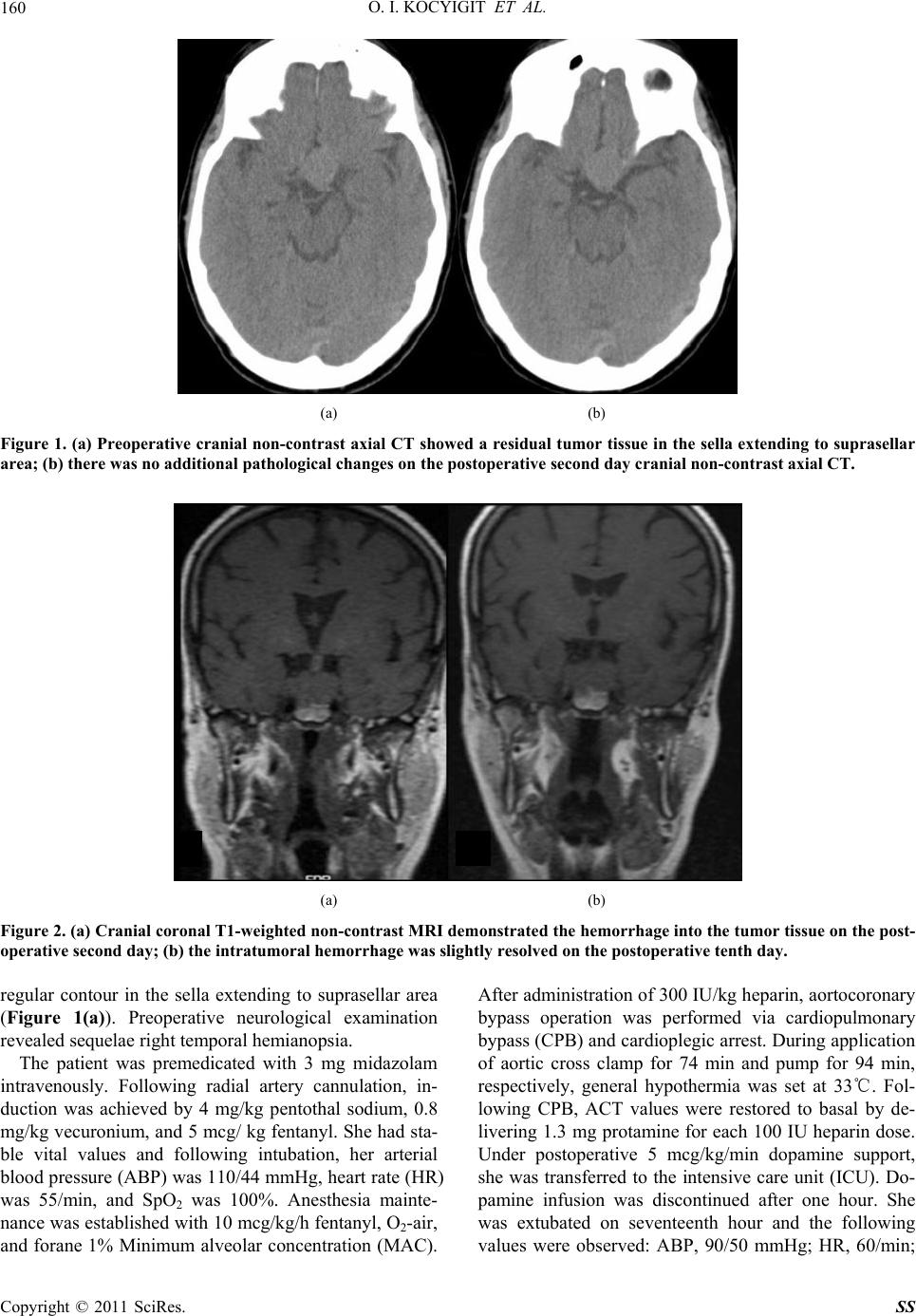 O. I. KOCYIGIT ET AL. 160 (a) (b) Figure 1. (a) Preoperative cranial non-contrast axial CT showed a residual tumor tissue in the sella extending to suprasellar area; (b) there was no additional pathological changes on the postoperative second day cranial non-contrast axial CT. (a) (b) Figure 2. (a) Cranial coronal T1-weighted non-contrast MRI demonstrated the hemorrhage into the tumor tissue on the post- operative second day; (b) the intratumoral hemorrhage was slightly resolved on the postoperative tenth day. regular contour in the sella extending to suprasellar area (Figure 1(a)). Preoperative neurological examination revealed sequelae right temporal hemianopsia. The patient was premedicated with 3 mg midazolam intravenously. Following radial artery cannulation, in- duction was achieved by 4 mg/kg pentothal sodium, 0.8 mg/kg vecuronium, and 5 mcg/ kg fentanyl. She had sta- ble vital values and following intubation, her arterial blood pressure (ABP) was 110/44 mmHg, heart rate (HR) was 55/min, and SpO2 was 100%. Anesthesia mainte- nance was established with 10 mcg/kg/h fentanyl, O2-air, and forane 1% Minimum alveolar concentration (MAC). After administration of 300 IU/kg heparin, aortocoronary bypass operation was performed via cardiopulmonary bypass (CPB) and cardioplegic arrest. During application of aortic cross clamp for 74 min and pump for 94 min, respectively, general hypothermia was set at 33℃. Fol- lowing CPB, ACT values were restored to basal by de- livering 1.3 mg protamine for each 100 IU heparin dose. Under postoperative 5 mcg/kg/min dopamine support, she was transferred to the intensive care unit (ICU). Do- pamine infusion was discontinued after one hour. She was extubated on seventeenth hour and the following values were observed: ABP, 90/50 mmHg; HR, 60/min; Copyright © 2011 SciRes. SS  O. I. KOCYIGIT ET AL. 161 SpO2, 99%; body temperature, 36.6℃; central venous pressure, 10 mmHg, respectively. No additional pathol- ogy was found on the cranial control CT of the patient admitted to the service on postoperative second day; however, aggravation of visual field defect was deter- mined as bitemporal hemianopsia at this time frame (Figure 1(b)). We therefore performed cranial magnetic resonance imaging (MRI) demonstrating hemorrhage into the tumor tissue (Figure 2(a)). We decided to fol- low-up the patient who exhibited no additional symp- toms and was discharged well on the fifth day following signs of resolution of hemorrhage. The control cranial MRI performed on the postoperative tenth day, showed a slight resolution of intratumoral hemorrhage and neuro- logical examination exhibited a decrease in the visual field defect (Figure 2(b)). Follow-up controls of the pa- tient at sixth and twelfth months demonstrated normal hormone levels without any additional clinical com- plaints. 3. Discussion Hypophyseal apoplexy occurs as a result of the sudden enlargement of adenoma tissue due to edema, hemor- rhage and/or infarction. There are many theories about pituitary apoplexy but the pathogenesis still remains un- clear. Pituitary tumors are known to bleed 5 times more than other intracranial tumors. According to a theory, enlarged pituitary tumor presses the vascular supply, brings out the ischemia and necrosis both the gland and the tumor. Another theory clarifies that the critical perfu- sion pressure of the adenomas is below normal gland arterial pressure, so instantaneous changes in perfusion pressure causes the adenoma infarction. Some other the- ory suggests that the outgrowing of the blood supply be- cause of the tumor enlargement results the ischemic ne- crosis and secondary hemorrhage [7,8]. Symptoms and signs show up because of compression on parasellar structures, disr uptio n of hypo ph yseal tissue, and meningeal irritation of blood or necrotic tissues [9,10]. In this syndrome, the following clinical symptoms can occur: unilateral or bilateral visual field defect, oph- talmoplegia, endocrinologic imbalance, serious headache, and coma [11]. In cardiac surgery, hemorrhage and in- farction may occur in the hypophyseal adenoma during CPB [12]. In the current case, postoperatively presented bitemporal hemianopsia was found to be increased as a result of hemorrhage in the residual tissue. This result was thought to be occurring due to influence of edema, hypoperfusion, and ischemia associated with nonpulsa- tile flow, on hypophyseal tissue. Cooper et al. reported the specific causes of apoplexy occurring after cardiac surgery as low degree of flow with anticoagulation-re- lated hemorrhage, embolization, hemodilution, ischemia and infarction associated with nonpulsatile flow [11]. The edema of the tissue developed after the CPB, sup- presses blood perfusion, damages surrounding tissues, and leads to necrosis. Combination of one or more of those factors may result in development of hypophyseal apoplexy. The vascular lesions of our patient were not suitable for off-pump surgery; therefore, operation was carried out with CPB. However, off-pump surgery involves use of heparin, and th e he modyn amic stability is no t as stab le as the systemic perfusion pressure established by CPB. Not only CPB, but also heparin administration at low levels during presurgical period, can also lead to this re- sult. Anticoagulation is known as a precipitating factor but, Levy et al thought that, despite the large number of pa- tients treated with anticoagulation, pituitary apoplexy remained a rare finding [13]. So we all agree that the number of nonsurgical patients who received anticoagu- lation is much larger than the patients who had Coronary artery By-pass surgery with CPB (cardiopulmonary by- pass). In our case, the residual tumor tissue was present, pre- operative hormone concentrations were within normal limits, and drug use was at regular basis. The CBP was well-tolerated, while no hypotensive, hypertensive, or hypoxic condition was observed. The postoperative medical treatment was continued. Regarding neuroanes- thesia principles, induction and recovery was achieved through obtaining lowest possible stress response. The cause behind long extubation duration was not a cardiac or hemodynamic problem, but was the aim to avoid any complication that might arise due to occurrence of awak- ening period in late hours of the day. Hypophyseal apoplexy cases may present with a fatal course. In order to save the patient and maintain the con- tinuity of visual functions, transsphenoidal decompres- sion may be required [3,9]. Previous studies have shown that the outcome of the medical treatment in pituitary apoplexy in stable patients without neuro-ophthalmic deficits is similar to those underwen t surgery, but if pro- gressive visual loss is existed, emergent decompression should be considered after medical stabilization to pre- vent a mortal rebleeding [14]. In our case, surgery was not considered for the hemorrhage within the residual tumor tissue observed on the cranial MRI. Thus, by tenth day, the hemorrhage was started to be resolved and vis- ual field defect was found to be reduced. 4. Conclusions The patients with coronary artery disease are exposed to Copyright © 2011 SciRes. SS  O. I. KOCYIGIT ET AL. Copyright © 2011 SciRes. SS 162 low or high dose anticoagulation during diagnostic or surgical procedures. In cases with hypophyseal adenoma background which are scheduled for graft surgery as per related surgical indications, complications can be minimal- ized and prevented earlier by performing endocrinologic control in the preoperative period, minimalization of stress associated with perioperative surgery and anesthe- sia, and close monitoring of the neurological status post- operatively. The physician should be awake for pituitary adenoma infarction after open cardiac surgery and should remem- ber that, without proper treatment, it can be fatal or cause permanent neurological damage. Surgical and endocrine treatment can be life saving. The outcome depends on early diagnosis and proper intervention. 5. References [1] M. B. Pliam, M. Cohen, L. Cheng, M. Spaenle, M. H. Bronstein and T. W. Atkin, “Pituitary Adenomas Com- plicating Cardiac Surgery: Summary and Review of 11 Cases,” Journal of Cardiac Surgery, Vol. 10, No. 2, March 1995, pp. 125-132. doi:10.1111/j.1540-8191.1995.tb01230.x [2] M. Wiesmann, J. Gliemroth, U. Kehler and U. Missler, “Pituitary Apoplexy after Cardiac Surgery Presenting as Deep Coma with Dilat ed Pupils,” Acta Anaesthesiologica Scandinavica, Vol. 43, No. 2, February 1999, pp. 236- 238. doi:10.1034/j.1399-6576.1999.430222.x [3] P. Maccagnan, C. L. Macedo, M. J. Kayath, R. G. No- gueira and J. Abucham, “Conservative Management of Pituitary Apoplexy: A Prospective Study,” Journal of Clinical Endocrinology & Metabolism, Vol. 80, No. 7, July1995, pp. 2190-2197. doi:10.1210/jc.80.7.2190 [4] L. C. Glass, “Images in Clinical Medicine. Pituitary Apo- plexy,” New England Journal of Medicine, Vol. 349, No- vember 2003, p. 2034. [5] G. Liberale, G. Bruninx, B. Vanderkelen, E. Dubois, E. Vandueren and G. Verhelst, “Pituitary Apoplexy after Aortic Abdominal Aneurysm Surgery: A Case Report,” Acta Chirurgica Belgica, Vol 106, No. 1, 2006, pp. 77-80. [6] Z. Chen, A. W. Murray and J. J. Quinlan, “Pituitary Apo- plexy Presenting as Unilateral Third Cranial Nerve Palsy after Coronary Artery Bypass Surgery,” Anesthesia & Analgesia, Vol. 98, No. 1, January 2004, pp. 46-48. doi:10.1213/01.ANE.0000093233.84896.27 [7] S. Murad-Kejbou and E. Eggenberger, “Pituitary Apo- plexy: Evaluation, Management, and Prognosis,” Current Opinion in Ophthalmology, Vol. 20, No. 6, November 2009, pp. 456-461. doi:10.1097/ICU.0b013e3283319061 [8] R. N. Nawar, D. AbdelMannan, W. R. Selman and B. M. Arafah, “Pituitary Tumor Apoplexy: A Review,” Journal of Intensive Care Medicine, Vol. 23, No. 2, March-April 2008, pp. 75-90. doi:10.1177/0885066607312992 [9] D. C. Bills, F. B. Meyer, E. R. Laws, D. H. Davis, M. J. Ebersold, B. W. Scheithauer, D. M. Ilstrup and C. F. Ab- boud, “A Retrospective Analysis of Pituitary Apoplexy,” Neurosurgery, Vol. 33, No. 4, October 1993, pp. 602-608. doi:10.1227/00006123-199310000-00007 [10] E. R. Cardoso and E. W. Peterson, “Pituitary Apoplexy. A Review,” Neurosurgery, Vol.14, No. 3, March 1984, pp. 363-373. doi:10.1227/00006123-198403000-00021 [11] M. L. Slavin and M. Budubin, “Pituitary Apoplexy Asso- ciated with Cardiac Surgery,” American Journal of Oph- thalmology, Vol. 98, No. 3, September1984, pp. 291-296. [12] D. M. Cooper, M. G. Bazaral, A. J. Furlan, E. Sevilla, M. A. Ghattas, L. R. Sheeler, J. R. Little, J. F. Hahn, W. C. Sheldon and F. D. Loop, “Pituitary Apoplexy: A Compli- cation of Cardiac Surgery,” Annals of Thoracic Surgery, Vol. 41, No. 5, May 1986, pp. 547-550. doi:10.1016/S0003-4975(10)63039-1 [13] E. Levy, A. Korach, G. Merin, M. Feinsod and B. Glen- ville, “Pituitary Apoplexy and CABG: Should We Change Our Strategy?” Annals of Thoracic Surgery, Vol. 84, No. 4, October 2007, pp. 1388-1390. doi:10.1016/j.athoracsur.2007.05.017 [14] R. E. Warwar, S. S. Bhullar, R. J. Pelstring and R. J. Fadell, “Sudden Death from Pituitary Apoplexy in a Pa- tient Presenting with an Isolated Sixth Cranial Nerve Palsy,” Journal of Neuro-Ophthalmology, Vol. 26, No. 2, June 2006, pp. 95-97. doi:10.1097/01.wno.0000223270.01813.57 |