

Paper Menu >>
Journal Menu >>
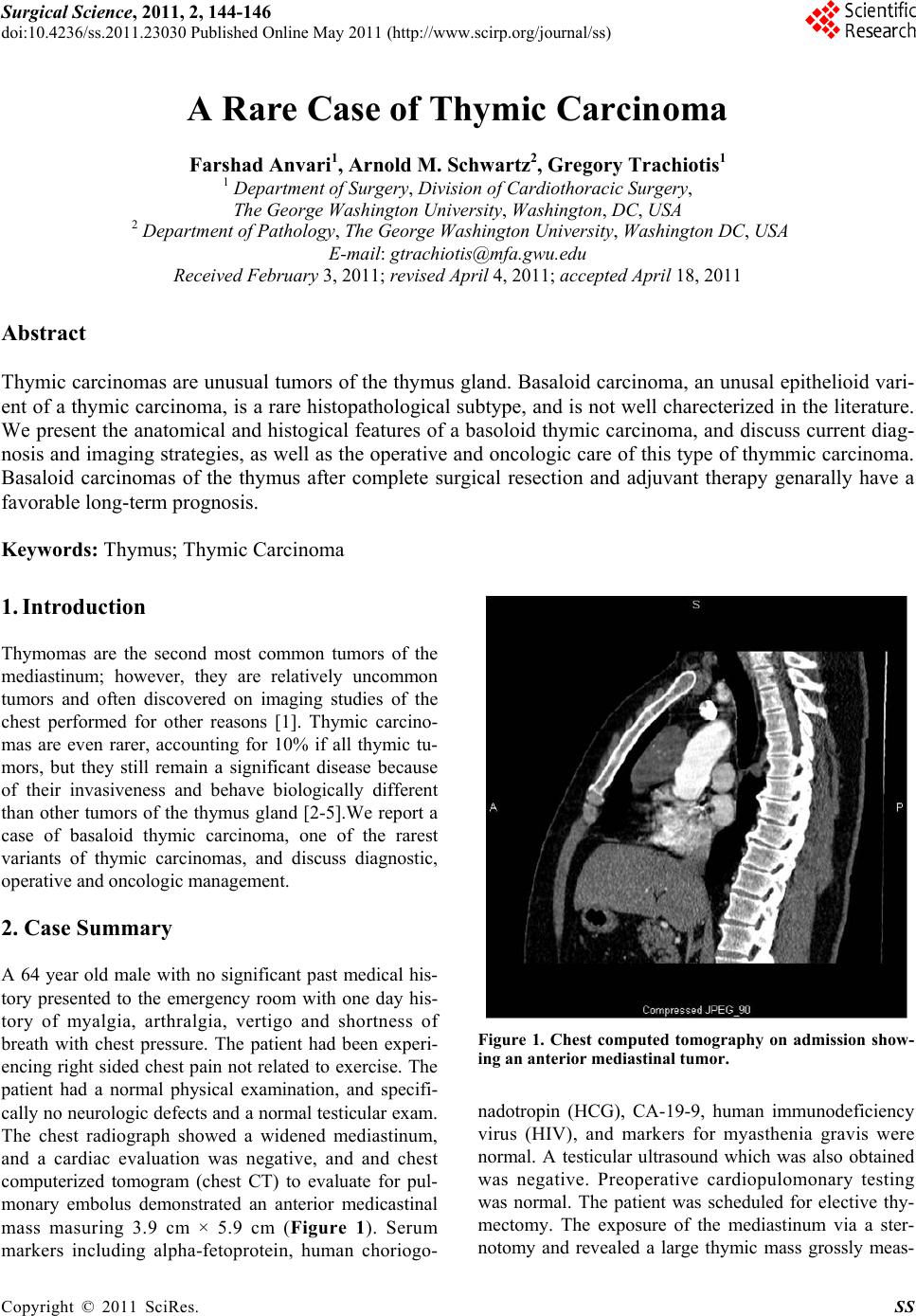 Surgical Science, 2011, 2, 144-146 doi:10.4236/ss.2011.23030 Published Online May 2011 (http://www.scirp.org/journal/ss) Copyright © 2011 SciRes. SS A Rare Case of Thymic Carcinoma Farshad Anvari1, Arnold M. Schwartz2, Gregory Trachiotis1 1 Department of Surgery, Division of Cardiothoracic Surgery, The George Washington University, Washington, DC, USA 2 Department of Pathology, The George Washington University, Washington DC, USA E-mail: [email protected] u Received February 3, 2011; revised April 4, 2011; accepted April 18, 2011 Abstract Thymic carcinomas are unusual tumors of the thymus gland. Basaloid carcinoma, an unusal epithelioid vari- ent of a thymic carcinoma, is a rare histopathological subtype, and is not well charecterized in the literature. We present the anatomical and histogical features of a basoloid thymic carcinoma, and discuss current diag- nosis and imaging strategies, as well as the operative and oncologic care of this type of thymmic carcinoma. Basaloid carcinomas of the thymus after complete surgical resection and adjuvant therapy genarally have a favorable long-term prognosis. Keywords: Thymus; Thymic Carcinoma 1. Introduction Thymomas are the second most common tumors of the mediastinum; however, they are relatively uncommon tumors and often discovered on imaging studies of the chest performed for other reasons [1]. Thymic carcino- mas are even rarer, accounting for 10% if all thymic tu- mors, but they still remain a significant disease because of their invasiveness and behave biologically different than other tumors of the thymus gland [2-5].We report a case of basaloid thymic carcinoma, one of the rarest variants of thymic carcinomas, and discuss diagnostic, operative and oncologic management. 2. Case Summary A 64 year old male with no significant past medical his- tory presented to the emergency room with one day his- tory of myalgia, arthralgia, vertigo and shortness of breath with chest pressure. The patient had been experi- encing right sided chest pain not related to exercise. The patient had a normal physical examination, and specifi- cally no neurologic defects and a normal testicular exam. The chest radiograph showed a widened mediastinum, and a cardiac evaluation was negative, and and chest computerized tomogram (chest CT) to evaluate for pul- monary embolus demonstrated an anterior medicastinal mass masuring 3.9 cm × 5.9 cm (Figure 1). Serum markers including alpha-fetoprotein, human choriogo- Figure 1. Chest computed tomography on admission show- ing an anterior mediastinal tumor. nadotropin (HCG), CA-19-9, human immunodeficiency virus (HIV), and markers for myasthenia gravis were normal. A testicular ultrasound which was also obtained was negative. Preoperative cardiopulomonary testing was normal. The patient was scheduled for elective thy- mectomy. The exposure of the mediastinum via a ster- notomy and revealed a large thymic mass grossly meas- 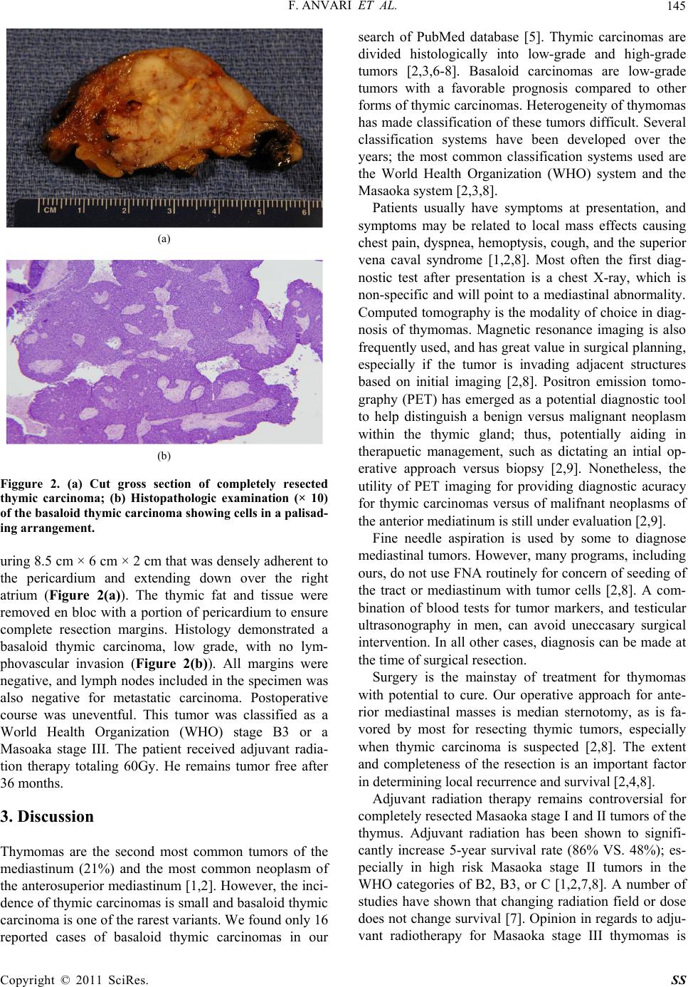 F. ANVARI ET AL. 145 (a) (b) Figgure 2. (a) Cut gross section of completely resected thymic carcinoma; (b) Histopathologic examination (× 10) of the basaloid thymic carcinoma showing cells in a palisad- ing arrangement. uring 8.5 cm × 6 cm × 2 cm that was densely adherent to the pericardium and extending down over the right atrium (Figure 2(a)). The thymic fat and tissue were removed en bloc with a portion of pericardium to ensure complete resection margins. Histology demonstrated a basaloid thymic carcinoma, low grade, with no lym- phovascular invasion (Figure 2(b)). All margins were negative, and lymph nodes included in the specimen was also negative for metastatic carcinoma. Postoperative course was uneventful. This tumor was classified as a World Health Organization (WHO) stage B3 or a Masoaka stage III. The patient received adjuvant radia- tion therapy totaling 60Gy. He remains tumor free after 36 months. 3. Discussion Thymomas are the second most common tumors of the mediastinum (21%) and the most common neoplasm of the anterosuperior mediastinum [1,2]. However, the inci- dence of thymic carcinomas is small and basaloid thymic carcinoma is one of the rarest variants. We found only 16 reported cases of basaloid thymic carcinomas in our search of PubMed database [5]. Thymic carcinomas are divided histologically into low-grade and high-grade tumors [2,3,6-8]. Basaloid carcinomas are low-grade tumors with a favorable prognosis compared to other forms of thymic carcinomas. Heterogeneity of thymomas has made classification of these tumors difficult. Several classification systems have been developed over the years; the most common classification systems used are the World Health Organization (WHO) system and the Masaoka system [2,3,8]. Patients usually have symptoms at presentation, and symptoms may be related to local mass effects causing chest pain, dyspnea, hemoptysis, cough, and the superior vena caval syndrome [1,2,8]. Most often the first diag- nostic test after presentation is a chest X-ray, which is non-specific and will point to a mediastinal abnormality. Computed tomography is the modality of choice in diag- nosis of thymomas. Magnetic resonance imaging is also frequently used, and has great value in surgical planning, especially if the tumor is invading adjacent structures based on initial imaging [2,8]. Positron emission tomo- graphy (PET) has emerged as a potential diagnostic tool to help distinguish a benign versus malignant neoplasm within the thymic gland; thus, potentially aiding in therapuetic management, such as dictating an intial op- erative approach versus biopsy [2,9]. Nonetheless, the utility of PET imaging for providing diagnostic acuracy for thymic carcinomas versus of malifnant neoplasms of the anterior mediatinum is still under evaluation [2,9]. Fine needle aspiration is used by some to diagnose mediastinal tumors. However, many programs, including ours, do not use FNA routinely for concern of seeding of the tract or mediastinum with tumor cells [2,8]. A com- bination of blood tests for tumor markers, and testicular ultrasonography in men, can avoid uneccasary surgical intervention. In all other cases, diagnosis can be made at the time of surgical resection. Surgery is the mainstay of treatment for thymomas with potential to cure. Our operative approach for ante- rior mediastinal masses is median sternotomy, as is fa- vored by most for resecting thymic tumors, especially when thymic carcinoma is suspected [2,8]. The extent and completeness of the resection is an important factor in determining local recurren ce and survival [2,4,8]. Adjuvant radiation therapy remains controversial for completely resected Masaoka stage I and II tumors of the thymus. Adjuvant radiation has been shown to signifi- cantly increase 5-year survival rate (86% VS. 48%); es- pecially in high risk Masaoka stage II tumors in the WHO categories of B2, B3, or C [1,2,7,8]. A number of studies have shown that changing radiation field or dose does not change survival [7]. Opinion in regards to adju- vant radiotherapy for Masaoka stage III thymomas is Copyright © 2011 SciRes. SS  F. ANVARI ET AL. Copyright © 2011 SciRes. SS 146 divided with some studies showing no significant sur- vival difference between surgery alone and surgery with radiation [4,8]. However, other studies have demon- strated that adjuvant radiotherapy for advanced thymo- mas, particularly Masaoka stage III, has long-term bene- fits with complete or partial remission [2,8]. Chemotherapy is not considered a treatment of choice when complete resection can be achieved [5]. However, cisplatin-based protocols have been used in patients with unresectable disease or gross residual disease [3]. Cur- rently there are no standardized regimens. In this case, we were able to achieve a complete resec- tion of a basaloid thymic carcinoma, which as single therapy has a 5 year survival rate of 92.9% for a Ma- saoka stage III tumor [4]. Although the low-grade nature of basaloid thymic carcinoma, based on macroscopic invasion of the pericardium (Masaoka stage III) and his- tology features (WHO B3) in this case, adjuvant radio- therapy was added, and should enhance the overall fa- vorable prognosis of this unusu a l thymic tumor. 4. References [1] T. K. Varghese Jr. and C. L. Lau, “The Mediastinum,” In: C. M. Townsend Jr., R. D. Beauchamp, B. M. Evers, K. L. Mattox, Sabiston Text Book of Surgery, 18th Edition, Saunders, Philadelphia, 2008. [2] V. Federico, A. Marco, D. Daniele, V. Domenico, A. R. Erino, G. de Tiziano, F. Federico and C. G. Furio, “Thy- moma and Thymic Carcinoma,” European Journal of Cardio-thoracic Surgery, Vol. 37, 2010, pp. 13-25. [3] D. J. Kim, W. I. Yang, S. S. Choi, K. D. Kim and K. Y. Chung, “Prognostic and Clinical Relevance of the World Health Organization Schema for the Classification of Thymic Epithelial Tumors: A Clinicopathologic Study of 108 Patients and Literature Review,” Chest, Vol. 127, No. 3, 2005, pp. 755-761. doi:10.1378/chest.127.3.755 [4] K. Kondo and Y. Monden, “Therapy for Thymic Epithe- lial Tumors: A Clinical Study of 1320 Patients from Ja- pan,” Annals of Thoracic Surgery, Vol. 76, No. 3, 2003, pp. 878-884. doi:10.1016/S0003-4975(03)00555-1 [5] P. J. Loehrer Sr., M. Jiroutek, S. Aisner, et al., “Com- bined Etoposide, Ifosfamide, and Cisplatin in the Treat- ment of Patients with Advanced Thymoma and Thymic Carcinoma: An Intergroup Trial,” Cancer, Vol. 91, No. 11, 2001, pp. 2010-2015. doi:10.1002/1097-0142(20010601)91:11<2010::AID- CNCR1226>3.3.CO;2-U [6] Y. Morisaki, K. Takagi, S. Sano, et al., “Basaloid Carci- noma of the Thymus: Report of a Case,” Surgery Today, Vol. 36, No. 1, 2006, pp. 68-70. doi:10.1007/s00595-005-3072-x [7] K. Ogawa, T. Uno, T. Toita, et al., “Postoperative Radio- therapy for Patients with Completely Resected Thymoma: A Multi-Institutional, Retrospective Review of 103 Pa- tients,” Cancer, Vol. 94, No. 5, 2002, pp. 1405-1413. doi:10.1002/cncr.10373 [8] S. Tomaszek, D. A. Wigle, S. Keshavjee and S. Fischer, “Thymomas: Review of Current Clinical Practice,” An- nals of Thoracic Surgery, Vol. 87, No. 6, 2009, pp. 1973- 1980. doi:10.1016/j.athoracsur.2008.12.095 [9] L. Luzzi, A. Campione, A. Gorla, G. Vassallo, A. Bianchi, A. Biggi and A. Terz i, “Role of Flu orine- Flurode oxygluc ose Positron Emission Tomography/Computed Tomography in Preoperative Assessment of Anterior Mediastinal Mas- ses,” European Journal of Cardio-Thoracic Surgery, Vol. 36, No. 3, 2009, pp. 475-479. doi:10.1016/j.ejcts.2009.03.055 |