

Paper Menu >>
Journal Menu >>
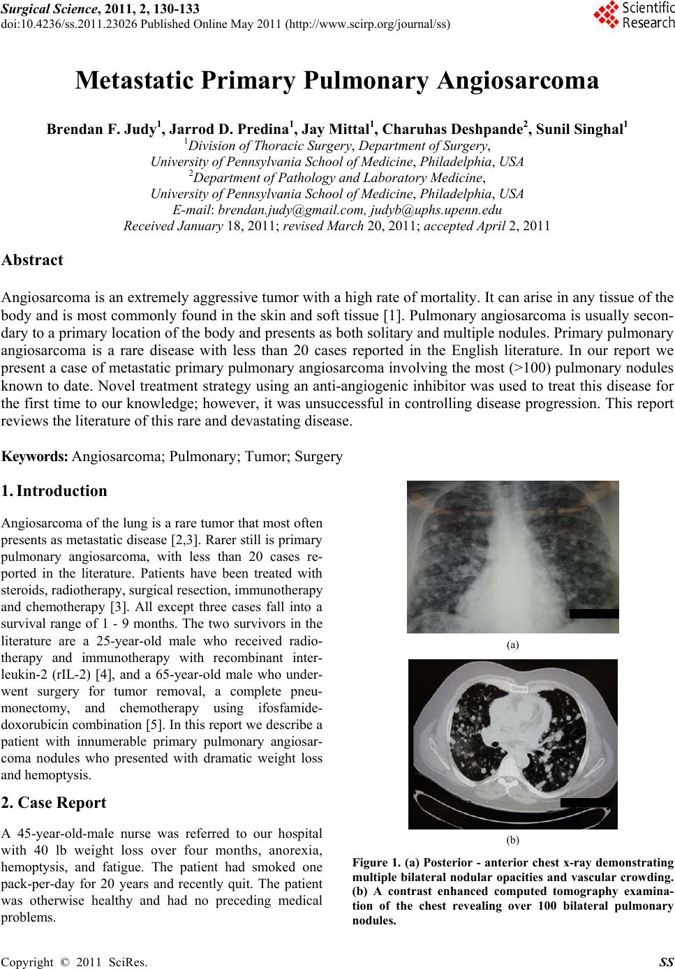 Surgical Science, 2011, 2, 130-133 doi:10.4236/ss.2011.23026 Published Online May 2011 (http://www.scirp.org/journal/ss) Copyright © 2011 SciRes. SS Metastatic Primary Pulmonary Angiosarcoma Brendan F. Judy1, Jarrod D. Predina1, Jay Mittal1, Charuhas Deshpande2, Sunil Singhal1 1Division of Thoracic Surgery, Department of Surgery, University of Penn syl va ni a School of Medici ne, Philadelphia, USA 2Department of Pathology and Laboratory Medicine, University of Penn syl va ni a School of Medici ne, Philadelphia, USA E-mail: [email protected], [email protected] Received January 18, 2011; revised March 20, 201 1; accepted April 2, 2011 Abstract Angiosarcoma is an extremely aggressive tumor with a high rate of mortality. It can arise in any tissue of the body and is most commonly found in the skin and soft tissue [1]. Pulmonary angiosarcoma is usually secon- dary to a primary location of the body and presents as both solitary and multiple nodules. Primary pulmonary angiosarcoma is a rare disease with less than 20 cases reported in the English literature. In our report we present a case of metastatic primary pulmonary angiosarcoma involving the most (>100) pulmonary nodules known to date. Novel treatment strategy using an anti-angiogenic inhibitor was used to treat this disease for the first time to our knowledge; however, it was unsuccessful in controlling disease progression. This report reviews the literature of this rare and devastating disease. Keywords: Angiosarcoma; Pulmonary; Tumor; Surgery 1. Introduction Angiosarcoma of the lung is a rare tumor that most often presents as metastatic disease [2 ,3]. Rarer still is primary pulmonary angiosarcoma, with less than 20 cases re- ported in the literature. Patients have been treated with steroids, radiotherapy, surgical resection, immunotherapy and chemotherapy [3]. All except three cases fall into a survival range of 1 - 9 months. The two survivors in the literature are a 25-year-old male who received radio- therapy and immunotherapy with recombinant inter- leukin-2 (rIL-2) [4], and a 65-year-old male who under- went surgery for tumor removal, a complete pneu- monectomy, and chemotherapy using ifosfamide- doxorubicin combination [5]. In this report we describe a patient with innumerable primary pulmonary angiosar- coma nodules who presented with dramatic weight loss and hemoptysis. 2. Case Report A 45-year-old-male nurse was referred to our hospital with 40 lb weight loss over four months, anorexia, hemoptysis, and fatigue. The patient had smoked one pack-per-day for 20 years and recently quit. The patient was otherwise healthy and had no preceding medical problems. (a) (b) Figure 1. (a) Posterior - anterior chest x-ray demonstrating multiple bilateral nodular opacities and vascular crowding. (b) A contrast enhanced computed tomography examina- tion of the chest revealing over 100 bilateral pulmonary nodules. 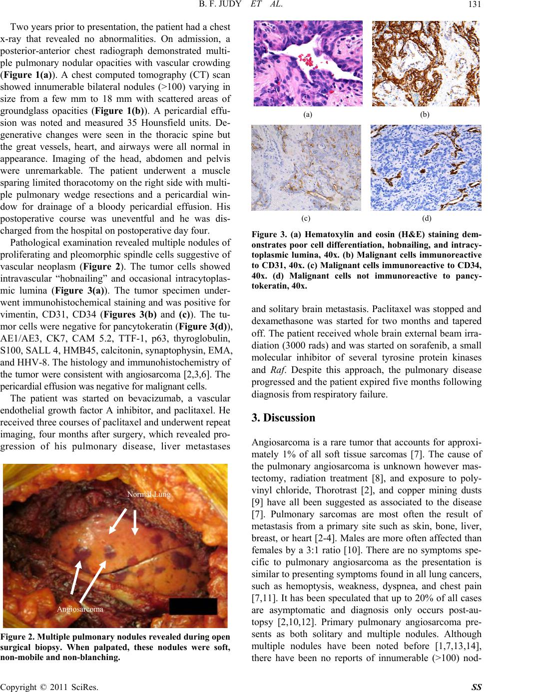 B. F. JUDY ET AL.131 Two years prior to presentation, th e patient had a chest x-ray that revealed no abnormalities. On admission, a posterior-anterior chest radiograph demonstrated multi- ple pulmonary nodular opacities with vascular crowding (Figure 1(a)). A chest computed tomography (CT) scan showed innumerable bilateral nodules (>100) varying in size from a few mm to 18 mm with scattered areas of groundglass opacities (Figure 1(b)). A pericardial effu- sion was noted and measured 35 Hounsfield units. De- generative changes were seen in the thoracic spine but the great vessels, heart, and airways were all normal in appearance. Imaging of the head, abdomen and pelvis were unremarkable. The patient underwent a muscle sparing limited thoracotomy on the right side with multi- ple pulmonary wedge resections and a pericardial win- dow for drainage of a bloody pericardial effusion. His postoperative course was uneventful and he was dis- charged from the hospital on postoperative day four. Pathological examination revealed multiple nodules of proliferating and pleomorphic spindle cells suggestive of vascular neoplasm (Figure 2). The tumor cells showed intravascular “hobnailing” and occasional intracytoplas- mic lumina (Figure 3(a)). The tumor specimen under- went immunohistochemical staining and was positive for vimentin, CD31, CD34 (Figures 3(b) and (c)). The tu- mor cells were negative for pancytokeratin (Figure 3(d)), AE1/AE3, CK7, CAM 5.2, TTF-1, p63, thyroglobulin, S100, SALL 4, HMB45, calcitonin, synaptophysin, EMA, and HHV-8. The histology and immunohistochemistry of the tumor were consistent with angiosarcoma [2,3,6]. The pericardial effusion was negative for malignant cells . The patient was started on bevacizumab, a vascular endothelial growth factor A inhibitor, and paclitaxel. He received three courses of paclitaxel and underwent repeat imaging, four months after surgery, which revealed pro- gression of his pulmonary disease, liver metastases Angiosarcoma Normal Lung Figure 2. Multiple pulmonary nodules re vealed dur ing open surgical biopsy. When palpated, these nodules were soft, non-mobile and non-blanching. (a) (b) (c) (d) Figure 3. (a) Hematoxylin and eosin (H&E) staining dem- onstrates poor cell differentiation, hobnailing, and intracy- toplasmic lumina, 40x. (b) Malignant cells immunoreactive to CD31, 40x. (c) Malignant cells immunoreactive to CD34, 40x. (d) Malignant cells not immunoreactive to pancy- tokeratin, 40x. and solitary brain metastasis. Paclitaxel was stopped and dexamethasone was started for two months and tapered off. The patient received whole brain external beam irra- diation (3000 rads) and was started on sorafenib, a small molecular inhibitor of several tyrosine protein kinases and Raf. Despite this approach, the pulmonary disease progressed and the patient expired five months following diagnosis from respiratory failure. 3. Discussion Angiosarcoma is a rare tumor that accounts for approxi- mately 1% of all soft tissue sarcomas [7]. The cause of the pulmonary angiosarcoma is unknown however mas- tectomy, radiation treatment [8], and exposure to poly- vinyl chloride, Thorotrast [2], and copper mining dusts [9] have all been suggested as associated to the disease [7]. Pulmonary sarcomas are most often the result of metastasis from a primary site such as skin, bone, liver, breast, or heart [2-4]. Males are more often affected than females by a 3:1 ratio [10]. There are no symptoms spe- cific to pulmonary angiosarcoma as the presentation is similar to presenting symptoms found in all lu ng cancers, such as hemoptysis, weakness, dyspnea, and chest pain [7,11]. It has been speculated that up to 20% of all cases are asymptomatic and diagnosis only occurs post-au- topsy [2,10,12]. Primary pulmonary angiosarcoma pre- sents as both solitary and multiple nodules. Although multiple nodules have been noted before [1,7,13,14], there have been no reports of innumerable (>100) nod- ( A Copyright © 2011 SciRes. SS 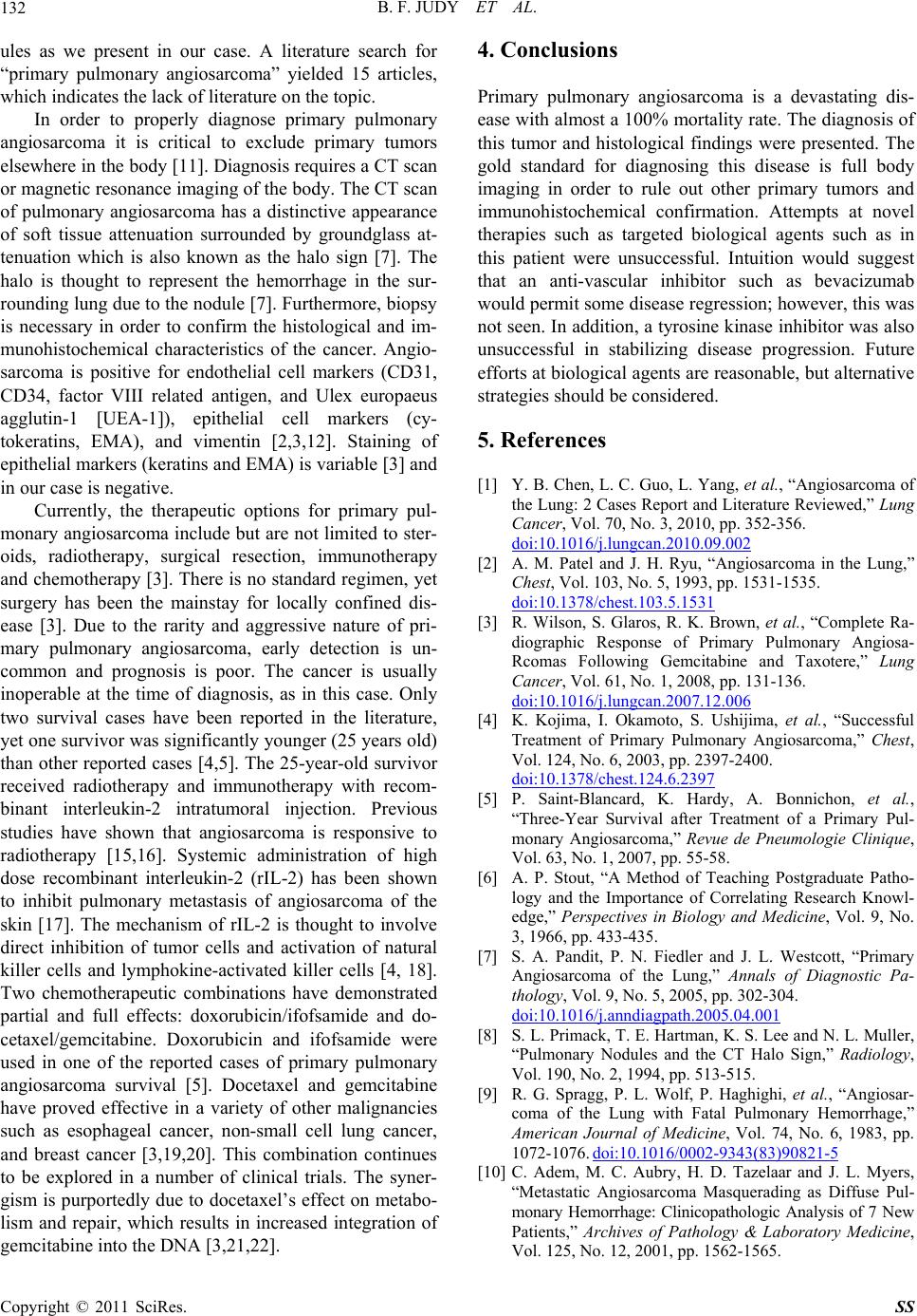 B. F. JUDY ET AL. 132 ules as we present in our case. A literature search for “primary pulmonary angiosarcoma” yielded 15 articles, which indicates the lack of literature on the topic. In order to properly diagnose primary pulmonary angiosarcoma it is critical to exclude primary tumors elsewhere in the body [11]. Diagnosis requires a CT scan or magnetic resonance imaging of the body. The CT scan of pulmonary angiosarcoma has a distinctive appearance of soft tissue attenuation surrounded by groundglass at- tenuation which is also known as the halo sign [7]. The halo is thought to represent the hemorrhage in the sur- rounding lung du e to the nodule [7]. Fur thermore, biopsy is necessary in order to confirm the histological and im- munohistochemical characteristics of the cancer. Angio- sarcoma is positive for endothelial cell markers (CD31, CD34, factor VIII related antigen, and Ulex europaeus agglutin-1 [UEA-1]), epithelial cell markers (cy- tokeratins, EMA), and vimentin [2,3,12]. Staining of epithelial markers (keratins and EMA) is variable [3] and in our case is negative. Currently, the therapeutic options for primary pul- monary angiosarcoma include but are not limited to ster- oids, radiotherapy, surgical resection, immunotherapy and chemotherapy [3]. There is no standard regimen, yet surgery has been the mainstay for locally confined dis- ease [3]. Due to the rarity and aggressive nature of pri- mary pulmonary angiosarcoma, early detection is un- common and prognosis is poor. The cancer is usually inoperable at the time of diagnosis, as in this case. Only two survival cases have been reported in the literature, yet one survivor was significantly younger (25 years old) than other reported cases [4,5]. The 25-year-old survivor received radiotherapy and immunotherapy with recom- binant interleukin-2 intratumoral injection. Previous studies have shown that angiosarcoma is responsive to radiotherapy [15,16]. Systemic administration of high dose recombinant interleukin-2 (rIL-2) has been shown to inhibit pulmonary metastasis of angiosarcoma of the skin [17]. The mechanism of rIL-2 is thought to involve direct inhibition of tumor cells and activation of natural killer cells and lymphokine-activated killer cells [4, 18]. Two chemotherapeutic combinations have demonstrated partial and full effects: doxorubicin/ifofsamide and do- cetaxel/gemcitabine. Doxorubicin and ifofsamide were used in one of the reported cases of primary pulmonary angiosarcoma survival [5]. Docetaxel and gemcitabine have proved effective in a variety of other malignancies such as esophageal cancer, non-small cell lung cancer, and breast cancer [3,19,20]. This combination continues to be explored in a number of clinical trials. The syner- gism is purportedly due to docetaxel’s effect on metabo- lism and repair, which results in increased integration of gemcitabine into the DNA [3,21,22]. 4. Conclusions Primary pulmonary angiosarcoma is a devastating dis- ease with almost a 100% mortality rate. The diagnosis of this tumor and histological findings were presented. The gold standard for diagnosing this disease is full body imaging in order to rule out other primary tumors and immunohistochemical confirmation. Attempts at novel therapies such as targeted biological agents such as in this patient were unsuccessful. Intuition would suggest that an anti-vascular inhibitor such as bevacizumab would permit some disease regression; however, this was not seen. In addition, a tyrosine kinase inhibitor was also unsuccessful in stabilizing disease progression. Future efforts at biological agents are reasonable, but alternative strategies should be considered. 5. References [1] Y. B. Chen, L. C. Guo, L. Yang, et al., “Angiosarcoma of the Lung: 2 Cases Report and Literature Reviewed,” Lung Cancer, Vol. 70, No. 3, 2010, pp. 352-356. doi:10.1016/j.lungcan.2010.09.002 [2] A. M. Patel and J. H. Ryu, “Angiosarcoma in the Lung,” Chest, Vol. 103, No. 5, 1993, pp. 1531-1535. doi:10.1378/chest.103.5.1531 [3] R. Wilson, S. Glaros, R. K. Brown, et al., “Complete Ra- diographic Response of Primary Pulmonary Angiosa- Rcomas Following Gemcitabine and Taxotere,” Lung Cancer, Vol. 61, No. 1, 2008, pp. 131-136. doi:10.1016/j.lungcan.2007.12.006 [4] K. Kojima, I. Okamoto, S. Ushijima, et al., “Successful Treatment of Primary Pulmonary Angiosarcoma,” Chest, Vol. 124, No. 6, 2003, pp. 2397-2400. doi:10.1378/chest.124.6.2397 [5] P. Saint-Blancard, K. Hardy, A. Bonnichon, et al., “Three-Year Survival after Treatment of a Primary Pul- monary Angiosarcoma,” Revue de Pneumologie Clinique, Vol. 63, No. 1, 2007, pp. 55-58. [6] A. P. Stout, “A Method of Teaching Postgraduate Patho- logy and the Importance of Correlating Research Knowl- edge,” Perspectives in Biology and Medicine, Vol. 9, No. 3, 1966, pp. 433-435. [7] S. A. Pandit, P. N. Fiedler and J. L. Westcott, “Primary Angiosarcoma of the Lung,” Annals of Diagnostic Pa- thology, Vol. 9, No. 5, 2005, pp. 302-304. doi:10.1016/j.anndiagpath.2005.04.001 [8] S. L. Primack, T. E. Hartman, K. S. Lee and N. L. Muller, “Pulmonary Nodules and the CT Halo Sign,” Radiology, Vol. 190, No. 2, 1994, pp. 513-515. [9] R. G. Spragg, P. L. Wolf, P. Haghighi, et al., “Angiosar- coma of the Lung with Fatal Pulmonary Hemorrhage,” American Journal of Medicine, Vol. 74, No. 6, 1983, pp. 1072-1076. doi:10.1016/0002-9343(83)90821-5 [10] C. Adem, M. C. Aubry, H. D. Tazelaar and J. L. Myers, “Metastatic Angiosarcoma Masquerading as Diffuse Pul- monary Hemorrhage: Clinicopathologic Analysis of 7 New Patients,” Archives of Pathology & Laboratory Medicine, Vol. 125, No. 12, 2001, pp. 1562-1565. Copyright © 2011 SciRes. SS  B. F. JUDY ET AL. Copyright © 2011 SciRes. SS 133 [11] E. A. Bacha, C. D. Wright, H. C. Grillo, et al., “Surgical Treatment of Primary Pulmonary Sarcomas,” European Journal of Cardio-Thoracic Surgery, Vol. 15, No. 4, 1999, pp. 456-460. doi:10.1016/S1010-7940(99)00045-7 [12] G. A. Carillo, M. A. Carretero, J. E. Vazquez, et al., “Epi- thelioid Angiosarcoma of the Lung with Pleural Metasta- ses: A Rare Cause of Haemoptysis Clinicopathological Conference,” Heart Lung Circ, Vol. 19, No. 10, pp. 624-628. [13] S. L. Segal, G. S. Lenchner, A. V. Cichelli, et al., “An- giosarcoma Presenting as Diffuse Alveolar Hemorrhage,” Chest, Vol. 94, No. 1, 1988, pp. 214-216. doi:10.1378/chest.94.1.214 [14] S. A. Yousem, “Angiosarcoma Presenting in the Lung,” Archives of Pathology & Laboratory Medicine, Vol. 110, No. 2, 1986, pp. 112-115. [15] C. A. Holden, M. F. Spittle and E. W. Jones, “Angiosar- coma of the Face and Scalp, Prognosis and Treatment,” Cancer, Vol. 59, No. 5, 1987, pp. 1046-1057. doi:10.1002/1097-0142(19870301)59:5<1046::AID-CNC R2820590533>3.0.CO;2-6 [16] R. Sasaki, T. Soejima, K. Kishi, et al., “Angiosarcoma Treated with Radiotherapy: Impact of Tumor Type and Size on Outcome,” International Journal of Radiation Oncology Biology Physics, Vol. 52, No. 4, 2002, pp. 1032-1040. doi:10.1016/S0360-3016(01)02753-5 [17] M. Masuz awa, N. Mochida, T. Amano, et al., “Evaluation of Recombinant Interleukin-2 Immunotherapy for Human Hemangiosarcoma in a SCID Mice Model (WB-SCID),” Journal of Dermatological Science, Vol. 27, No. 2, 2001, pp. 88-94. doi:10.1016/S0923-1811(01)00103-7 [18] H. Ihda, Y. Tokura, M. Fushimi, et al., “Malignant He- mangioendothelioma,” International Journal of Derma- tology, Vol. 34, No. 11, 1995, pp. 811-816. doi:10.1111/j.1365-4362.1995.tb04406.x [19] C. Atasoy, S. Fitoz, H. Yigit, et al., “Radiographic, CT, and MRI Findings in Primary Pulmonary Angiosarcoma,” Clinical Imaging, Vol. 25, No. 5, 2001, pp. 337-340. doi:10.1016/S0899-7071(01)00302-3 [20] K. M. Leu, L. J. Ostruszka, D. Shewach, et al., “Labora- tory and Clinical Evidence of Synergistic Cytotoxicity of Sequential Treatment with Gemcitabine Followed by Do- cetaxel in the Treatment of Sarcoma,” Journal of Clinical Oncology, Vol. 22, No. 9, 2004, pp. 1706-1712. doi:10.1200/JCO.2004.08.043 [21] K. Okamoto, M. Ocker, D. Neureiter, et al., “Bcl-2-Spe- cific Sirnas Restore Gemcitabine Sensitivity in Human Pancreatic Cancer Cells,” Journal of Cellular and Mo- lecular Medicine, Vol. 11, No. 2, 2007, pp. 349-361. doi:10.1111/j.1582-4934.2007.00013.x [22] L. Ricotti, A. Tesei, F. De Paola, et al., “In Vitro Schedule -Dependent Interaction between Docetaxel and Gemcit- abine in Human Gastric Cancer Cell Lines,” Clinical Can- cer Research, Vol. 9, No. 2, 2003, pp. 900-905. |