

Paper Menu >>
Journal Menu >>
 Open Journal of Urology, 2011, 1, 19-24 doi:10.4236/oju.2011.12006 Published Online May 2011 (http://www.SciRP.org/journal/oju) Copyright © 2011 SciRes. OJU Cadaveric Assessment of Synthetic Mid-Urethral Sling Placement Scott Serels Bladder Control Center of Norwalk, Norwalk CT, USA E-mail:[email protected] Received March 5, 2011; revised April 25, 2011; accepted Ma y 5, 2011 Abstract Purpose: To determine if 3 types of mid-urethral synthetic slings are visually the same. Materials and Methods: A retropubic, obturator, and single incision sling was individually placed in three cadavers. Tension was set using a spacer (obturator and retropubic). Single incision sling (SIS) tension was set by visual inspection. Thirty physicians were asked to determine placement method, tension, and location of the 3 slings. Results: Physicians were composed of 5 urolo- gists, 7 urogynecologist, and 18 general gynecologists, with an average of 53 slings performed per year. Conclusions: This study showed that after placement of a sling it is hard to tell how the sling was placed and that most physicians felt the SIS was tensioned the best and most likely at the mid-urethra. Retropubic Obturator SIS % correctly identified sling 40% 43% 23% % thought tension was just right 33% 47% 73% % thought sling was at mid-urethra 50% 67% 83% Keywords: Stress Incontin ence, Surgical Correction of Incontinence, Single Incision Slings 1. Introduction Urinary incontinence is a common problem in adult women, with a prevalence of approximately 35% [1]. Stress urinary incontinence (SUI), the most common form of urinary incontinence in women, is an involuntary loss of urine that occurs during physical activity, such as coughing, sneezing, laughing, or exercise. Treatment options for SUI in women are designed to prevent the involuntary loss of urine from the urethra during increases in intra-abdominal pressure. Nonsurgical therapies include behavioral therapy (e.g. bladder training, fluid and dietary modification) and l imit ed drug t herapy. Surgical therapy for this condition has existed for over 100 years. The suburethral sling was first described in 1907 by von Giordano, and consisted of pl acing autol ogous tissue underneath the bladder neck and suspending it superiorly. Since then, over 200 different surgical procedures have been described. Sling material has also evolved from autologous fascia lata, muscle, and rectus fascia to synthetic material. The majority of midurethral slings now available u se type I polypropylene mesh. The development of the mid-urethral slings and corresponding placement methodologies have evolved to improve efficacy, safety, cost-effectiveness, and to minimize invasiveness. Currently, the three main approaches to placement of synthetic mid urethral sling include retropubic, transobturator, and the single incision mini sling. The Integral Theory of Female Urinary Incontinence by Ulmsten and Petros in the early 1990’s challenged our understanding of incontinence significantly and led to the market introduction of the minimally invasive retropubic midurethral sling known as tension-free vaginal tape (TVT) [2]. The TVT procedure initiated in 1996 revolutionized the surgical manag ement of female SUI in terms of its efficacy and minimal invasiveness. Although 80% - 89% cure rates, with durable cure rates up to 5 years post-procedure, have been reported, blind passage through the retropubic space poses considerable risks. [3,4] Pot enti al compli cati ons i ncl ude bowel i njury , bl adder injury, major vascular injury, and nerve injury. Bladder perforation in retropubic slings is a common complication with a recent meta-analysis reporting rates of 3.2%, [5] 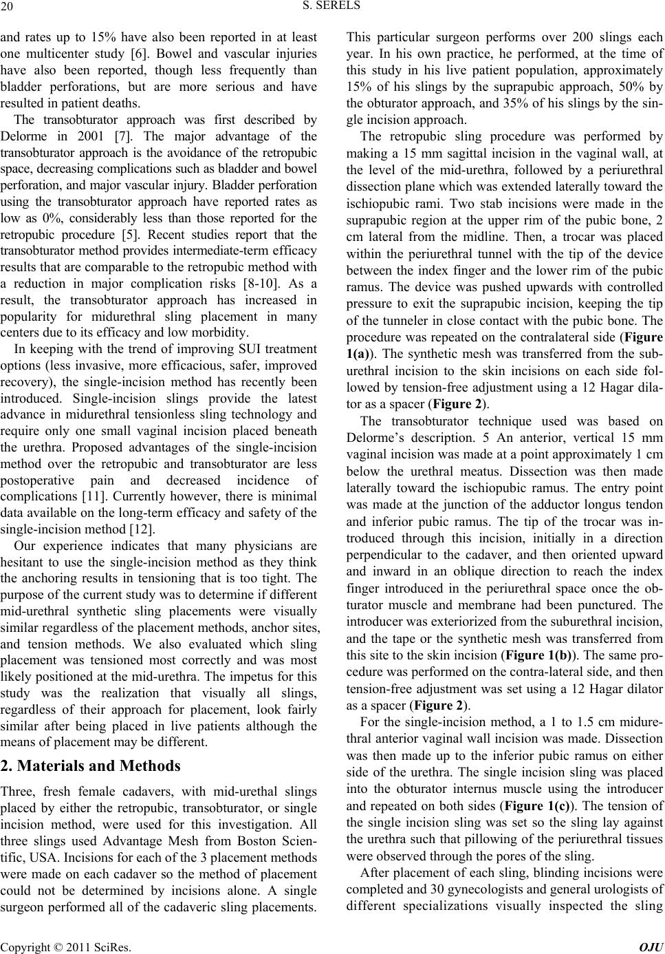 20 S. SERELS and rates up to 15% have also been reported in at least one multicenter study [6]. Bowel and vascular injuries have also been reported, though less frequently than bladder perforations, but are more serious and have resulted in patient deaths. The transobturator approach was first described by Delorme in 2001 [7]. The major advantage of the transobturator approach is the avoidance of the retropubic space, decreasing complicatio ns such as bladder and bowel perforation, and major vascular injury. Bladder perforation using the transobturator approach have reported rates as low as 0%, considerably less than those reported for the retropubic procedure [5]. Recent studies report that the transobturator method provides intermediate-term efficacy results that are comparable to the retropubic method with a reduction in major complication risks [8-10]. As a result, the transobturator approach has increased in popularity for midurethral sling placement in many centers due to its efficacy and low morbidity. In keeping with the trend of improving SUI treatment options (less invasive, more efficacious, safer, improved recovery), the single-incision method has recently been introduced. Single-incision slings provide the latest advance in midurethral tensionless sling technology and require only one small vaginal incision placed beneath the urethra. Proposed advantages of the single-incision method over the retropubic and transobturator are less postoperative pain and decreased incidence of complications [11]. Currently however, there is minimal data available on the long-term efficacy and safety of the single-incisio n m e t hod [12]. Our experience indicates that many physicians are hesitant to use the single-incision method as they think the anchoring results in tensioning that is too tight. The purpose of the current study was to determine if different mid-urethral synthetic sling placements were visually similar regardless of the placement methods, anchor sites, and tension methods. We also evaluated which sling placement was tensioned most correctly and was most likely positioned at th e mid-urethr a. The impetus for this study was the realization that visually all slings, regardless of their approach for placement, look fairly similar after being placed in live patients although the means of placement may be different. 2. Materials and Methods Three, fresh female cadavers, with mid-urethal slings placed by either the retropubic, transobturator, or single incision method, were used for this investigation. All three slings used Advantage Mesh from Boston Scien- tific, USA. Incisions for each of the 3 placement methods were made on each cadaver so the method of placement could not be determined by incisions alone. A single surgeon performed all of the cadaveric sling placements. This particular surgeon performs over 200 slings each year. In his own practice, he performed, at the time of this study in his live patient population, approximately 15% of his slings by the suprapubic approach, 50% by the obturator approach, and 35% of his slings by the sin- gle incision approach. The retropubic sling procedure was performed by making a 15 mm sagittal incision in the vaginal wall, at the level of the mid-urethra, followed by a periurethral dissection plane which was extended laterally toward the ischiopubic rami. Two stab incisions were made in the suprapubic region at the upper rim of the pubic bone, 2 cm lateral from the midline. Then, a trocar was placed within the periurethral tunnel with the tip of the device between the index finger and the lower rim of the pubic ramus. The device was pushed upwards with controlled pressure to exit the suprapubic incision, keeping the tip of the tunneler in close contact with the pubic bone. The procedure was repeated on the contralateral side (Figure 1(a)). The synthetic mesh was transferred from the sub- urethral incision to the skin incisions on each side fol- lowed by tension-free adjustment using a 12 Hagar dila- tor as a spacer (Figure 2). The transobturator technique used was based on Delorme’s description. 5 An anterior, vertical 15 mm vaginal incision was made at a point approximately 1 cm below the urethral meatus. Dissection was then made laterally toward the ischiopubic ramus. The entry point was made at the junction of the adductor longus tendon and inferior pubic ramus. The tip of the trocar was in- troduced through this incision, initially in a direction perpendicular to the cadaver, and then oriented upward and inward in an oblique direction to reach the index finger introduced in the periurethral space once the ob- turator muscle and membrane had been punctured. The introducer was exteriorized from the suburethral incision, and the tape or the synthetic mesh was transferred from this site to the skin incision (Figure 1(b)). The same pro- cedure was performed on the contra-lateral side, and then tension-free adjustment was set using a 12 Hagar dilator as a spacer (Figure 2). For the single-incision method, a 1 to 1.5 cm midure- thral anterior vaginal wall incision was made. Dissection was then made up to the inferior pubic ramus on either side of the urethra. The single incision sling was placed into the obturator internus muscle using the introducer and repeated on both sides (Figure 1(c)). The tension of the single incision sling was set so the sling lay against the urethra such that pillowing of the periurethral tissues were observ ed through the por es of the sling. After placement of each sling, blinding incisions were complet ed and 30 gynecologis t s and general urologists of ifferent specializations visually inspected the sling d Copyright © 2011 SciRes. OJU 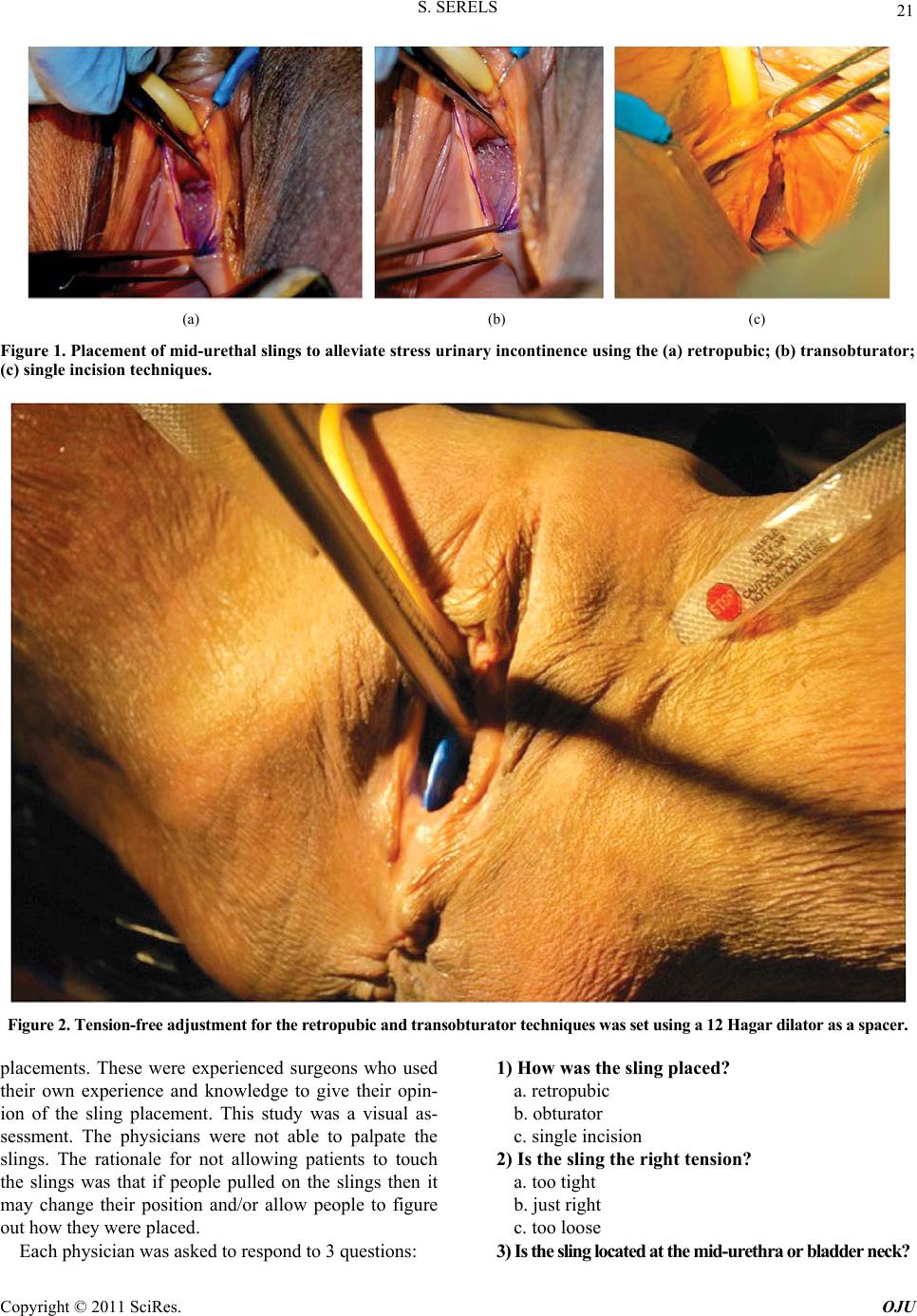 S. SERELS Copyright © 2011 SciRes. OJU 21 (a) (b) (c) Figure 1. Placement of mid-urethal slings to alle viate stre ss urinary incontinence using the (a) re tropubic; (b) tr ansobturat or; (c) single incision techniques. Figure 2. Tension-free adjustment for the retropub ic and transobtu rator techniques was set using a 12 Hagar dilator as a spacer. 1) How was the sling placed? placements. These were experienced surgeons who used their own experience and knowledge to give their opin- ion of the sling placement. This study was a visual as- sessment. The physicians were not able to palpate the slings. The rationale for not allowing patients to touch the slings was that if people pulled on the slings then it may change their position and/or allow people to figure out how they wer e placed. a. retropubic b. obturator c. single incision 2) Is the sling the right tension? a. too tight b. just right c. too loose 3) Is the sling located at the mid-urethra or bladder neck? Each physician was asked to respond to 3 questions: 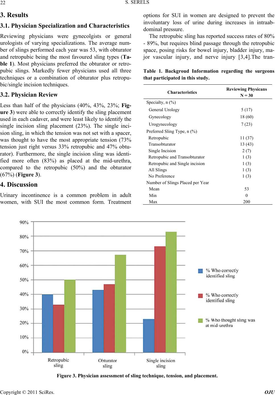 22 S. SERELS 3. Results 3.1. Physician Specialization and Characteristics Reviewing physicians were gynecolgists or general urologists of varying specializations. The average num- ber of slings performed each year was 53, with obturator and retropubic being the most favoured sling types (Ta- ble 1). Most physicians preferred the obturator or retro- pubic slings. Markedly fewer physicians used all three techniques or a combination of obturator plus retropu- bic/single incision techniques. 3.2. Physician Review Less than half of the physicians (40%, 43%, 23%; Fig- ure 3) were able to correctly identify the sling placement used in each cad aver, and were least likely to id entify th e single incision sling placement (23%). The single inci- sion sling, in which the tension was not set with a spacer, was thought to have the most appropriate tension (73% tension just right versus 33% retropubic and 47% obtu- rator). Furthermore, the single incision sling was identi- fied more often (83%) as placed at the mid-urethra, compared to the retropubic (50%) and the obturator (67%) (Figure 3). 4. Discussion Urinary incontinence is a common problem in adult women, with SUI the most common form. Treatment options for SUI in women are designed to prevent the involuntary loss of urine during increases in intraab- dominal pressure. The retropubic sling has reported success rates of 80% - 89%, but requires blind passage through the retropubic space, posing risks for bowel injury, bladder injury, ma- jor vascular injury, and nerve injury [3,4].The tran- Table 1. Backgroud Information regarding the surgeons that participated in this study. Characteristics Reviewing Physicans N = 30 Specialty, n (%) General Urology 5 (17) Gynecology 18 (60) Urogynecology 7 (23) Preferred Sling Type, n (%) Retropubic 11 (37) Transobturator 13 (43) Single Incision 2 (7) Retropubic and Transobturator 1 (3) Retropubic and Single incisi o n 1 (3) All Slings 1 (3) No Preference 1 (3) Number of Slings Placed per Year Mean 53 Min 0 Max 200 Figure 3. Physician assessment of sling tec hnique , tension, and placement. Copyright © 2011 SciRes. OJU 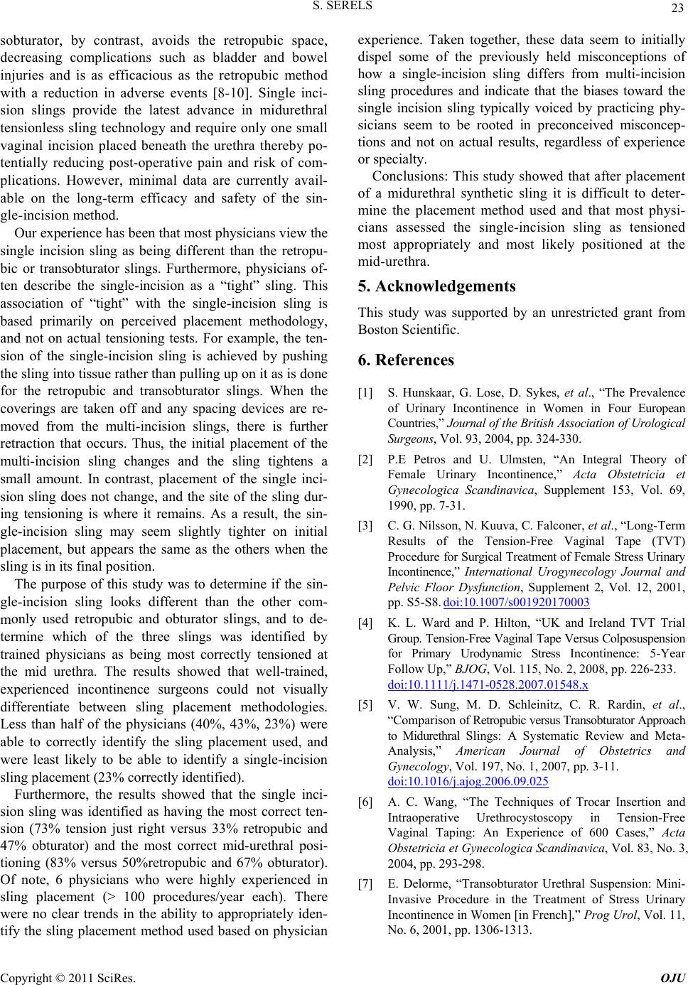 S. SERELS 23 sobturator, by contrast, avoids the retropubic space, decreasing complications such as bladder and bowel s being different than the retropu- bi ferent than the other c m ten- si experience. Taken together, these data seem to initially dispel some of the previously held misconceptions of d the single-incision sling as tensioned m . Lose, D. Sykes, et al., “The Prevalence continence in Women in Four European he British Association of Urological 93, 2004, pp. 324-330. ternational Urogynecology Journal and injuries and is as efficacious as the retropubic method with a reduction in adverse events [8-10]. Single inci- sion slings provide the latest advance in midurethral tensionless sling technology and require only one small vaginal incision placed beneath the urethra thereby po- tentially reducing post-operative pain and risk of com- plications. However, minimal data are currently avail- able on the long-term efficacy and safety of the sin- gle-incision method. Our ex per ie nce h as b een tha t most physicians view the single incision sling a c or transobturator slings. Furthermore, physicians of- ten describe the single-incision as a “tight” sling. This association of “tight” with the single-incision sling is based primarily on perceived placement methodology, and not on actual tensioning tests. For example, the ten- sion of the single-incision sling is achieved by pushing the sling into tissue rather than pulling up on it as is do ne for the retropubic and transobturator slings. When the coverings are taken off and any spacing devices are re- moved from the multi-incision slings, there is further retraction that occurs. Thus, the initial placement of the multi-incision sling changes and the sling tightens a small amount. In contrast, placement of the single inci- sion sling does not change, and the site of the sling dur- ing tensioning is where it remains. As a result, the sin- gle-incision sling may seem slightly tighter on initial placement, but appears the same as the others when the sling is in its final position. The purpose of this study was to determine if the sin- gle-incision sling looks difom- only used retropubic and obturator slings, and to de- termine which of the three slings was identified by trained physicians as being most correctly tensioned at the mid urethra. The results showed that well-trained, experienced incontinence surgeons could not visually differentiate between sling placement methodologies. Less than half of the physicians (40%, 43%, 23%) were able to correctly identify the sling placement used, and were least likely to be able to identify a single-incision sling placement (23% correctly identified). Furthermore, the results showed that the single inci- sion sling was identified as having the most correct on (73% tension just right versus 33% retropubic and 47% obturator) and the most correct mid-urethral posi- tioning (83% versus 50%retropubic and 67% obturator). Of note, 6 physicians who were highly experienced in sling placement (> 100 procedures/year each). There were no clear trends in the ability to appropriately iden- tify the sling placement method used based on physician how a single-incision sling differs from multi-incision sling procedures and indicate that the biases toward the single incision sling typically voiced by practicing phy- sicians seem to be rooted in preconceived misconcep- tions and not on actual results, regardless of experience or specialty. Conclusions: This study showed that after placement of a midurethral synthetic sling it is difficult to deter- mine the placement method used and that most physi- cians assesse ost appropriately and most likely positioned at the mid-urethra. 5. Acknowledgements This study was supported by an unrestricted grant from Boston Scientific. 6. References [1] S. Hunskaar, G of Urinary In Countries,” Journal of t Surgeons, Vol. [2] P.E Petros and U. Ulmsten, “An Integral Theory of Female Urinary Incontinence,” Acta Obstetricia et Gynecologica Scandinavica, Supplement 153, Vol. 69, 1990, pp. 7-31. [3] C. G. Nilsson, N. Kuuva, C. Falconer, et al., “Long-Term Results of the Tension-Free Vaginal Tape (TVT) Pr oc ed ur e for Surgical Treatme nt of Female Stress Urinary Incontinence,” In Pelvic Floor Dysfunction, Supplement 2, Vol. 12, 2001, pp. S5-S8. doi:10.1007/s001920170003 [4] K. L. Ward and P. Hilton, “UK and Ireland TVT Trial Group. Tension-Free Vaginal Tape Versus Colposuspension for Primary Urodynamic Stress Incontinence: 5-Year Follow Up,” BJOG, Vol. 115, No. 2, 2008, pp. 226-233. doi:10.1111/j.1471-0528.2007.01548.x [5] V. W. Sung, M. D. Schleinitz, C. R. Rardin, et al., “Com parison of Retropubic versus Transobturator Approach to Midurethral Slings: A Systematic Review and Meta- Analysis,” American Journal of Obstetrics and Gynecology, Vol. 197, No. 1, 2007, pp. 3-11. doi:10.1016/j.ajog.2006.09.025 [6] A. C. Wang, “The Techniques of Trocar Insertion and Intraoperative Urethrocystoscopy in Tension-Free Vaginal Taping: An Experience of 600 Ca Obstetricia et Gynecologica Scases,” Acta ndinavica, Vol. 83, No. 3, 6-1313. 2004, pp. 293-298. [7] E. Delorme, “Transobturator Urethral Suspension: Mini- Invasive Procedure in the Treatment of Stress Urinary Incontinence in Women [in French],” Prog Urol, Vol. 11, No. 6, 2001, pp. 130 Copyright © 2011 SciRes. OJU  24 9/000099337 S. SERELS [8] W. Al-Singary, I. S. Shergill, S. E. Allen, et al., “Transobturator Tape for Incontinence: A 3-Year Follow-Up,” Urologia Internationalis, Vol. 78, No. 3, 2007, pp. 198-201. doi:10.115 mplications,” [9] P. M. Latthe, R. Foon and P. Toozs-Hobson, “Transobturator and Retropubic Tape Procedures in Stress Urinary Incontinence: A Systematic Review and Meta-Analysis of Effectiveness and Co BJOG, Vol. 114, No. 5, 2007, pp. 522-531. doi:10.1111/j.1471-0528.2007.01268.x [10] P. Grise, S. Droupy, C. Saussine, et al., “Transobturator Tape Sling for Female Stress Incontinence with Polypropy-Lene Tape and Outside-In Procedure: Prospective Study with 1 Year of Min and Review of Transob-Turator Tape Sling,” Urology, Vol. 68, 2006, pp. 759-763. doi:10.1016/j.urology.2006.04.020 [11] R. Moore, T. Erickson, S. Serels, Review of Early Experience Usinet al., “Retrospective g the AMS MiniArc atment for Female Stress Urinary Inconti- Single Incision Sling,” Journal of Minimally Invasive Gynecology, Vol. 14, No. 6, 2007, pp. S129-130 (Abst.357). [12] R. D. Moore, S. R. Serels and G. W. Davila, “Minimally Invasive Tre nence,” Expert Review of Obstetrics & Gynecology, Vol. 3, No. 2, 2008, pp. 257-272. doi:10.1586/17474108.3.2.257 imal Follow-Up Copyright © 2011 SciRes. OJU |