

Paper Menu >>
Journal Menu >>
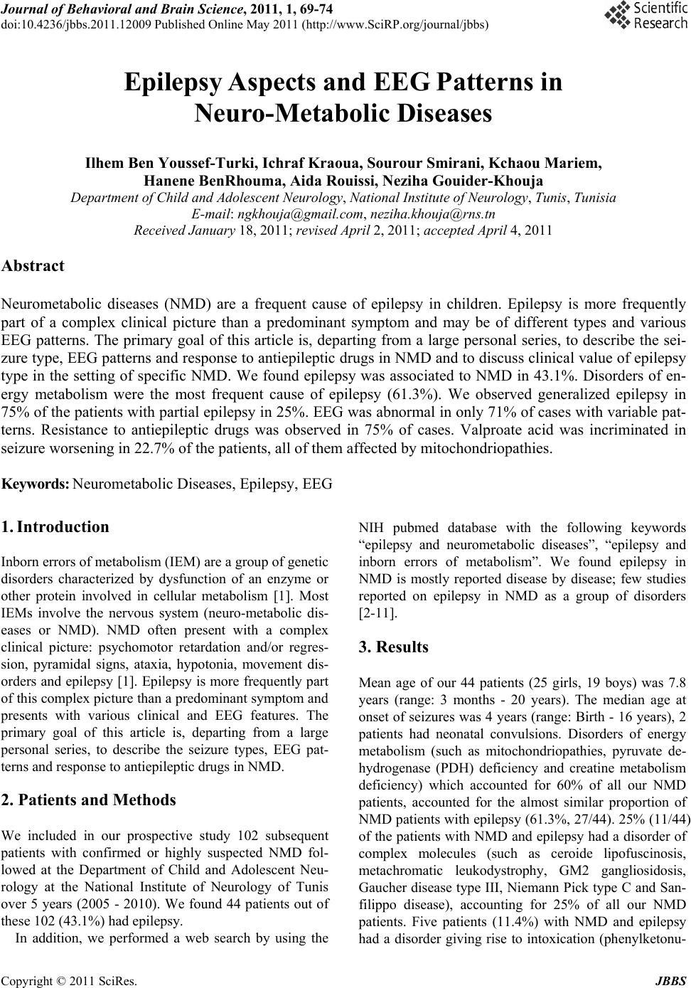 Journal of Behavioral and Brain Science, 2011, 1, 69-74 doi:10.4236/jbbs.2011.12009 Published Online May 2011 (http://www.SciRP.org/journal/jbbs) Copyright © 2011 SciRes. JBBS Epilepsy Aspects and EEG Patterns in Neuro-Metabolic Diseases Ilhem Ben Youssef-Turki, Ichraf Kraoua, Sourour Smirani, Kchaou Mariem, Hanene BenRhouma, Aida Rouissi, Neziha Gouider-Khouja Department of Chi l d an d Ad ol escent Neurology , National Institute of Neu rology, Tunis, Tunisia E-mail: [email protected], [email protected] Received January 18, 2011; revised April 2, 2011; accepted April 4, 2011 Abstract Neurometabolic diseases (NMD) are a frequent cause of epilepsy in children. Epilepsy is more frequently part of a complex clinical picture than a predominant symptom and may be of different types and various EEG patterns. The primary goal of this article is, departing from a large personal series, to describe the sei- zure type, EEG patterns and response to antiepileptic drugs in NMD and to discuss clinical value of epilepsy type in the setting of specific NMD. We found epilepsy was associated to NMD in 43.1%. Disorders of en- ergy metabolism were the most frequent cause of epilepsy (61.3%). We observed generalized epilepsy in 75% of the patients with partial epilepsy in 25%. EEG was abnormal in only 71% of cases with variable pat- terns. Resistance to antiepileptic drugs was observed in 75% of cases. Valproate acid was incriminated in seizure worsening in 22.7% of the patients, all of them affected by mitochondriopathies. Keywords: Neurometabolic Diseases, Epilepsy, EEG 1. Introduction Inborn errors of metabolism (IEM) are a group of genetic disorders characterized by dysfunction of an enzyme or other protein involved in cellular metabolism [1]. Most IEMs involve the nervous system (neuro-metabolic dis- eases or NMD). NMD often present with a complex clinical picture: psychomotor retardation and/or regres- sion, pyramidal signs, ataxia, hypotonia, movement dis- orders and epilepsy [1]. Epilepsy is more frequently part of this complex picture than a predominant symptom and presents with various clinical and EEG features. The primary goal of this article is, departing from a large personal series, to describe the seizure types, EEG pat- terns and response to antiepileptic drugs in NMD. 2. Patients and Methods We included in our prospective study 102 subsequent patients with confirmed or highly suspected NMD fol- lowed at the Department of Child and Adolescent Neu- rology at the National Institute of Neurology of Tunis over 5 years (2005 - 2010). We found 44 patients out of these 102 (43.1%) had epilepsy. In addition, we performed a web search by using the NIH pubmed database with the following keywords “epilepsy and neurometabolic diseases”, “epilepsy and inborn errors of metabolism”. We found epilepsy in NMD is mostly reported disease by disease; few studies reported on epilepsy in NMD as a group of disorders [2-11]. 3. Results Mean age of our 44 patients (25 girls, 19 boys) was 7.8 years (range: 3 months - 20 years). The median age at onset of seizures was 4 years (range: Birth - 16 years), 2 patients had neonatal convulsions. Disorders of energy metabolism (such as mitochondriopathies, pyruvate de- hydrogenase (PDH) deficiency and creatine metabolism deficiency) which accounted for 60% of all our NMD patients, accounted for the almost similar proportion of NMD patients with epilepsy (61.3%, 27/44). 25% (11/44) of the patients with NMD and epilepsy had a disorder of complex molecules (such as ceroide lipofuscinosis, metachromatic leukodystrophy, GM2 gangliosidosis, Gaucher disease type III, Niemann Pick type C and San- filippo disease), accounting for 25% of all our NMD patients. Five patients (11.4%) with NMD and epilepsy had a disorder giving rise to intoxication (phenylketonu- 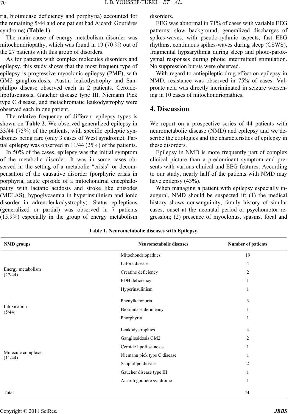 70 I. B. YOUSSEF-TURKI ET AL. ria, biotinidase deficiency and porphyria) accounted for the remaining 5/44 and one patient had Aicardi Goutières syndrome) (Table 1). The main cause of energy metabolism disorder was mitochondriopathy, which was found in 19 (70 %) out of the 27 patients with this group of disorders. As for patients with complex molecules disorders and epilepsy, this study shows that the most frequent type of epilepsy is progressive myoclonic epilepsy (PME), with GM2 gangliosidosis, Austin leukodystrophy and San- philipo disease observed each in 2 patients. Ceroide- lipofuscinosis, Gaucher disease type III, Niemann Pick type C disease, and metachromatic leukodystrophy were observed each in one patient. The relative frequency of different epilepsy types is shown on Table 2. We observed generalized epilepsy in 33/44 (75%) of the patients, with specific epileptic syn- dromes being rare (only 3 cases of West syndrome). Par- tial epilepsy was observed in 11/44 (25%) of the patients. In 50% of the cases, epilepsy was the initial symptom of the metabolic disorder. It was in some cases ob- served in the setting of a metabolic “crisis” or decom- pensation of the causative disorder (porphyric crisis in porphyria, acute episode of a mitochondrial encephalo- pathy with lactatic acidosis and stroke like episodes (MELAS), hypoglycaemia in hyperinsulinism and ionic disorder in adrenoleukodystrophy). Status epilepticus (generalized or partial) was observed in 7 patients (15.9%) especially in the group of energy metabolism disorders. EEG was abnormal in 71% of cases with variable EEG patterns: slow background, generalized discharges of spikes-waves, with pseudo-rythmic aspects, fast EEG rhythms, continuous spikes-waves during sleep (CSWS), fragmental hypsarythmia during sleep and photo-parox- ysmal responses during photic intermittent stimulation. No suppression bursts were observed. With regard to antiepileptic drug effect on epilepsy in NMD, resistance was observed in 75% of cases. Val- proate acid was directly incriminated in seizure worsen- ing in 10 cases of mitochondriopathies. 4. Discussion We report on a prospective series of 44 patients with neurometabolic disease (NMD) and epilepsy and we de- scribe the etiologies and the characteristics of epilepsy in these disorders. Epilepsy in NMD is more frequently part of complex clinical picture than a predominant symptom and pre- sents with various clinical and EEG features. According to our study, nearly half of the patients with NMD may have epilepsy (43%). When managing a patient with epilepsy especially in- augural, NMD should be suspected if: (1) the medical history shows consanguinity, family history of similar cases, onset at the neonatal period or psychomotor re- gression; (2) presence of myoclonus, spasms, focal and Table 1. Neurometabolic diseases with Epilepsy. NMD groups Neurometabolic diseases Numbe r of patients Mitochondriopathies 19 Lafora disease 4 Creatine deficiency 2 PDH deficiency 1 Energy metabolism (27/44) Hyperinsulinism 1 Phenylketonuria 3 Biotinidase deficiency 1 Intoxication (5/44) Phorphyria 1 Leukodystrophies 4 Gangliosidosis GM2 2 Ceroide lipofuscinosis 1 Niemann pick type C disease 1 Sanphilipo disease 2 Gaucher disease type III 1 Molecule complexe (11/44) Aicardi goutière syndrome 1 Total 44 Copyright © 2011 SciRes. JBBS 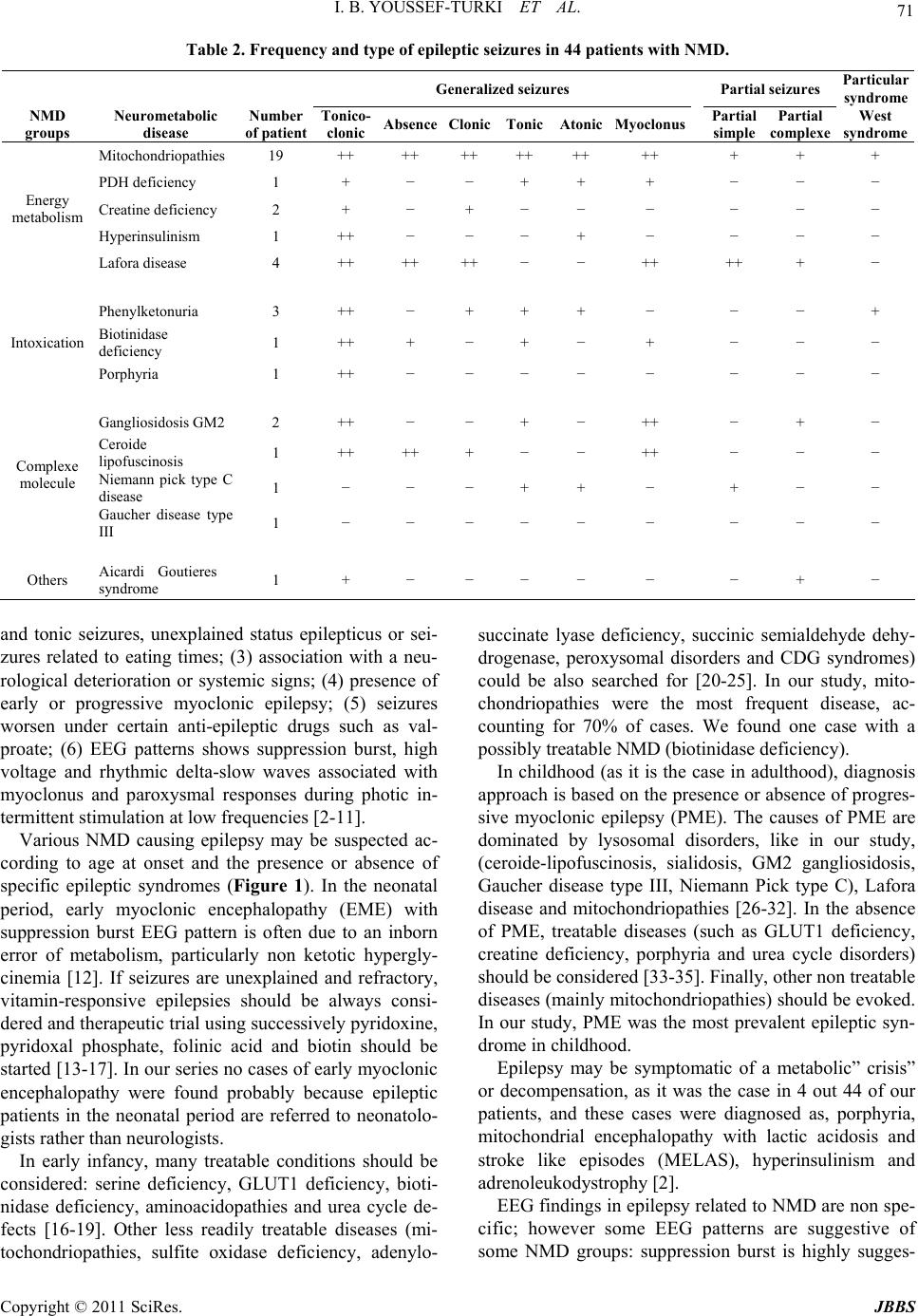 I. B. YOUSSEF-TURKI ET AL. 71 Table 2. Frequency and type of epileptic seizures in 44 patients with NMD. Generalized seizures Partial seizures Particular syndrome NMD groups Neurometabolic disease Number of patient Tonico- clonic AbsenceClonicTonicAtonicMyoclonus Partial simple Partial complexe West syndrome Mitochondriopathies19 ++ ++ ++ ++ ++ ++ + + + PDH deficiency 1 + − − + + + − − − Creatine deficiency 2 + − + − − − − − − Hyperinsulinism 1 ++ − − − + − − − − Energy metabolism Lafora disease 4 ++ ++ ++ − − ++ ++ + − Phenylketonuria 3 ++ − + + + − − − + Biotinidase deficiency 1 ++ + − + − + − − − Intoxication Porphyria 1 ++ − − − − − − − − Gangliosidosis GM22 ++ − − + − ++ − + − Ceroide lipofuscinosis 1 ++ ++ + − − ++ − − − Niemann pick type C disease 1 − − − + + − + − − Complexe molecule Gaucher disease type III 1 − − − − − − − − − Others Aicardi Goutieres syndrome 1 + − − − − − − + − and tonic seizures, unexplained status epilepticus or sei- zures related to eating times; (3) association with a neu- rological deterioration or systemic signs; (4) presence of early or progressive myoclonic epilepsy; (5) seizures worsen under certain anti-epileptic drugs such as val- proate; (6) EEG patterns shows suppression burst, high voltage and rhythmic delta-slow waves associated with myoclonus and paroxysmal responses during photic in- termittent stimulation at low frequencies [2-11]. Various NMD causing epilepsy may be suspected ac- cording to age at onset and the presence or absence of specific epileptic syndromes (Figure 1). In the neonatal period, early myoclonic encephalopathy (EME) with suppression burst EEG pattern is often due to an inborn error of metabolism, particularly non ketotic hypergly- cinemia [12]. If seizures are unexplained and refractory, vitamin-responsive epilepsies should be always consi- dered and therapeutic trial using successively pyridoxine, pyridoxal phosphate, folinic acid and biotin should be started [13-17]. In our series no cases of early myoclonic encephalopathy were found probably because epileptic patients in the neonatal period are referred to neonatolo- gists rather than neurologists. In early infancy, many treatable conditions should be considered: serine deficiency, GLUT1 deficiency, bioti- nidase deficiency, aminoacidopathies and urea cycle de- fects [16-19]. Other less readily treatable diseases (mi- tochondriopathies, sulfite oxidase deficiency, adenylo- succinate lyase deficiency, succinic semialdehyde dehy- drogenase, peroxysomal disorders and CDG syndromes) could be also searched for [20-25]. In our study, mito- chondriopathies were the most frequent disease, ac- counting for 70% of cases. We found one case with a possibly treatable NMD (biotinidase deficiency). In childhood (as it is the case in adulthood), diagnosis approach is based on the presence or absence of progres- sive myoclonic epilepsy (PME). The causes of PME are dominated by lysosomal disorders, like in our study, (ceroide-lipofuscinosis, sialidosis, GM2 gangliosidosis, Gaucher disease type III, Niemann Pick type C), Lafora disease and mitochondriopathies [26-32]. In the absence of PME, treatable diseases (such as GLUT1 deficiency, creatine deficiency, porphyria and urea cycle disorders) should be considered [33-35]. Finally, other non treatable diseases (mainly mitochondriopathies) should be evoked. In our study, PME was the most prevalent epileptic syn- drome in childhood. Epilepsy may be symptomatic of a metabolic” crisis” or decompensation, as it was the case in 4 out 44 of our patients, and these cases were diagnosed as, porphyria, mitochondrial encephalopathy with lactic acidosis and stroke like episodes (MELAS), hyperinsulinism and adrenoleukodystrophy [2]. EEG findings in epilepsy related to NMD are non spe- cific; however some EEG patterns are suggestive of some NMD groups: suppression burst is highly sugges- Copyright © 2011 SciRes. JBBS 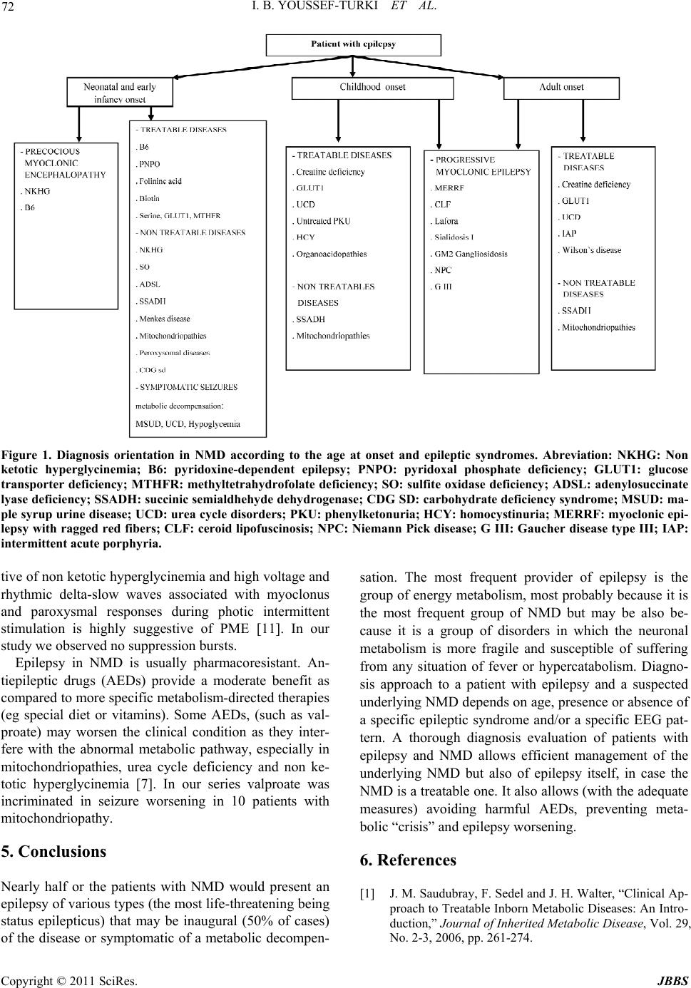 I. B. YOUSSEF-TURKI ET AL. Copyright © 2011 SciRes. JBBS 72 Figure 1. Diagnosis orientation in NMD according to the age at onset and epileptic syndromes. Abreviation: NKHG: Non ketotic hyperglycinemia; B6: pyridoxine-dependent epilepsy; PNPO: pyridoxal phosphate deficiency; GLUT1: glucose transporter deficiency; MTHFR: methyltetrahydrofolate deficiency; SO: sulfite oxidase deficiency; ADSL: adenylosuccinate lyase deficiency; SSADH: succ inic semialdhehyde dehydr ogenase; CDG SD: carbohydrate deficiency syndrome; MSUD: ma- ple syrup urine disease; UCD: urea cycle disorders; PKU: phenylketonuria; HCY: homocystinuria; MERRF: myoclonic epi- lepsy with ragged red fibers; CLF: ceroid lipofuscinosis; NPC: Niemann Pick disease; G III: Gaucher disease type III; IAP: intermittent acute porphyria. tive of non ketotic hyperglycinemia and high voltage and rhythmic delta-slow waves associated with myoclonus and paroxysmal responses during photic intermittent stimulation is highly suggestive of PME [11]. In our study we observed no suppression bursts. Epilepsy in NMD is usually pharmacoresistant. An- tiepileptic drugs (AEDs) provide a moderate benefit as compared to more specific metabolism-directed therapies (eg special diet or vitamins). Some AEDs, (such as val- proate) may worsen the clinical condition as they inter- fere with the abnormal metabolic pathway, especially in mitochondriopathies, urea cycle deficiency and non ke- totic hyperglycinemia [7]. In our series valproate was incriminated in seizure worsening in 10 patients with mitochondriopathy. 5. Conclusions Nearly half or the patients with NMD would present an epilepsy of various types (the most life-threatening being status epilepticus) that may be inaugural (50% of cases) of the disease or symptomatic of a metabolic decompen- sation. The most frequent provider of epilepsy is the group of energy metabolism, most probably because it is the most frequent group of NMD but may be also be- cause it is a group of disorders in which the neuronal metabolism is more fragile and susceptible of suffering from any situation of fever or hypercatabolism. Diagno- sis approach to a patient with epilepsy and a suspected underlying NMD depends on age, presence or absence of a specific epileptic syndrome and/or a specific EEG pat- tern. A thorough diagnosis evaluation of patients with epilepsy and NMD allows efficient management of the underlying NMD but also of epilepsy itself, in case the NMD is a treatable one. It also allows (with the adequate measures) avoiding harmful AEDs, preventing meta- bolic “crisis” and epilepsy worsening. 6. References [1] J. M. Saudubray, F. Sedel and J. H. Walter, “Clinical Ap- proach to Treatable Inborn Metabolic Diseases: An Intro- duction,” Journal of Inherited Metabolic Disease, Vol. 29, No. 2-3, 2006, pp. 261-274.  I. B. YOUSSEF-TURKI ET AL. 73 3. doi:10.1007/s10545-006-0358-0 [2] N. Bahi-Buisson, K. Mention, P. L. Léger, et al., “Neona- tal Epilepsy and Inborn Errors of Metabolism,” Archives de Pédiatrie, Vol. 13, No. 3, 2006, pp. 284-292. [3] D. C. De Vivo, “Inherited Metabolic Disorders and Sei- zures in Infancy,” Journal of Child Neurology, Vol. 17, 2002, pp. 3S1-3S2. [4] D. R. Jr. Nordli and D. C. De Vivo, “Classification of Infantile Seizures: Implications for Identification and Treatment of Inborn Errors of Metabolism,” Journal of Child Neurology, Vol. 17, 2002, pp. 3S3-3S7. [5] J. M. Pascual, J. Campistol and A. Gil-Nagel, “Epilepsy in Inherited Metabolic Disorders,” Neurologist, Vol. 14, 2008, pp. S2-S14. [6] P. L. Pearl, H. D. Bennett and Z. Khademian, “Seizures and Metabolic Disease,” Current Neurology and Neuro- science Reports, Vol. 5, No. 2, 2005, pp. 127-133. [7] F. Sedel, I. Gourfinkel-An, O. Lyon-Caen, M. Baulac, J. M. Saudubray and V. Navarro, “Epilepsy and Inborn Er- rors of Metabolism in Adults: A Diagnostic Approach,” Journal of Inherited Metabolic Disease, Vol. 30, No. 6, 2007, pp. 846-854. doi:10.1007/s10545-007-0723-7 [8] S. Stöckler-Ipsiroglu and B. Plecko, “Metabolic Epilepsies: Approaches to a Diagnostic Challenge,” Canadian Jour- nal of Neurological Sciences, Vol. 36, 2009, pp. S67-S72. [9] F. Vigevano and A. Bartuli, “Infantile Epileptic Syn- dromes and Metabolic Etiologies,” Journal of Child Neurology, Vol. 17, 2002, pp. S9-S1 [10] N. I. Wolf, T. Bast and R. Surtees, “Epilepsy in Inborn Errors of Metabolism,” Epileptic Di sorders, Vol. 7, No. 2, 2005, pp. 67-81. [11] N. I. Wolf, A. García-Cazorla and G. F. Hoffmann, “Epi- lepsy and Inborn Errors of Metabolism in Children,” Journal of Inherited Metabolic Disease, Vol. 32, No. 5, 2009, pp. 609-617. [12] D. A. Applegarth and J. R Toone, “Glycine Encepha- Lopathy (Nonketotic Hyperglycinaemia): Review and Update,” Journal of Inherited Metabolic Disease, Vol. 27, No. 3, 2004, pp. 417-422. [13] P. Baxter, P. Griffiths, T. Kelly, et al., “Pyridoxine-depen- dent Seizures: Demographic, Clinical, MRI and Psycho- metric Features, and Effect of Dose on Intelligence Quo- tient,” Developmental Medicine & Child Neurology, Vol. 38, No. 11, 1996, pp. 998-1006. [14] M. F. Kuo and H. S. Wang, “Pyridoxal Phosphate-res- ponsive Epilepsy with Resistance to Pyridoxine,” Pedia- tric Neurology, Vol. 26, No. 2, 2002, pp. 146-147. doi:10.1016/S0887-8994(01)00357-5 [15] O. A. Torres, V. S. Miller, N. M. Buist, et al., “Folinic Acid-responsive Neonatal Seizures,” Journal of Child Neurology, Vol. 14, 1999, pp. 529-532. doi:10.1177/088307389901400809 [16] J. E. Collins, N. S. Nicholson, N. Dalton, et al., “Biotini- dase Deficiency: Early Neurological Presentation,” De- velopmental Medicine & Child Neurology, Vol. 36, 1994, pp. 268-270. [17] B. A. Salbert, J. M. Pellock and B. Wolf, “Characteriza- tion of Seizures Associated with Biotinidase Deficiency,” Neurology, Vol. 43, No. 7, 1993, pp.1351-1355. [18] T. J. De Koning and L. W. Klomp, “Serine-deficiency Syndromes,” Current Opinion in Neurology, Vol. 17, No. 2, 2004, pp. 197-204. doi:10.1097/00019052-200404000-00019 [19] K. Brockmann, D. Wang, C. G. Korenke, et al., “Auto- somal Dominant Glut-1 Deficiency Syndrome and Familial Epilepsy,” Annals of Neurology, Vol. 50, 2001, pp. 476-485. doi:10.1002/ana.1222 [20] J. Finsterer, “Central Nervous System Manifestations of Mitochondrial Disorders,” Acta Neurologica Scandi- navica, Vol. 114, 2006, pp. 217-238. [21] A. B. Dublin, J. K. Hald and S. L. Wootton-Gorges, “Isolated Sulfite Oxidase Deficiency: MR Imaging Features,” American Journal of Neuroradiology, Vol. 23, No. 3, 2002, pp. 484-485. [22] G. Van den Berghe, M. F. Vincent and J. Jaeken, “Inborn Errors of the Purine Nucleotide Cycle: Adenylosuccinase Deficiency,” Journal of Inherited Metabolic Disease, Vol. 20, No. 2, 1997, pp.193-202. doi:10.1023/A:1005304722259 [23] P. L. Pearl, K. M. Gibson, M. T. Acosta, et al., “Clinical Spectrum of Succinic Semialdehyde Dehydrogenase Defi- ciency,” Neurology, Vol. 60, No. 9, 2003, pp.1413-1417. [24] Y. Takahashi, Y. Suzuki, K. Kumazaki, et al., “Epilepsy in Peroxisomal Diseases,” Epilepsia, Vol. 38, No. 2, 1997, pp. 182-188. doi:10.1111/j.1528-1157.1997.tb01095.x [25] T. Marquardt and J. Denecke, “Congenital Disorders of Glycosylation: Review of Their Molecular Bases, Clinical Presentations and Specific Therapies,” European Journal of Pediatrics, Vol. 162, No. 6, 2003, pp. 359-379. [26] A. Shahwan, M. Farrell and N. Delanty, “Progressive Myoclonic Epilepsies: A Review of Genetic and Thera- peutic Aspects,” The Lancet Neurology, Vol. 4, No. 4, 2005, pp. 239-248. doi:10.1016/S1474-4422(05)70043-0 [27] K. E. Wisniewski, N. Zhong and M. Philippart, “Pheno/genotypic Correlations of Neuronal Ceroid Lipo- fuscinoses,” Neurology, Vol. 57, No. 4 , 2001, pp. 576-581. [28] J. A. Lowden and J. S. O’Brien, “Sialidosis: A Review of Human Neuraminidase Deficiency,” American Journal of Human Genetics, Vol. 31, No. 1, 1979, pp. 1-18. [29] A. Federico, S. Battistini, S. G. Ciacci, et al., “Cherry-red Spot Myoclonus Syndrome (Type I Sialidosis),” Develop- mental Neuroscience, Vol. 13, 1991, pp. 320-326. [30] J. K. Park, E. Orvisky, N. Tayebi, et al., “Myoclonic Epilepsy in Gaucher Disease: Genotype-phenotype Insights from a Rare Patient Subgroup,” Pediatric Research, Vol. 53, No. 3, 2003, pp. 387-395. [31] M. Sevin, G. Lesca, N. Baumann, et al., “The Adult form of Niemann-Pick Disease Type C,” Brain, Vol. 130, No. 1, 2007, pp.120-133. doi:10.1093/brain/awl260 [32] S. Ganesh, R. Puri, S. Singh, S. Mittal and D. Dubey, “Recent Advances in the Molecular Basis of Lafora’s Progressive Myoclonus Epilepsy,” Journal of Human Genetics, Vol. 51, No. 1, 2006, pp. 1-8. [33] V. Leuzzi, “Inborn Errors of Creatine Metabolism and Copyright © 2011 SciRes. JBBS  I. B. YOUSSEF-TURKI ET AL. Copyright © 2011 SciRes. JBBS 74 Epilepsy: Clinical Features, Diagnosis, and Treatment,” Journal of Child Neurology, Vol. 17, 2002, pp. S89-S97. [34] A. S. Winkler, T. J. Peters and R. D. Elwes, “Neuropsy- chiatric Porphyria in Patients with Refractory Epilepsy: Report of Three Cases,” Journal of Neurology, Neuro- surgery & Psychiatry, Vol. 76, No. 3, 2005, pp. 380-383. [35] G. M. Enns, “Neurologic Damage and Neurocognitive Dysfunction in Urea Cycle Disorders,” Seminars in Pedi- atric Neurology, Vol. 15, No. 3, 2008, pp. 132-139. doi:10.1016/j.spen.2008.05.007 |