

Paper Menu >>
Journal Menu >>
 Advances in Molecular Imaging, 2011, 1, 12-15 doi:10.4236/ami.2011.11002 Published Online April 2011 (http://www.SciRP.o rg/journal/ami) Copyright © 2011 SciRes . AMI Incidental Uptake of In-111 Ibritumomab Tiuxetan in Surgically Tre ated Fracture Hidenari Hirata1, Makoto Nakagawa1, Koichiro Abe2, Takashi Okafuji1, Kenji Shinozaki1, Ilseung Choi3, Naokuni Uike3, Shuji Sakai1 1Department of Radiology, National Kyushu Cancer Center, Fukuoka, Japan 2Department of Clinical Radiology, Graduate School of Medical Sciences, Kyushu Universit y, Fukuoka, Japan 3Department of Hematology, National Kyushu Cancer Center, Fukuoka, Japan E-mail: k-[email protected]-u.ac.jp Received March 5, 2011; Revised April 5, 2011; Accepted April 30, 2011 Abstract In-111 ibritumomab tiuxetan pretherapy imaging is performed prior to radioimmunotherapy with Y-90 ibri- tumomab tiuxetan in patients with lymphoma in order to estimate the biodistribution of the radiolabeled an- tibodies. We report the case of a 61-year-old woman with relapsed follicular lymphoma. In-111 ibritumomab tiuxetan pretherapy imaging showed tracer accumulation in a surgically treated fracture of the left calcaneus as well as in the involved lesions. The patient had fractured her left calcaneus 6 months before the radioim- munotherapy, and fracture of the left calcaneus without lymphoma involvement had been revealed opera- tively at that time. Clinical features and magnetic resonance imaging (MRI) indicated an inflammatory re- sponse in the left heel. We concluded that this inflammation played an important role in the uptake on pre- therapy imaging. It should be kept in mind that the inflammation is a differential diagnosis of lymphoma in- volvement in In-111 ibritumomab tiuxetan pretherapy imaging. To the best of our knowledge, this is the first case report of In-111 ibritumomab tiuxetan uptake in a surgically treated fracture. Keywords: In-111, Ibritumomab, Fracture, Lymphoma 1. Introduction In January 2008, radioimmunotherapy with Y-90 ibritu- momab tiu xeta n was a ppr ove d in J apa n. T his is a n ef fec- tive treatment for refractory or relapsed non-Hodgkin’s lymphoma [1]. It is composed of a radionuclide conju- gated to the monoclonal antibody targeting CD20 on the surface of lymphocytes. The beta emission from Y-90 induces cellular damage in the target. In ad dition to that, radiation damage can be achieved in neighboring cells that not express the antigen or that compose poorly vas- cularized bulky tumors. The main toxicity is myelosup- pression. In order to predict toxicities, estimating the biodistribution of the radiolabeled antibodies is impor- tant. The gamma emitter In-111-labeled ibritumomab tiuxetan is generally administered prior to the pure beta emitter Y-90-labeled ibritumomab tiuxetan because of the impossibility of obtaining a drug distribution image by the beta emitter. Patients with altered biodistribution (e.g. prominent bone marrow uptake) on In-111 ibritu- momab tiuxetan pretherapy imaging cannot receive Y-90 ibritumomab tiuxetan. Here we report a patient who underwent radioimmu- notherapy with Y-90 ibritumomab tiuxetan for relapsed follicular lymphoma. In this patient, pretherapy imaging using In-111 ibritumomab tiuxetan demonstrated uptake not only in the involved lesions, but also in a surgically treated fracture. To the best of our knowledge, this is the first case report of In-111 ibritumomab tiuxetan uptake in a surgically treated fracture. 2. Case Report A 61-year-old woman was diagnosed with stage IVA follicular lymphoma, and complete remission was at- tained after eight cycles of R-CHOP (rituximab, cyclo- phosphamide, doxorubicin, vincristine, and prednisolone) chemotherapy in another hospital. Four years after fi- nishing R-CHOP chemotherapy, computed tomography (CT) demonstrated enlargement of her mesenteric and left internal iliac lymph nodes. During a 5-month “wait- and-see” period, CT showed an increase in the size of 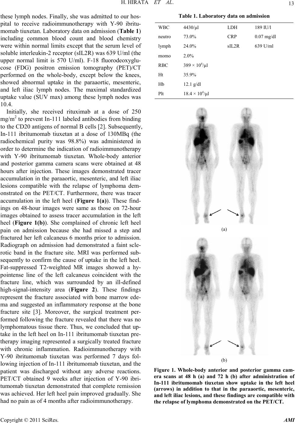 H. HIRATA ET AL. Copyright © 2011 SciRes . AMI 13 these l ymph no des. Finall y, she was ad mitted to our ho s- pital to receive radioimmunotherapy with Y-90 ibritu- momab tiuxetan. Laboratory data on admission (Tab le 1 ) including common blood count and blood chemistry were within normal limits except that the serum level of solub le i nte rl e uki n -2 receptor ( sI L2 R) was 639 U/ml (the upper normal limit is 570 U/ml). F-18 fluorodeoxyglu- cose (FDG) positron emission tomography (PET)/CT performed on the whole-body, except below the knees, showed abnormal uptake in the paraaortic, mesenteric, and left iliac lymph nodes. The maximal standardized uptake value (SUV max) among these lymph nodes was 10.4. Initially, she received rituximab at a dose of 250 mg/ m 2 t o pr event In -111 labeled antibodies from binding to the CD20 antigens of normal B cells [2]. Subsequently, In-111 ibritumomab tiuxetan at a dose of 130MBq (the radiochemical purity was 98.8%) was administered in order to determine the indication of radioimmunotherapy with Y-90 ibritumomab tiuxetan. Whole-body anterior and posterior gamma camera scans were obtained at 48 hours after injection. These images demonstrated tracer accumulation in the paraaortic, mesenteric, and left iliac lesions compatible with the relapse of lymphoma dem- onstrated on the PET/CT. Furthermore, there was tracer accumulation in the left heel (Figure 1(a)). These find- ings on 48-hour images were same as those on 72-hour images obtained to assess tracer accumulation in the left heel (Figure 1(b)). She complained of chronic left heel pain on admission because she had missed a step and fractured her left calcaneus 6 months prior to admission. Radiograph on admission had demonstrated a faint scle- rotic band in the fracture site. MRI was performed sub- sequently to confirm the cause of uptake in the left heel. Fat-suppressed T2-weighted MR images showed a hy- pointense line of the left calcaneus coincident with the fracture line, which was surrounded by an ill-defined high -s i g na l-intensity area (Figure 2). These findings represent the fracture associated with bone marrow ede- ma and suggested an inflammatory response at the bone fracture site [3]. Moreover, the surgical treatment per- formed following the fracture revealed that there was no lymphomatous tis sue there. Thus, we concluded t hat up- take i n the le ft heel o n In-111 ibritumomab tiuxetan pre- therapy imaging represented a surgically treated fracture with chronic inflammation. Radioimmunotherapy with Y-90 ibritumomab tiuxetan was performed 7 days fol- lo wing i nje ctio n of I n-111 ibritumomab tiuxetan, and the patient was discharged without any adverse reactions. PET/CT obtained 9 weeks after injection of Y-90 ibri- tumomab tiuxetan demonstrated that complete remission was achieved. Her left heel pain improved gradually. She had no pain as of 4 months after radioimmunotherapy. Table 1. L ab orat ory data on ad miss io n WBC 4430 /μl LDH 189 IU/l neutro 73.0% CRP 0.07 mg/dl lymph 24.0% sIL2R 639 U/ml momo 2.0% RBC 389 × 104/μl Ht 35. 9% Hb 12.1 g/dl Plt 1 8. 4 × 10 4/μl (a) (b) Figure 1. Whole-body anterior and posterior gamma cam- era scans at 48 h (a) and 72 h (b) after administration of In-111 ibritumomab tiuxetan show uptake in the left heel (arrows) in addition to that in the paraaortic, mesenteric, and le ft i liac lesi on s, a nd t hese fi nding s are co mpat ibl e w ith the relapse of lymphoma demonstrated on the PET/CT. 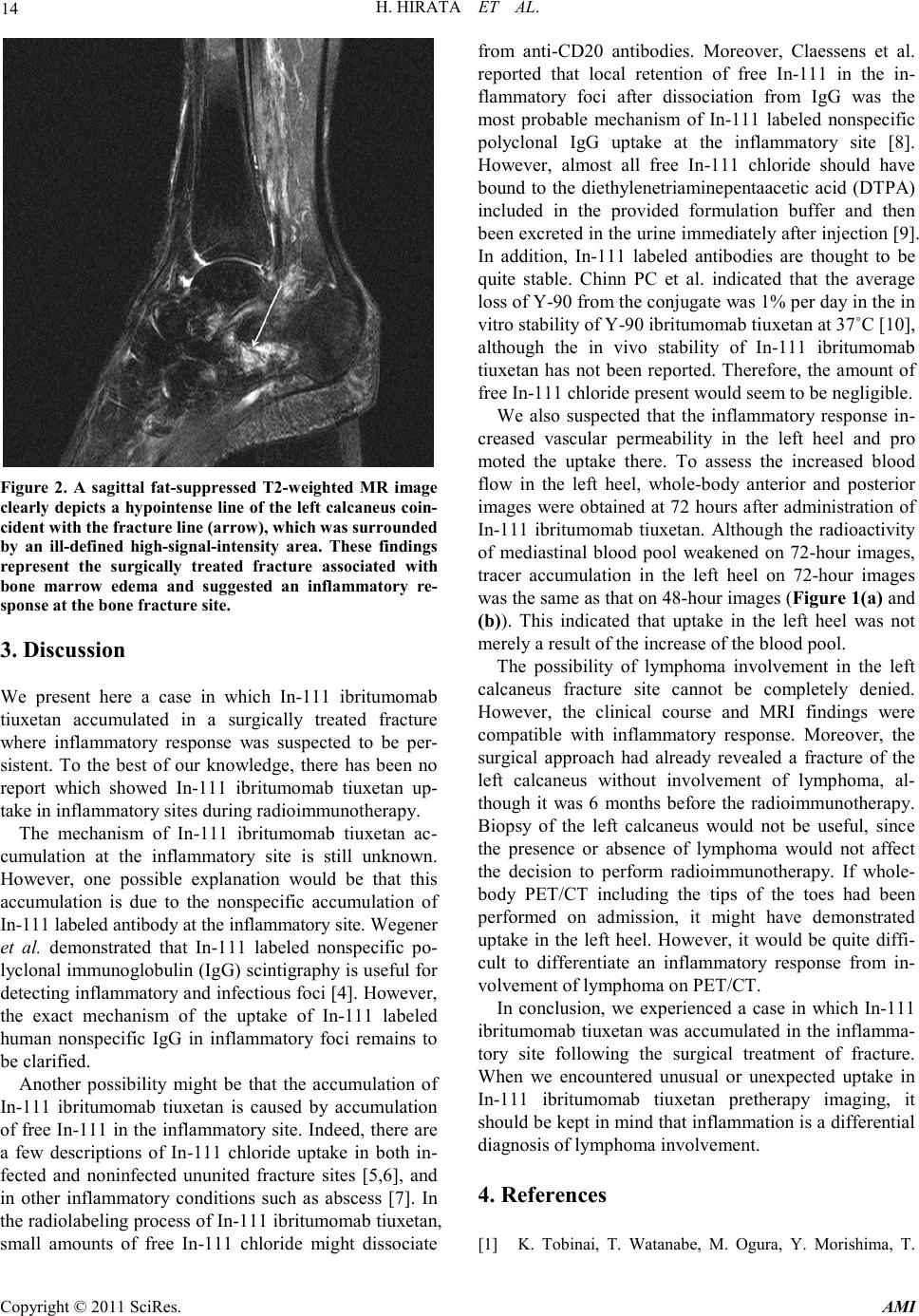 H. HIRATA ET AL. Copyright © 2011 SciRes . A MI 14 Figure 2. A sagittal fat-suppressed T2-weighted MR image clearly depicts a hypointense line of the left cal caneus coin- cident with the fracture line (arr o w), w hich was surrounded by an ill-defined high-signal-intensity area. These findings represent the surgically treated fracture associated with bone marrow edema and suggested an inflammatory re- sponse at the bone fracture site. 3. Discussion We present here a case in which In-111 ibritumomab tiuxetan accumulated in a surgically treated fracture where inflammatory response was suspected to be per- sistent. To the best of our knowledge, there has been no report which showed In-111 ibritumomab tiuxetan up- take i n in fl ammato ry sites during ra dio immu not herapy. The mechanism of In-111 ibritumomab tiuxetan ac- cumulation at the inflammatory site is still unknown. However, one possible explanation would be that this accumulation is due to the nonspecific accumulation of In-111 labeled antibody at the inflammatory site. Wegener et al. demonstrated that In-111 labeled nonspecific po- lyclonal immunoglobulin (IgG) scintigraphy is useful for detecting infla mmator y and infectiou s foci [4] . However, the exact mechanism of the uptake of In-111 labeled human nonspecific IgG in inflammatory foci remains to be clarified. Another possibility might be that the accumulation of In-111 ibritumomab tiuxetan is caused by accumulation of fr ee In-111 in the inflammatory site. Indeed, there are a few descriptions of In-111 chloride uptake in both in- fected and noninfected ununited fracture sites [5,6], and in other inflammatory conditions such as abscess [7]. In the radiolabeling process of In-111 ibritumomab tiuxetan, small amounts of free In-111 chloride might dissociate from anti-CD20 antibodies. Moreover, Claessens et al. reported that local retention of free In-111 in the in- flammatory foci after dissociation from IgG was the most probable mechanism of In-111 labeled nonspecific polyclonal IgG uptake at the inflammatory site [8]. However, almost all free In-111 chloride should have bound to the diethylenetriaminepentaacetic acid (DTPA) included in the provided formulation buffer and then been excreted in the urine immediately after injection [9]. In addition, In-111 labeled antibodies are thought to be quite stable. Chinn PC et al. indicated that the average loss of Y -90 from the conjugate was 1% per day in the in vitro stabilit y o f Y-90 ibritumomab tiuxetan at 37˚C [10], although the in vivo stability of In-111 ibritumomab tiuxetan has not been reported. Therefore, the amount of free In-111 chloride present would seem to be negligible. We also suspected that the inflammatory response in- creased vascular permeability in the left heel and pro moted the uptake there. To assess the increased blood flow in the left heel, whole-body anterior and posterior images were obtained at 72 hours after administration of In-111 ibritumomab tiuxetan. Although the radioactivity of mediastinal blood pool weakened on 72-hour images, tracer accumulation in the left heel on 72-hour images was the same as that on 48-hour i ma ges (Figure 1(a) and (b)). This indicated that uptake in the left heel was not merely a result of the increase of the blood pool. The possibility of lymphoma involvement in the left calcaneus fracture site cannot be completely denied. However, the clinical course and MRI findings were compatible with inflammatory response. Moreover, the surgical approach had already revealed a fracture of the left calcaneus without involvement of lymphoma, al- though it was 6 months before the radioimmunotherapy. Biopsy of the left calcaneus would not be useful, since the presence or absence of lymphoma would not affect the decision to perform radioimmunotherapy. If whole- body PET/CT including the tips of the toes had been performed on admission, it might have demonstrated uptake in the left heel. However, it would be quite diffi- cult to differentiate an inflammatory response from in- volvement of lymphoma o n PET/CT. In conclusion, we experienced a case in which In-111 ibritumomab tiuxetan was accumulated in the inflamma- tory site following the surgical treatment of fracture. When we encountered unusual or unexpected uptake in In-111 ibritumomab tiuxetan pretherapy imaging, it should be kep t in mind that inflammation is a differe ntial diagnosis of lymphoma involvement. 4. Referen ces [1] K. Tobinai, T. Watanabe, M. Ogura, Y. Morishima, T.  H. HIRATA ET AL. Copyright © 2011 SciRes . AMI 15 Hotta, K. Ishizawa, K. Itoh, S. Okamoto, M. Taniwaki, N. Tsukamoto, H. Okumura, T. Terauchi, S. Nawano, M. Matsusako, Y. Matsuno, S. Nakamura, S. Mori, Y. Oha- shi, M. Hayashi and K. Endo, “Japanese Phase II Study of 90Y-Ibritumomab Tiuxetan in Patients with Relapsed or Refractory Indolent B-Cell Lymphoma,” Cancer Scien ce, Vol. 100, No. 1, 2009, pp. 158-164. doi:10.1111/j.1349-7006.2008.00999.x [2] S. J. Knox, M. L. Goris, K. Trisler, R. Negrin, T. Davis, T. M. Liles, A. Grillo-López, P. Chinn, C. Varns, S. C. Ning, S. Fowler, N. Deb, M. Becker, C. Marquez and R. Levy, “Yttrium-90-Labeled Anti-CD20 Monoclonal An- tibody Therapy of Recurrent B-Cell Lymphoma,” Clini- cal Can cer Research , Vol. 2, No. 3, 1996, pp. 457-470. [3] J. A. Narváez, J. Narváez, R. Ortega, C. Aguilera, A. Sánch ez and E. And ía, “Pain ful Heel: MR Imaging Find- ings,” Radiographics, Vol. 20, No. 2, 2000, pp. 333-352. [4] W. A. Wegener, M. G. Velchik, D. Weiss, S. Ter, A. Byars, M. Neptune and A. Alavi, “Infectious Imaging with Indium-111-Labeled Non specific Polyclon al Human Immunoglobulin,” The Journal of Nuclear Medicine, Vol. 32, No. 11, 1991, pp. 2079-2085. [5] B. A. Sayle, H . D . F awcet t , W. M . Yud t , S . C . Wan g, J. T. Mader an d G. Cierny 3rd, “Indium-111 Chloride Imaging with Ununited Fractures,” Clinical Nuclear Medicine, Vol. 12, No. 3, 1987 , pp. 20 8-209. doi:10.1097/00003072-198703000-00010 [6] H. Oht a, “In-111 Chloride Uptake in Ununited Fractures: A Case Report,” Kaku Igaku (Japanese Journal of Nuc- lear Med icine), Vol. 37, No. 2, 20 00, pp. 121-124. [7] B. A. S ayle, S. B alachand ran an d C. A. Rogers, “Indium- 111 Chloride Imaging in Patients with Suspected Ab- scesses: Concise Communication,” The Journal of Nuc- lear Med icine, Vol. 24, No. 12, 1983, pp. 1114-1118. [8] R. A. Claessens, E. B. Koenders, O. C. Boerman, W. J. Oyen, G. F. Bor m, J. W. van d er Meer an d F. H. Corstens, “Dissociation of Indium from Indium-111-Labelled Die- thylene Triamine Penta-Acetic Acid Conjugated Non- Specific Polyclonal Human Immunoglobulin G in In- flammatory Foci,” European Journal of Nuclear Medi- cine, Vol. 22, No. 3, 1995, pp. 212-219. doi:10.1007/BF01081515 [9] M. Cremonesi, M. Ferrari, C. M. Grana, A. Vanazzi, M. Stabin, M. Bartolomei, S. Papi, G. Prisco, G. Martinelli, G. Paganelli and P. F. Ferrucci, “High-Dose Radioim- munotherapy with 90Y-Ibritumomab Tiuxetan: Compara- tive Dosimetric Study for Tailored Treatment,” The Journal of Nuclear Medicine, Vol. 48, No. 11, 2007, pp. 1871-1879. doi:10.2967/jnumed.107.044016 [10] P. C. Chinn, J. E. Leonard, J. Rosenberg, N. Hanna and D. R. Anderson, “Preclinical Evaluation of 90Y-Labeled Anti-CD20 Monoclonal Antibody for Treatment of Non-Hodgkin ’s Lymphoma,” International Journal of Oncology, Vol. 15, No. 5, 1999, pp. 1017-1025. |