

Paper Menu >>
Journal Menu >>
 Vol.3, No.4, 295-300 (2011) Natural Science http://dx.doi.org/10.4236/ns.2011.34038 Copyright © 2011 SciRes. OPEN ACCESS Possible mechanism of increasing resistance of the myocardium during combination of post infarction remodeling and diabetes mellitus Margarita V. Egorova*, Sergey A. Afanasiev, Dina S. Kondratyeva, Boris N. Kozlov, Sergey V. Popov Laboratory of Molecular Cell Pathology and Genetic Diagnosis, Research Institute of Cardiology, Siberian Branch of Russian Academy of Medical Science, Tomsk, Russia; *Corresponding Author: [email protected] Received 2 February 2011; revised 21 February 2011; accepted 13 March 2011. ABSTRACT It was shown that the energy metabolism of the heart mitochondria of experimental animals and patients is more resistant to damage at com- bined postinfarction cardiosclerosis and dia- betes in comparison with the individual pathol- ogy. We found that the changes of free fatty acid content and conjugation of the processes of oxidation and phosphorylation in heart mito- chondria are components of the metabolic sta- bility of myocardium at the combined develop- ment of postinfarction cardiosclerosis and dia- betes mellitus. Our data demonstrate a direct link between the violations of the processes of oxidative phosphorylation and accumulation of free fatty acids owing to change in activity of endogenous phospholipases, in particularly, mi- tochondrial phospholipase A2. Similar results were obtained for intraoperative biopsy speci- mens of patients’ hearts, and of adult Wistar rats’ hearts. We hypothesized that the preserva- tion of energy metabolism is a manifestation of summing up of compensatory processes at de- velopment of nonspecific response of cells to damage at the early stages of pathological pro- cess. Keywords: Postinfarction Remodeling; Diabetes; Heart Mitochondria; Fatty Acids; Phospholipase A2 1. INTRODUCTION It is well known that many pathologies of the cardio- vascular system are accompanied by increased activity of endogenous phospholipases and, as a consequence, by accumulation of free fatty acids [1] which, in turn, pro- vokes uncoupling of the processes of oxidation and phosphorylation in mitochondria [2]. A decrease of insu- lin action on adipose tissue at diabetes mellitus (DM) leads to the increased content of fatty acids in blood and their intake into the myocardial cells. Myocardial con- sequences of DM even in conditions of adequate oxygen supply of myocardium resemble metabolic imbalance in myocardium of patients with severe coronary heart dis- ease (CHD) [3,4]. At DM, as at CHD, the corresponding complex of pathophysiological changes appears either because of the inability of mitochondria to oxidize the entire volume of incoming fatty acids, or because of critical lowering of coronary blood flow (at CHD), or because of the enhanced transport of fatty acids in the cytosol (at DM) [3,4]. It is logical to suppose that hemodynamic changes on the DM background can in- crease the probability of lethal outcome that is confirmed by clinical studies [4,5]. However, there is data of manifestation of myocar- dium resistance to ischemia (in vivo and in vitro) of animals with short term of induced diabetes [6-8]: the induction of diabetes on the background of postinfarc- tion cardiosclerosis preserves, paradoxically, myocar- dium contractile properties. Animals with combined pa- thology are characterized by less pronounced changes in glucose levels, body weight and heart [9,10]. The purpose of this study was evaluation of influence of diabetes mellitus and postinfarction remodeling on ability to oxidative phosphorylation of isolated heart mi- tochondria of animals and humans both in the case of in- dividual pathologies and in the case of their combination. 2. EXPERIMENTAL PART 2.1. Materials The work was accomplished on mature male rats of Wistar and intraoperative biopsy samples of patients. 2.1.1. Animals 5 groups for 8 rats were formed from animals: 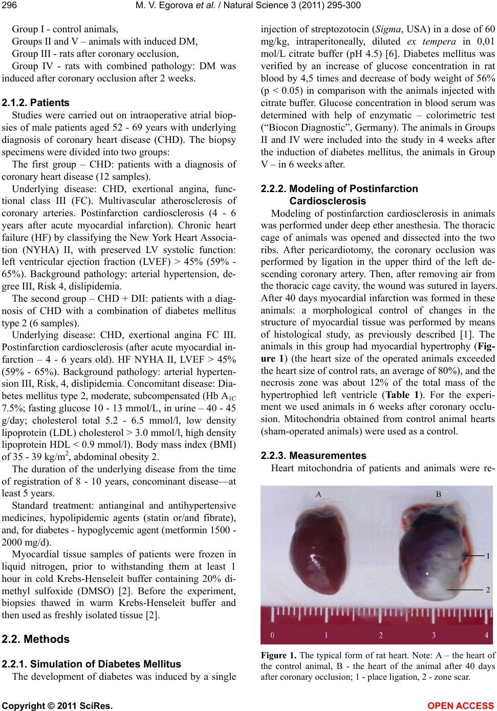 M. V. Egorova et al. / Natural Science 3 (2011) 295-300 Copyright © 2011 SciRes. OPEN ACCESS 296 Group I - control animals, Groups II and V – animals with induced DM, Group III - rats after coronary occlusion, Group IV - rats with combined pathology: DM was induced after coronary occlusion after 2 weeks. 2.1.2. Patients Studies were carried out on intraoperative atrial biop- sies of male patients aged 52 - 69 years with underlying diagnosis of coronary heart disease (CHD). The biopsy specimens were divided into two groups: The first group – CHD: patients with a diagnosis of coronary heart disease (12 samples). Underlying disease: CHD, exertional angina, func- tional class III (FC). Multivascular atherosclerosis of coronary arteries. Postinfarction cardiosclerosis (4 - 6 years after acute myocardial infarction). Chronic heart failure (HF) by classifying the New York Heart Associa- tion (NYHA) II, with preserved LV systolic function: left ventricular ejection fraction (LVEF) > 45% (59% - 65%). Background pathology: arterial hypertension, de- gree III, Risk 4, dislipidemia. The second group – CHD + DII: patients with a diag- nosis of CHD with a combination of diabetes mellitus type 2 (6 samples). Underlying disease: CHD, exertional angina FC III. Postinfarction cardiosclerosis (after acute myocardial in- farction – 4 - 6 years old). HF NYHA II, LVEF > 45% (59% - 65%). Background pathology: arterial hyperten- sion III, Risk, 4, dislipidemia. Concomitant disease: Dia- betes mellitus type 2, moderate, subcompensated (Hb A1C 7.5%; fasting glucose 10 - 13 mmol/L, in urine – 40 - 45 g/day; cholesterol total 5.2 - 6.5 mmol/l, low density lipoprotein (LDL) cholesterol > 3.0 mmol/l, high density lipoprotein HDL < 0.9 mmol/l). Body mass index (BMI) of 35 - 39 kg/m2, abdominal obesity 2. The duration of the underlying disease from the time of registration of 8 - 10 years, concominant disease—at least 5 years. Standard treatment: antianginal and antihypertensive medicines, hypolipidemic agents (statin or/and fibrate), and, for diabetes - hypoglycemic agent (metformin 1500 - 2000 mg/d). Myocardial tissue samples of patients were frozen in liquid nitrogen, prior to withstanding them at least 1 hour in cold Krebs-Henseleit buffer containing 20% di- methyl sulfoxide (DMSO) [2]. Before the experiment, biopsies thawed in warm Krebs-Henseleit buffer and then used as freshly isolated tissue [2]. 2.2. Methods 2.2.1. Simulation of Diabetes Mellitus The development of diabetes was induced by a single injection of streptozotocin (Sigma, USA) in a dose of 60 mg/kg, intraperitoneally, diluted ex tempera in 0,01 mol/L citrate buffer (pH 4.5) [6]. Diabetes mellitus was verified by an increase of glucose concentration in rat blood by 4,5 times and decrease of body weight of 56% (p < 0.05) in comparison with the animals injected with citrate buffer. Glucose concentration in blood serum was determined with help of enzymatic – colorimetric test (“Biocon Diagnostic”, Germany). The animals in Groups II and IV were included into the study in 4 weeks after the induction of diabetes mellitus, the animals in Group V – in 6 weeks after. 2.2.2. Modeling of Postinfarction Cardiosclerosis Modeling of postinfarction cardiosclerosis in animals was performed under deep ether anesthesia. The thoracic cage of animals was opened and dissected into the two ribs. After pericardiotomy, the coronary occlusion was performed by ligation in the upper third of the left de- scending coronary artery. Then, after removing air from the thoracic cage cavity, the wound was sutured in layers. After 40 days myocardial infarction was formed in these animals: a morphological control of changes in the structure of myocardial tissue was performed by means of histological study, as previously described [1]. The animals in this group had myocardial hypertrophy (Fig- ure 1) (the heart size of the operated animals exceeded the heart size of control rats, an average of 80%), and the necrosis zone was about 12% of the total mass of the hypertrophied left ventricle (Table 1). For the experi- ment we used animals in 6 weeks after coronary occlu- sion. Mitochondria obtained from control animal hearts (sham-operated animals) were used as a control. 2.2.3. Measurementes Heart mitochondria of patients and animals were re- Figure 1. The typical form of rat heart. Note: A – the heart of the control animal, B - the heart of the animal after 40 days after coronary occlusion; 1 - place ligation, 2 - zone scar. 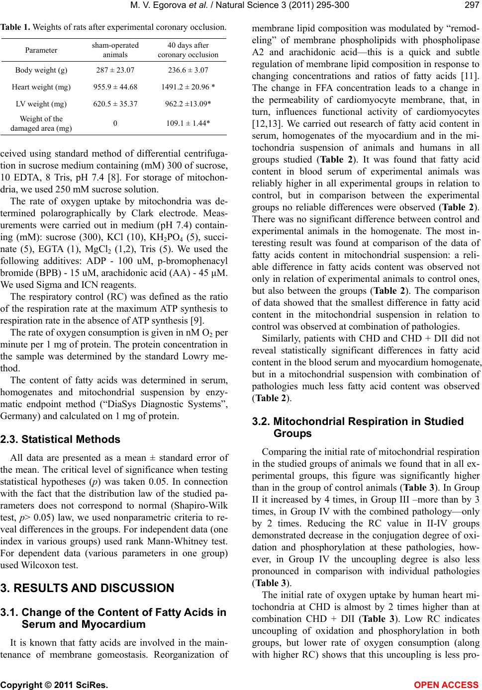 M. V. Egorova et al. / Natural Science 3 (2011) 295-300 Copyright © 2011 SciRes. OPEN ACCESS 297 Table 1. Weights of rats after experimental coronary occlusion. Parameter sham-operated animals 40 days after coronary occlusion Body weight (g) 287 ± 23.07 236.6 ± 3.07 Heart weight (mg) 955.9 ± 44.68 1491.2 ± 20.96 * LV weight (mg) 620.5 ± 35.37 962.2 ±13.09* Weight of the damaged area (mg) 0 109.1 ± 1.44* ceived using standard method of differential centrifuga- tion in sucrose medium containing (mM) 300 of sucrose, 10 EDTA, 8 Tris, pH 7.4 [8]. For storage of mitochon- dria, we used 250 mM sucrose solution. The rate of oxygen uptake by mitochondria was de- termined polarographically by Clark electrode. Meas- urements were carried out in medium (pH 7.4) contain- ing (mM): sucrose (300), KCl (10), KH2PO4 (5), succi- nate (5), EGTA (1), MgCl2 (1,2), Tris (5). We used the following additives: ADP - 100 uM, p-bromophenacyl bromide (BPB) - 15 uM, arachidonic acid (AA) - 45 μM. We used Sigma and ICN reagents. The respiratory control (RC) was defined as the ratio of the respiration rate at the maximum ATP synthesis to respiration rate in the absence of ATP synthesis [9]. The rate of oxygen consumption is given in nM O2 per minute per 1 mg of protein. The protein concentration in the sample was determined by the standard Lowry me- thod. The content of fatty acids was determined in serum, homogenates and mitochondrial suspension by enzy- matic endpoint method (“DiaSys Diagnostic Systems”, Germany) and calculated on 1 mg of protein. 2.3. Statistical Methods All data are presented as a mean ± standard error of the mean. The critical level of significance when testing statistical hypotheses (p) was taken 0.05. In connection with the fact that the distribution law of the studied pa- rameters does not correspond to normal (Shapiro-Wilk test, p> 0.05) law, we used nonparametric criteria to re- veal differences in the groups. For independent data (one index in various groups) used rank Mann-Whitney test. For dependent data (various parameters in one group) used Wilcoxon test. 3. RESULTS AND DISCUSSION 3.1. Change of the Content of Fatty Acids in Serum and Myocardium It is known that fatty acids are involved in the main- tenance of membrane gomeostasis. Reorganization of membrane lipid composition was modulated by “remod- eling” of membrane phospholipids with phospholipase A2 and arachidonic acid—this is a quick and subtle regulation of membrane lipid composition in response to changing concentrations and ratios of fatty acids [11]. The change in FFA concentration leads to a change in the permeability of cardiomyocyte membrane, that, in turn, influences functional activity of cardiomyocytes [12,13]. We carried out research of fatty acid content in serum, homogenates of the myocardium and in the mi- tochondria suspension of animals and humans in all groups studied (Table 2). It was found that fatty acid content in blood serum of experimental animals was reliably higher in all experimental groups in relation to control, but in comparison between the experimental groups no reliable differences were observed (Table 2). There was no significant difference between control and experimental animals in the homogenate. The most in- teresting result was found at comparison of the data of fatty acids content in mitochondrial suspension: a reli- able difference in fatty acids content was observed not only in relation of experimental animals to control ones, but also between the groups (Table 2). The comparison of data showed that the smallest difference in fatty acid content in the mitochondrial suspension in relation to control was observed at combination of pathologies. Similarly, patients with CHD and CHD + DII did not reveal statistically significant differences in fatty acid content in the blood serum and myocardium homogenate, but in a mitochondrial suspension with combination of pathologies much less fatty acid content was observed (Table 2). 3.2. Mitochondrial Respiration in Studied Groups Comparing the initial rate of mitochondrial respiration in the studied groups of animals we found that in all ex- perimental groups, this figure was significantly higher than in the group of control animals (Table 3). In Group II it increased by 4 times, in Group III –more than by 3 times, in Group IV with the combined pathology—only by 2 times. Reducing the RC value in II-IV groups demonstrated decrease in the conjugation degree of oxi- dation and phosphorylation at these pathologies, how- ever, in Group IV the uncoupling degree is also less pronounced in comparison with individual pathologies (Table 3). The initial rate of oxygen uptake by human heart mi- tochondria at CHD is almost by 2 times higher than at combination CHD + DII (Table 3). Low RC indicates uncoupling of oxidation and phosphorylation in both groups, but lower rate of oxygen consumption (along with higher RC) shows that this uncoupling is less pro- 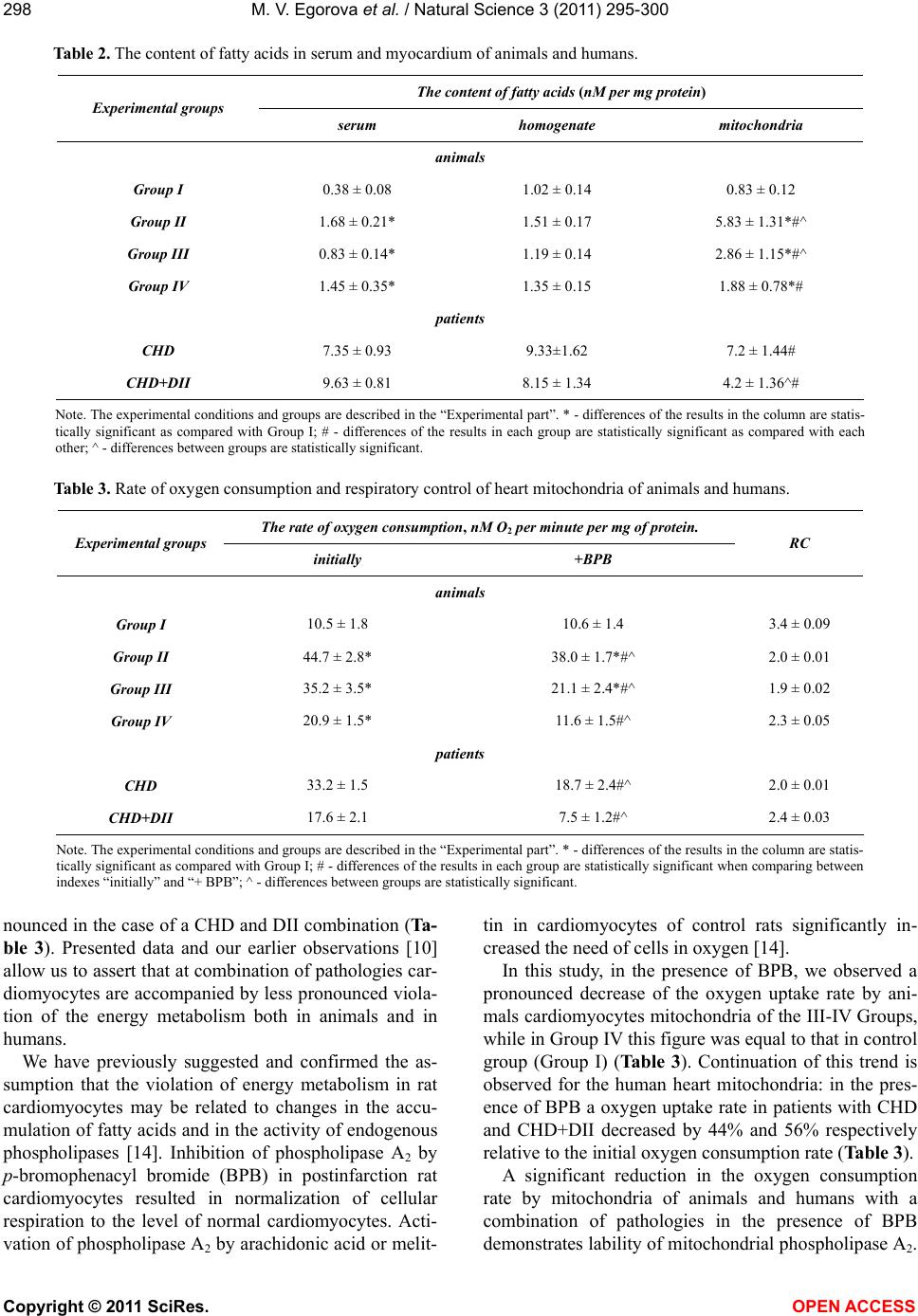 M. V. Egorova et al. / Natural Science 3 (2011) 295-300 Copyright © 2011 SciRes. OPEN ACCESS 298 Table 2. The content of fatty acids in serum and myocardium of animals and humans. The content of fatty acids (nM per mg protein) Experimental groups serum homogenate mitochondria animals Group I 0.38 ± 0.08 1.02 ± 0.14 0.83 ± 0.12 Group II 1.68 ± 0.21* 1.51 ± 0.17 5.83 ± 1.31*#^ Group III 0.83 ± 0.14* 1.19 ± 0.14 2.86 ± 1.15*#^ Group IV 1.45 ± 0.35* 1.35 ± 0.15 1.88 ± 0.78*# patients CHD 7.35 ± 0.93 9.33±1.62 7.2 ± 1.44# CHD+DII 9.63 ± 0.81 8.15 ± 1.34 4.2 ± 1.36^# Note. The experimental conditions and groups are described in the “Experimental part”. * - differences of the results in the column are statis- tically significant as compared with Group I; # - differences of the results in each group are statistically significant as compared with each other; ^ - differences between groups are statistically significant. Table 3. Rate of oxygen consumption and respiratory control of heart mitochondria of animals and humans. The rate of oxygen consumption, nM O2 per minute per mg of protein. Experimental groups initially +BPB RC animals Group I 10.5 ± 1.8 10.6 ± 1.4 3.4 ± 0.09 Group II 44.7 ± 2.8* 38.0 ± 1.7*#^ 2.0 ± 0.01 Group III 35.2 ± 3.5* 21.1 ± 2.4*#^ 1.9 ± 0.02 Group IV 20.9 ± 1.5* 11.6 ± 1.5#^ 2.3 ± 0.05 patients CHD 33.2 ± 1.5 18.7 ± 2.4#^ 2.0 ± 0.01 CHD+DII 17.6 ± 2.1 7.5 ± 1.2#^ 2.4 ± 0.03 Note. The experimental conditions and groups are described in the “Experimental part”. * - differences of the results in the column are statis- tically significant as compared with Group I; # - differences of the results in each group are statistically significant when comparing between indexes “initially” and “+ BPB”; ^ - differences between groups are statistically significant. nounced in the case of a CHD and DII combination (Ta- ble 3). Presented data and our earlier observations [10] allow us to assert that at combination of pathologies car- diomyocytes are accompanied by less pronounced viola- tion of the energy metabolism both in animals and in humans. We have previously suggested and confirmed the as- sumption that the violation of energy metabolism in rat cardiomyocytes may be related to changes in the accu- mulation of fatty acids and in the activity of endogenous phospholipases [14]. Inhibition of phospholipase A2 by p-bromophenacyl bromide (BPB) in postinfarction rat cardiomyocytes resulted in normalization of cellular respiration to the level of normal cardiomyocytes. Acti- vation of phospholipase A2 by arachidonic acid or melit- tin in cardiomyocytes of control rats significantly in- creased the need of cells in oxygen [14]. In this study, in the presence of BPB, we observed a pronounced decrease of the oxygen uptake rate by ani- mals cardiomyocytes mitochondria of the III-IV Groups, while in Group IV this figure was equal to that in control group (Group I) (Table 3). Continuation of this trend is observed for the human heart mitochondria: in the pres- ence of BPB a oxygen uptake rate in patients with CHD and CHD+DII decreased by 44% and 56% respectively relative to the initial oxygen consumption rate (Table 3). A significant reduction in the oxygen consumption rate by mitochondria of animals and humans with a combination of pathologies in the presence of BPB demonstrates lability of mitochondrial phospholipase A2. 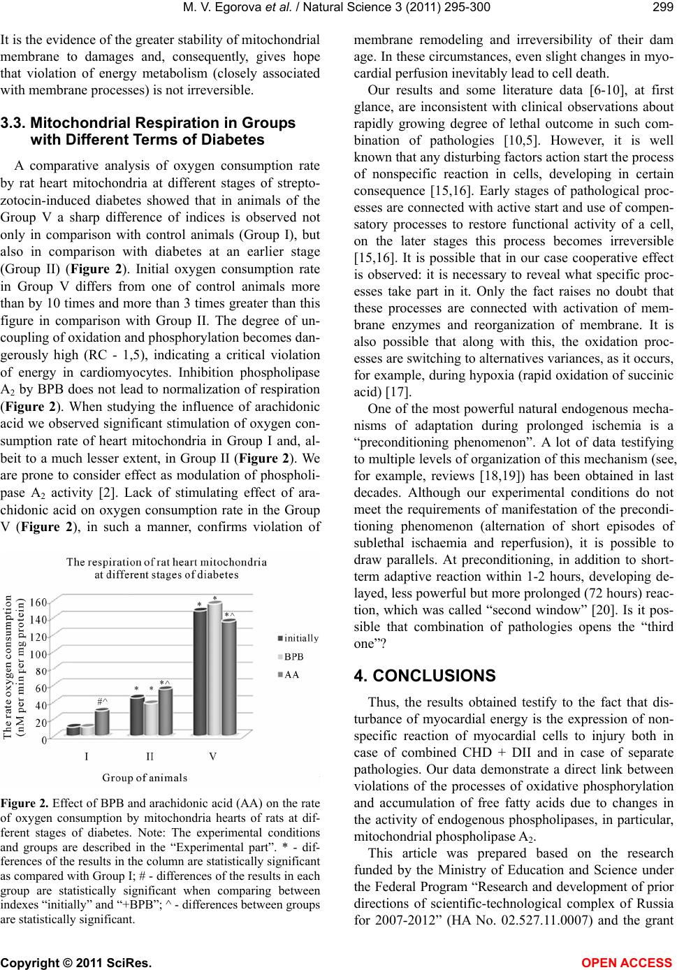 M. V. Egorova et al. / Natural Science 3 (2011) 295-300 Copyright © 2011 SciRes. OPEN ACCESS 299 It is the evidence of the greater stability of mitochondrial membrane to damages and, consequently, gives hope that violation of energy metabolism (closely associated with membrane processes) is not irreversible. 3.3. Mitochondrial Respiration in Groups with Different Terms of Diabetes A comparative analysis of oxygen consumption rate by rat heart mitochondria at different stages of strepto- zotocin-induced diabetes showed that in animals of the Group V a sharp difference of indices is observed not only in comparison with control animals (Group I), but also in comparison with diabetes at an earlier stage (Group II) (Figure 2). Initial oxygen consumption rate in Group V differs from one of control animals more than by 10 times and more than 3 times greater than this figure in comparison with Group II. The degree of un- coupling of oxidation and phosphorylation becomes dan- gerously high (RC - 1,5), indicating a critical violation of energy in cardiomyocytes. Inhibition phospholipase A2 by BPB does not lead to normalization of respiration (Figure 2). When studying the influence of arachidonic acid we observed significant stimulation of oxygen con- sumption rate of heart mitochondria in Group I and, al- beit to a much lesser extent, in Group II (Figure 2). We are prone to consider effect as modulation of phospholi- pase A2 activity [2]. Lack of stimulating effect of ara- chidonic acid on oxygen consumption rate in the Group V (Figure 2), in such a manner, confirms violation of Figure 2. Effect of BPB and arachidonic acid (AA) on the rate of oxygen consumption by mitochondria hearts of rats at dif- ferent stages of diabetes. Note: The experimental conditions and groups are described in the “Experimental part”. * - dif- ferences of the results in the column are statistically significant as compared with Group I; # - differences of the results in each group are statistically significant when comparing between indexes “initially” and “+BPB”; ^ - differences between groups are statistically significant. membrane remodeling and irreversibility of their dam age. In these circumstances, even slight changes in myo- cardial perfusion inevitably lead to cell death. Our results and some literature data [6-10], at first glance, are inconsistent with clinical observations about rapidly growing degree of lethal outcome in such com- bination of pathologies [10,5]. However, it is well known that any disturbing factors action start the process of nonspecific reaction in cells, developing in certain consequence [15,16]. Early stages of pathological proc- esses are connected with active start and use of compen- satory processes to restore functional activity of a cell, on the later stages this process becomes irreversible [15,16]. It is possible that in our case cooperative effect is observed: it is necessary to reveal what specific proc- esses take part in it. Only the fact raises no doubt that these processes are connected with activation of mem- brane enzymes and reorganization of membrane. It is also possible that along with this, the oxidation proc- esses are switching to alternatives variances, as it occurs, for example, during hypoxia (rapid oxidation of succinic acid) [17]. One of the most powerful natural endogenous mecha- nisms of adaptation during prolonged ischemia is a “preconditioning phenomenon”. A lot of data testifying to multiple levels of organization of this mechanism (see, for example, reviews [18,19]) has been obtained in last decades. Although our experimental conditions do not meet the requirements of manifestation of the precondi- tioning phenomenon (alternation of short episodes of sublethal ischaemia and reperfusion), it is possible to draw parallels. At preconditioning, in addition to short- term adaptive reaction within 1-2 hours, developing de- layed, less powerful but more prolonged (72 hours) reac- tion, which was called “second window” [20]. Is it pos- sible that combination of pathologies opens the “third one”? 4. CONCLUSIONS Thus, the results obtained testify to the fact that dis- turbance of myocardial energy is the expression of non- specific reaction of myocardial cells to injury both in case of combined CHD + DII and in case of separate pathologies. Our data demonstrate a direct link between violations of the processes of oxidative phosphorylation and accumulation of free fatty acids due to changes in the activity of endogenous phospholipases, in particular, mitochondrial phospholipase A2. This article was prepared based on the research funded by the Ministry of Education and Science under the Federal Program “Research and development of prior directions of scientific-technological complex of Russia for 2007-2012” (HA No. 02.527.11.0007) and the grant 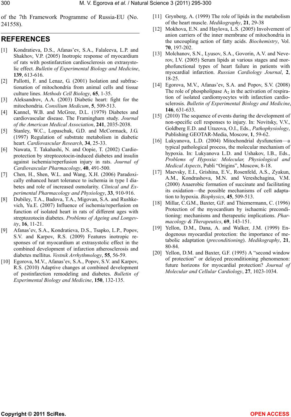 M. V. Egorova et al. / Natural Science 3 (2011) 295-300 Copyright © 2011 SciRes. OPEN ACCESS 300 of the 7th Framework Programme of Russia-EU (No. 241558). REFERENCES [1] Kondratieva, D.S., Afanas’ev, S.A., Falaleeva, L.P. and Shakhov, V.P. (2005) Inotropic response of myocardium of rats with postinfarction cardiosclerosis on extrasysto- lic effect. Bulletin of Experimental Biology and Medicine, 139, 613-616. [2] Pallotti, F. and Lenaz, G. (2001) Isolation and subfrac- tionation of mitochondria from animal cells and tissue culture lines. Methods Cell Biology, 65, 1-35. [3] Aleksandrov, A.A. (2003) Diabetic heart: fight for the mitochondria. Consilium Medicum, 5, 509-513. [4] Kannel, W.B. and McGree, D.L. (1979) Diabetes and cardiovascular disease. The Framingham study. Journal of the American Medical Association, 241, 2035-2038. [5] Stanley, W.C., Lopaschuk, G.D. and McCormack, J.G. (1997) Regulation of substrate metabolism in diabetic heart. Cardiovascular Research, 34, 25-33. [6] Nawata, T. Takahashi, N. and Oopie, T. (2002) Cardio- protection by streptozotocin-induced diabetes and insulin against ischemia/reperfusion injury in rats. Journal of Cardiovascular Pharmacology, 40, 491-500. [7] Chen, H., Shen, W.L. and Wang, X.H. (2006) Paradoxi- cally enhanced heart tolerance to ischemia in type I dia- betes and role of increased osmolarity. Clinical and Ex- perimental Pharmacology and Physiology, 33, 910-916. [8] Dubiley, T.A., Badova, T.A., Migovan, S.A. and Rushke- vich, Yu.E. (2007) Influence of ischemia/reperfusion on function of isolated heart in rats of different ages with streptozotocin diabetes. Problems of Ageing and Longev- ity, 16, 11-21. [9] Afanas’ev, S.A., Kondratieva, D.S., Tsapko, L.P., Popov, S.V. and Karpov, R.S. (2009) Features inotropic re- sponses of rat myocardium at extrasystolic effect in the combined development of infarction atherosclerosis and diabetes mellitus. Vestnik Arrhythmology, 55, 56-59. [10] Egorova, M.V., Afanas’ev, S.A., Popov, S.V. and Karpov, R.S. (2010) Adaptive changes at combined development of postinfarction remodeling and diabetes. Bulletin of Experimental Biology and Medicine, 150, 132-135. [11] Grynberg, A. (1999) The role of lipids in the metabolism of the heart muscle. Medikography, 21, 29-38 [12] Mokhova, E.N. and Haylova, L.S. (2005) Involvement of anion carriers of the inner membrane of mitochondria in the uncoupling action of fatty acids. Biochemistry, Vol. 70, 197-202. [13] Molchanov, S.N., Lyusov, S.A., Govorin, A.V. and Neve- rov, I.V. (2005) Serum lipids at various stages and mor- phofunctional types of heart failure in patients with myocardial infarction. Russian Cardiology Journal, 2, 18-25. [14] Egorova, M.V., Afanas’ev, S.A. and Popov, S.V. (2008) The role of phospholipase A2 in the activation of respira- tion of isolated cardiomyocytes with infarction cardio- sclerosis. Bulletin of Experimental Biology and Medicine, 146, 631-633. [15] (2010) The sequence of events during the development of non-specific cell responses to injury. In: Novitsky, V.V., Goldberg E.D. and Urazova, O.I., Eds., Pathophysiology, Publishing GEOTAR-Media, Moscow, 1, 59-62. [16] Lukyanova, L.D. (2004) Mitochondrial dysfunction—a typical pathological process, the molecular mechanism of hypoxia. In: Lukyanova L.D. and Ushakov, I.B., Eds., Problems of Hypoxia: Molecular, Physiological and Medical Aspects, Publi “Origins”, Moscow, 8-18. [17] Maevsky, E.I., Grishina, E.V., Rosenfeld, A.S., Zyakun, A.M., Kondrashova, M.N. and Vereshchagina, V.M. (2000) Anaerobic formation of succinate and facilitating its oxidation—the possible mechanisms of cell adapta- tion to hypoxia. Biophysics, 45, 509-513. [18] Millar, C.G.M., Baxter, G.F. and Thiemermann, C. (1996) Protection of the myocardium by ischaemic precondi- tioning: mechanisms and therapeutic implications. Phar- macology & Therapeutics, 69, 143-151. [19] Yellon, D.M., Dana, A. and Walker, J.M. (1999) En- dogenous myocardial protection: the importance of me- tabolic adaptation (preconditioning). Medikography, 21, 80-84. [20] Yellon, D.M. and Baxter, G.F. (1995) A “second window of protection” or delayed preconditioning phenomenon: future horizons for myocardial protection? Journal of Molecular and Cellular Cardiology, 27, 1023-1034. |