

Paper Menu >>
Journal Menu >>
 New Journal of Glass and Ceramics, 2011, 1, 7-12 doi:10.4236/njgc.2011.11002 Published Online April 2011 (http://www.SciRP.org/journal/njgc) Copyright © 2011 SciRes. NJGC 7 Effect of Aging on Chlorophyll Species Embedded in Silica Xerogels Matrix José R. Martínez1*, Erika Espericueta1,2, Gerardo Ortega-Zarzosa1 1Universidad Autónoma de San Luis Potosí, San Luis Potosí, México; 2Centro de Investigación en Materiales Avanzados, Chihuahua, México. Email: [email protected] Received February 16th, 2011; revised March 30th, 2011; accepted April 6th, 2011. ABSTRACT The spectral fluorescence characteristics of extracts leaves embedded in silica xerogel matrix promoted by aged time was studied. We obta in a higher PSII stability for extra ct of leaves embedded in xerogel matrix, with concen tration of 8 g / 2 0 ml and water to TEOS molar ratio o f 11, which remain bioactive over a very long period of time, whereas for a water to TEOS molar ratio of 5 a blue-shift fluor escence is observed indicating the PSII d enaturizing and formation of fluorescing aggre gat es associated with the act i vat i o n of carot e n oi ds rad i cal s. Keywords: Fluorescence Spectra, Sol-Gel, Chlorophyll, Extract Leaves, Photosystem II 1. Introduction Sol-gel has even been used to encapsulate and maintain the activity of biomolecules. However, currently investi- gation about organically-modified sol-gels in an effort to control overall dopant mobility, accessibility, and reac- tivity are made in the present. The number of potential applications of organic doped sol-gel glasses is very large; important possibilities in- clude optics and electro-optics. Development of these applications requires a good understanding of the struc- ture of the doped sol-gel matrices, the properties of the matrices on the molecular level and the conditions that the oxide network imposes on the optical properties of the dopant [1]. Sol-gel technique with the ability to trap photoactive substances in an inorganic gel glass, has led to some new applications in nonlinear optics, solid-state tunable lasers, photochemical hole burning, etc [2,3]. Metalloporphyrin complexes have received much at- tention owing to their important role in biology, solar energy conversion, photonics, and catalysis [4]. Spectro- scopic evidence confirmed stability of the dispersion over a period of three to four months. As an optical substance adsorbed into an inorganic matrix, chlorophyll and composites based in chlorophyll has been poorly studied, in spite of the interesting appli- cations of chlorophyll’s optical properties [5-7]. The higher plants are an efficient system to absorb and transfer the energy through photosynthetic apparatus. This situation can be used to take advantage to prepare materials based in extract of plants for eventually design efficient optical- and electrooptical-based sol-gel encap- sulated biomolecules [8]. The immobilization of organic pigments on inorganic media may be useful for the design of energy conversion devices [9]. Thus, it is necessary to understand the structural evo- lution of extracted components of leaves embedded in silica xerogel matrix, as well as the interaction between the pigments embedded into the inorganic matrix. The typical fluorescence spectrum of green plants, embedded or not embedded in xerogel matrix, is in the range from 650 nm to 780 nm, presenting two maximum of fluorescence located at about 670 nm and 730 nm corresponding to chlorophyll a and at about 650 nm and 710 nm corresponding to chlorophyll b [8,10]. All green plants contain chlorophyll a and chlorophyll b in their chloroplast. In higher plants, chlorophyll a is the major pigment and chlorophyll b is an accessory pigment, and the a/b ratio is generally around 3 to 1 [11]. Chlorophyll b differs from chlorophyll a by having an aldehyde (–CHO) group instead of a methyl group (–CH3). The differences in these structures cause that the two maximum of fluorescence of chlorophyll a are located at 670 nm and 730 nm, while the two maximum of fluores- cence of chlorophyll b are at 650 nm and 710 nm [10,12].  Effect of Aging on Chlorophyll Species Embedded in Silica Xerogels Matrix 8 The green plants contain the photosystem I and II (PSI, PSII). A photosystem consist of a peripheral antenna or ligh-harvesting complex, an inner antenna and the reac- tion center. The Photosystem II is the principal system in which the electron transfer in the photosynthesis process occur. Chlorophyll is present in the two photosystems, PSI and PSII, but only photosystem II has additional minor chlorophyll a/b binding antenna protein. Both photosys- tems differ in their absorption and fluorescence charac- teristic. The maximum of fluorescence intensity can be found for PSI at 735 nm and PSII at 683 nm. At room temperature it can be assumed that all variable fluores- cence originates from PSII [10,13]. The photosystem PSII can be monitoring by chloro- phyll a fluorescence. Thus the observed maximum inten- sity fluorescence, corresponding to excitation energy transfer to the photosystem II, is at about 683 nm [8]. Metal porphyrins are molecules present in nature, such leaves, for example the magnesium porphyrin in chloro- phyll. The components of the complex extracted of leaves includes the chlorophyll, which is the principal component in most green plants, but exist in the Photo- system II at least 25 different types of protein subunits, PSII is a large supramolecular pigment-protein complex embedded in the thylakoid membranes of green plants. In previous works we show that the samples with ex- tract of leaves embedded in silica xerogel matrix show that PSII chlorophyll fluorescence transients is non pre- sent and the samples show a higher PSII thermostability for leaves embedded in xerogel matrix than in the green leaves [8,14]. Under heat treatment, the original bands located at about 670 and 714 nm observed in the fluorescence spectrum, are substituted by a blue-shifted band at about 580 nm. These changes can be attributed to the tight en- vironment around the chlorophyll and to the different polarity of the environment inside the pores of the silica matrix, among the conversion of chlorophyll to protein species, as phaeophytene [15,16]. The large shift is typical for weakening of chlorophyll – protein interactions in pigment – protein complex (PPCs). One may assume that the observed spectral changes are associated with changes of spectral distortion in the pigments embedded in silica xeroge l matrix during heat treatment, due to the structural evolution of the ma- trix. In this work, by measurements of the PSII fluores- cence comportment, we report biostability of sol-gel amorphous bulk samples containing extract of spinach leaves which is sensitive to concentration of the aggre- gates. 2. Experimental Procedure The standard method to extract compound of leaves is by crude extraction from frozen leaves, washed with water, by simple grinding and mixing them with ethanol solvent. For the extraction three arbitrary concentrations, 4 g, 8 g and 50 g of leaves for 20 ml of ethan ol, corresponding to low, medium and very high concentration, were used, in order to obtain information abou t the effect of con centra- tion in the aging process. The solution of ethanol and extract was centrifuged at 3000 rpm for 5 minutes and then filtered with a Wattsman filter nr. 1. In order to obtain the organic composites of extract of leaves embedded in xerogel matrix, a precursor material composed of TEOS, water, and ethanol with pigments extract of leaves, of spinach was prepared. The set of samples were prepared using a constant ethanol to TEOS molar ratio of 4:1 and a molar ratio of water to TEOS (R) of R = 11 and R = 5. These quantities correspond to a high water/TEOS ratio, needed to enhance the hydrolysis to assure a close amorphous structure for the as-prepared SiO2 powder [17]. The TEOS was dissolved in the etha- nol with pigments using magnetic stirring for 15 min and then the water was added to the ethanol-TEOS solution using magnetic stirring for 10 minutes to form the start- ing material. The pH of the final solution was 5. Soft pieces of the gel were obtained after 48 hr. Those pieces were ground to form a fine powder. The emission spectra measurements were carried out in a fluorescence spectrometer by Ocean Optics Inc. model SF2000 using a reflectance diffuse 45˚ configura- tion using a double fiber optic component, excited with an Omnicrome argon ion laser of 532 nm and filter HNF-532-1.0, in order to obtain the fluorescence contri- bution due to the photosystem II of samples measured after gelling and for the same samples measured after 36 months of gelling. In order to get similar grain size the samples were ground to form fine powder; the experimental set up to obtain emission spectra measurement was regularly used for compared spectroscopy measurement, such NIR, Raman and fluorescence, obtained in the same experi- mental condition, in this case by relative intensity fluo- rescence. 3. Results and Discussion In order to investigate the structural and fluorescence change, the set of samples with different extract concen- tration were irradiate with 532 nm laser using the diffuse reflectance, the fluorescence results obtained are shown in the next figures. Figures 1-3, shows the fluorescence spectra for silica xerogel with 4 g, 8g and 50 g per 20 ml of extract spin- ach leave with R = 11, respectively. The as-prepared samples present the typical chlorophyll a fluorescence (range from 650 nm to 780 nm) in which no change of Copyright © 2011 SciRes. NJGC  Effect of Aging on Chlorophyll Species Embedded in Silica Xerogels Matrix9 fluorescence characteristic occurred with aged time in- creasing. For Figures 1 and 3, a blue-shift of the spectra is observed when the aged time is increased at 36 months and the maximum fluorescence shift from 678 nm to 669 nm, and 686 nm to 675 nm, after 36 month, respectively, whereas for Figure 2, the maximum fluorescence is un- changed maintenance at 678 nm. In the latest case we obtain a great biostability for su ch formation parameters. When the ratio R is reduced to R = 5, maintaining the concentration of 8 g / 20 ml the biostability is break ing as we can observed in Figure 4. For this sample the aged effect decomposes the chlorophyll contribution and a denaturalizati o n occu rs a s we di scuss in next paragra ph. 550 600 650700 750 0 month 36 month Normalized fl uor esc e nce in t ens ity Emission wavelength (nm) Figure 1. Fluorescent emission spectra of spinach leaves, with concentration of 4 g / 20 ml, embedded in silica xerogel matrix measured after gelling and after 36 months of gel- ling. 550 600 650700 750 36 month 0 month Emission wavelength (nm) Normalized fluorescence intensity Figure 2. Fluorescent emission spectra of spinach leaves, with concentration of 8 g / 20 ml, embedded in silica xerogel matrix measured after gelling and after 36 months of gel- ling. 550 600 650 700 750 0 month 36 month Normaliz ed fluo res cenc e intens i ty Emission wavelength (nm) Figure 3. Fluorescent emission spectra of spinach leaves, with concentration of 50 g / 20 ml, embedded in silica xe- rogel matrix measured after gelling and after 36 months of gelling. 550 600 650700 750 0 month 36 month Normalized fluorescence intensity Emission wavelength (nm) Figure 4. Fluorescent emission spectra of spinach leaves, with concentration of 4 g / 20 ml and water to TEOS molar ratio of 5, embedded in silica xerogel matrix measured after gelling and after 36 months of gelling. Figure 2 shows there is not significant change of fluorescence of chlorophyll entrapped in silica xerogel up to 36 months, revealing unchanged in chlorophyll struc- ture, in samples where extracted leaves of spinach were embedded in silica xerogel, thus the chlorophyll a mo l e - cules remain structurally same in silica xerogel over a very long period of time. In particular the maximum po- sition of fluorescence of the PSII unchanged in terms of sol-gel aging time. This time period is very long com- pared with time, about 3 months, reported in work where chlorophyll a molecules was entrapped in silica gel nanomatrix [16]. This results contrast with the case of barley leaves embedded in silica xerogel matrix where a 10 nm blue shift of the red emission maximum after aging for about 12 months was observed [8]. Copyright © 2011 SciRes. NJGC  Effect of Aging on Chlorophyll Species Embedded in Silica Xerogels Matrix 10 In conclusion extract of spinach leaves with concen- tration of 8 g / 20 ml and R = 11 embedded in silica xe- rogel matrix present a higher PSII biostability, remaining bioactive over very l ong periods of time. Thus the formation of biostable compound, for very long aged times, of chlorophyll species embedded in sil- ica xerogel can be obtained by controlling concentration of extract leave and ethanol to TEOS and water to TEOS molar ratios. In Figure 1 where a 9 nm shift of maximum fluores- cence is observed, which can be associated with changes of spectral distortion in the pigments embedded in silica xerogel matrix during the aged, due to the structural evolution of the matrix. The shift is attributed also to changes of the tight environment around the chlorophyll and to the different polarity of the environment insid e the pores of the silica matrix, among the conversion of chlo- rophyll to protein species, as phaeophytene [15,16]. In- dicate us that, among the previous assignment for the fluorescence blue shift, denaturations of chlorophyll spe- cies occur with a increasing of chlorophyll b respect to chlorophyll a. Figure 2, represent a biostabilizing system in which the chlorophyll a don’t decompose to phaeophytene and only a little increase of chlorophyll b is observed when the aged time increasing indicated by an increase in the fluorescent intensity with a wav e length shift of the band located at 726 nm to 713 nm. In Figure 3, we show the sample with a high extract leave concentratio n. In this case a pigment aggregation is present and then a red shift of maximum fluorescent, respect to 675 nm obtained for the samples with 4 g and 8 g per 20 ml of extract leave concentration. The maxi- mum fluorescence is located at 686 nm. The band located at 724 nm present a higher intensity compared with sam- ples of Figures 1 and 2, corresponding to low extract leave concentration. When the water to TEOS molar ratio is reduced the biostabilization is breaking as we can observe in Figure 4, where the fluorescent spectra for an extract leave con- centration of 8 g / 20 ml and water to TEOS molar ratio of 5 is shown. The fluorescent contribution between 630 nm to 750 nm corresponds to chlorophyll b with bands located at 668 nm and 710 nm characteristic of such chlorophyll specie. The aging effect produce a rapid denaturation of chlorophyll compound and a blue shift occur. The typical red fluorescence disappear and a blue-shifted band at about 580 nm is present [14], indicating the decomposi- tion of the photosyste m II. This situation is also ob served in samples of ortho-amnino-substituted porphyrin, H2T(o-NH2)PP, embedded in SiO2 matrix [15]. The large shift is typical for weakening of chlorophyll – protein interactions in pigment – protein complex (PPCs). The carotenoid radical cation formation play one important role in photosynthetic pigment-protein com- plexes, and quench chlorophyll fluorescence [18,19]. The chlorophyll molecule contain the phytyl group associated to carotenoid radical, the low water content retarding the gel formation and the denaturation of photosystem II active the carotenoid function of protect the photosyn- thetic apparatus by quenching chlorophyll triplet state which inhibits the formation of singlet state oxygen [20-22], and stabilize light-harvesting protein structures [23-25]. The active presence of carotenoid radicals is indicated by the blue shift band located at 583 nm [26]. The difference in structure due to concentration of ex- tract leave and water to TEOS molar ratio, are reflected on the fluorescence differences discussed previously. In Figure 5 we observe that, among the differences in rela- tive intensities for bands characteristic of chlorophyll a and b, related with the photosystems I and II, we observe that the position of maximum fluorescence of the band corresponding to PSII at about 680 nm, is red shifted conform the concentration is increased. This result is in agreement with results reported by Furukawa et al., for layered silica/surfactant mesostructured this films con- taining chlorophyllous pigments where the red shift in optical measurements indicates the aggregation of pig- ments [9]. The fluorescent bands, for samples labeled by C1 and C2 concentrations in Figure 5 are very similar, with a lighter red shift for sample C2 respect to C1. However for aged samples these bands present higher differences indicating that a higher biostabilisation is obtained for concentration C2, as we can observe in Figure 6. 550 600 650 700 750 Normalized fluorescence int ensity Em ission wavelen g th (nm) R5, C2 C3 C1 C2 Figure 5. Fluorescent emission spectra of spinach leaves, with concentration of 4 g / 20 ml (C1), 8 g / 20 ml (C2), 50 g / 20 ml (C3) and 8 g / 20 ml with water to TEOS molar ratio of 5 (R5,C2), embedded in silica xerogel matrix measured after gelling. Copyright © 2011 SciRes. NJGC 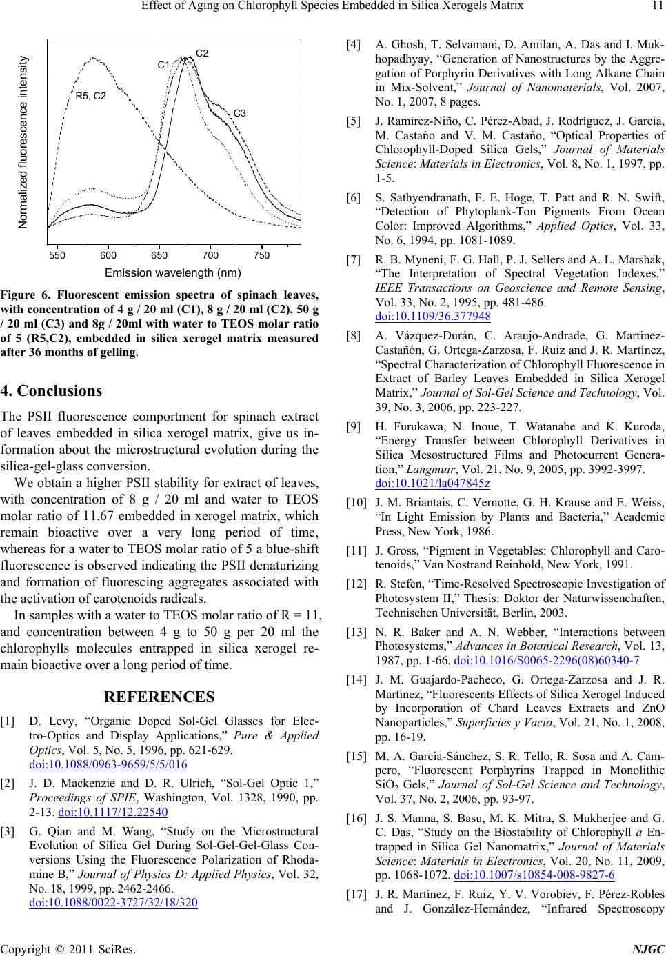 Effect of Aging on Chlorophyll Species Embedded in Silica Xerogels Matrix11 550 600650 700750 Normalized fluorescence intensity Emission wavelength (nm) R5, C2 C2 C1 C3 Figure 6. Fluorescent emission spectra of spinach leaves, with concentration of 4 g / 20 ml (C1), 8 g / 20 ml (C2), 50 g / 20 ml (C3) and 8g / 20ml with water to TEOS molar ratio of 5 (R5,C2), embedded in silica xerogel matrix measured after 36 months of gelling. 4. Conclusions The PSII fluorescence comportment for spinach extract of leaves embedded in silica xerogel matrix, give us in- formation about the microstructural evolution during the silica-gel-glass conversion. We obtain a higher PSII stability for extract of leaves, with concentration of 8 g / 20 ml and water to TEOS molar ratio of 11.67 embedded in xerogel matrix, which remain bioactive over a very long period of time, whereas for a water to TEOS molar ratio of 5 a blue-shift fluorescence is observed indicating the PSII denaturizing and formation of fluorescing aggregates associated with the activation of carotenoids radical s. In samples with a water to TEOS molar ratio of R = 11, and concentration between 4 g to 50 g per 20 ml the chlorophylls molecules entrapped in silica xerogel re- main bioactive ov er a long period of time. REFERENCES [1] D. Levy, “Organic Doped Sol-Gel Glasses for Elec- tro-Optics and Display Applications,” Pure & Applied Optics, Vol. 5, No. 5, 1996, pp. 621-629. doi:10.1088/0963-9659/5/5/016 [2] J. D. Mackenzie and D. R. Ulrich, “Sol-Gel Optic 1,” Proceedings of SPIE, Washington, Vol. 1328, 1990, pp. 2-13. doi:10.1117/12.22540 [3] G. Qian and M. Wang, “Study on the Microstructural Evolution of Silica Gel During Sol-Gel-Gel-Glass Con- versions Using the Fluorescence Polarization of Rhoda- mine B,” Journal of Physics D: Applied Physics, Vol. 32, No. 18, 1999, pp. 2462-2466. doi:10.1088/0022-3727/32/18/320 [4] A. Ghosh, T. Selvamani, D. Amilan, A. Das and I. Muk- hopadhyay, “Generation of Nanostructures by the Aggre- gation of Porphyrin Derivatives with Long Alkane Chain in Mix-Solvent,” Journal of Nanomaterials, Vol. 2007, No. 1, 2007, 8 pages. [5] J. Ramírez-Niño, C. Pérez-Abad, J. Rodríguez, J. García, M. Castaño and V. M. Castaño, “Optical Properties of Chlorophyll-Doped Silica Gels,” Journal of Materials Science: Materials in Electronics, Vol. 8, No. 1, 1997, pp. 1-5. [6] S. Sathyendranath, F. E. Hoge, T. Patt and R. N. Swift, “Detection of Phytoplank-Ton Pigments From Ocean Color: Improved Algorithms,” Applied Optics, Vol. 33, No. 6, 1994, pp. 1081-1089. [7] R. B. Myneni, F. G. Hall, P. J. Sellers and A. L. Marshak, “The Interpretation of Spectral Vegetation Indexes,” IEEE Transactions on Geoscience and Remote Sensing, Vol. 33, No. 2, 1995, pp. 481-486. doi:10.1109/36.377948 [8] A. Vázquez-Durán, C. Araujo-Andrade, G. Martínez- Castañón, G. Ortega-Zarzosa, F. Ruiz and J. R. Martínez, “Spectral Characterization of Chlorophyll Fluorescence in Extract of Barley Leaves Embedded in Silica Xerogel Matrix,” Journal of Sol-Gel Science and Technology, Vol. 39, No. 3, 2006, pp. 223-227. [9] H. Furukawa, N. Inoue, T. Watanabe and K. Kuroda, “Energy Transfer between Chlorophyll Derivatives in Silica Mesostructured Films and Photocurrent Genera- tion,” Langmuir, Vol. 21, No. 9, 2005, pp. 3992-3997. doi:10.1021/la047845z [10] J. M. Briantais, C. Vernotte, G. H. Krause and E. Weiss, “In Light Emission by Plants and Bacteria,” Academic Press, New York, 1986. [11] J. Gross, “Pigment in Vegetables: Chlorophyll and Caro- tenoids,” Van Nostrand Reinhold, New York, 1991. [12] R. Stefen, “Time-Resolved Spectroscopic Investigation of Photosystem II,” Thesis: Doktor der Naturwissenchaften, Technischen Universität, Berlin, 2003. [13] N. R. Baker and A. N. Webber, “Interactions between Photosystems,” Advances in Botanical Research, Vol. 13, 1987, pp. 1-66. doi:10.1016/S0065-2296(08)60340-7 [14] J. M. Guajardo-Pacheco, G. Ortega-Zarzosa and J. R. Martínez, “Fluorescents Effects of Silica Xerogel Induced by Incorporation of Chard Leaves Extracts and ZnO Nanoparticles,” Superficies y Vacio, Vol. 21, No. 1, 2008, pp. 16-19. [15] M. A. García-Sánchez, S. R. Tello, R. Sosa and A. Cam- pero, “Fluorescent Porphyrins Trapped in Monolithic SiO2 Gels,” Journal of Sol-Gel Science and Technology, Vol. 37, No. 2, 2006, pp. 93-97. [16] J. S. Manna, S. Basu, M. K. Mitra, S. Mukherjee and G. C. Das, “Study on the Biostability of Chlorophyll a En- trapped in Silica Gel Nanomatrix,” Journal of Materials Science: Materials in Electronics, Vol. 20, No. 11, 2009, pp. 1068-1072. doi:10.1007/s10854-008-9827-6 [17] J. R. Martínez, F. Ruiz, Y. V. Vorobiev, F. Pérez-Robles and J. González-Hernández, “Infrared Spectroscopy Copyright © 2011 SciRes. NJGC 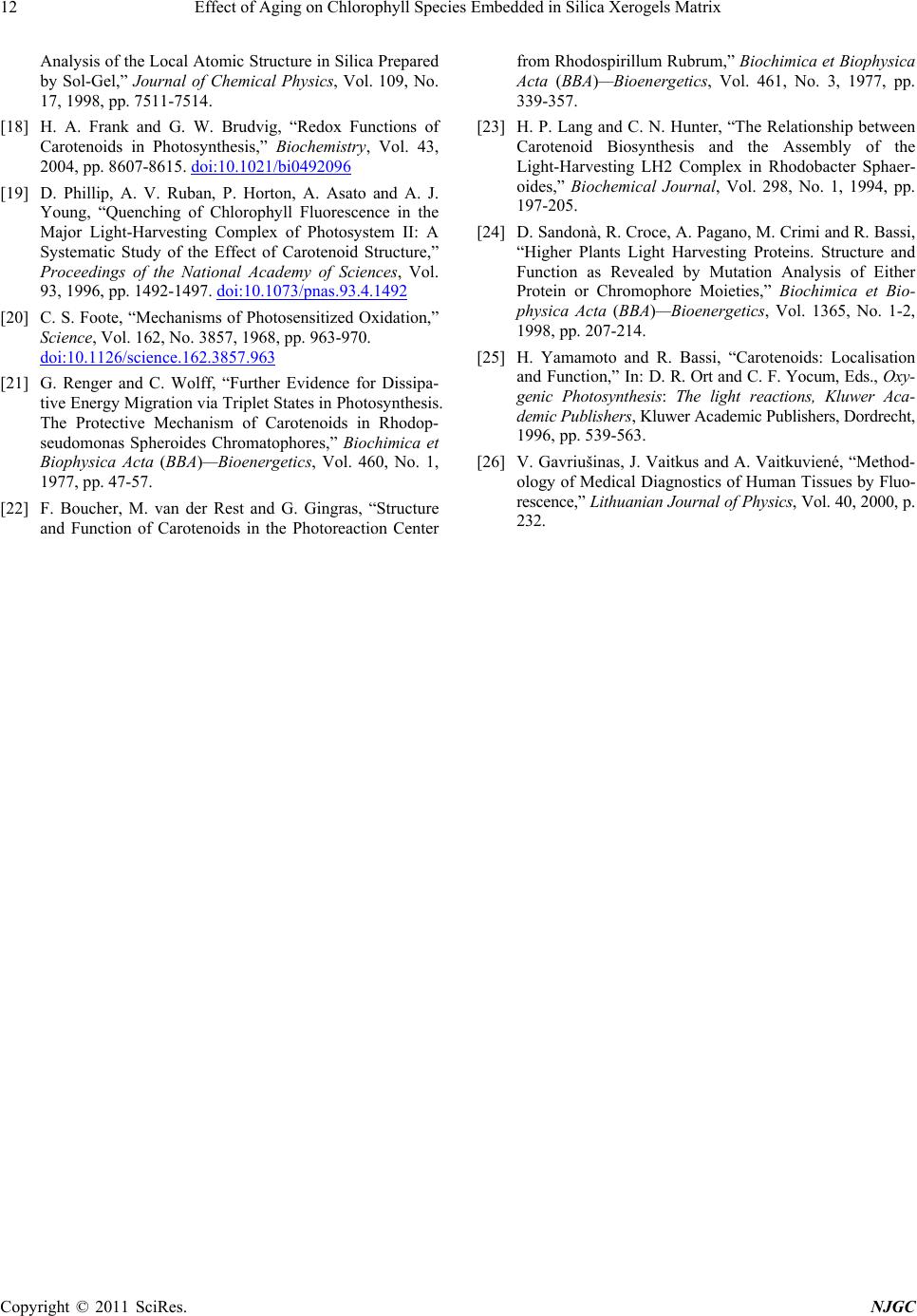 Effect of Aging on Chlorophyll Species Embedded in Silica Xerogels Matrix Copyright © 2011 SciRes. NJGC 12 Analysis of the Local Atomic Structure in Silica Prepared by Sol-Gel,” Journal of Chemical Physics, Vol. 109, No. 17, 1998, pp. 7511-7514. [18] H. A. Frank and G. W. Brudvig, “Redox Functions of Carotenoids in Photosynthesis,” Biochemistry, Vol. 43, 2004, pp. 8607-8615. doi:10.1021/bi0492096 [19] D. Phillip, A. V. Ruban, P. Horton, A. Asato and A. J. Young, “Quenching of Chlorophyll Fluorescence in the Major Light-Harvesting Complex of Photosystem II: A Systematic Study of the Effect of Carotenoid Structure,” Proceedings of the National Academy of Sciences, Vol. 93, 1996, pp. 1492-1497. doi:10.1073/pnas.93.4.1492 [20] C. S. Foote, “Mechanisms of Photosensitized Oxidation,” Science, Vol. 162, No. 3857, 1968, pp. 963-970. doi:10.1126/science.162.3857.963 [21] G. Renger and C. Wolff, “Further Evidence for Dissipa- tive Energy Migration via Triplet States in Photosynthesis. The Protective Mechanism of Carotenoids in Rhodop- seudomonas Spheroides Chromatophores,” Biochimica et Biophysica Acta (BBA)—Bioenergetics, Vol. 460, No. 1, 1977, pp. 47-57. [22] F. Boucher, M. van der Rest and G. Gingras, “Structure and Function of Carotenoids in the Photoreaction Center from Rhodospirillum Rubrum,” Biochimica et Biophysica Acta (BBA)—Bioenergetics, Vol. 461, No. 3, 1977, pp. 339-357. [23] H. P. Lang and C. N. Hunter, “The Relationship between Carotenoid Biosynthesis and the Assembly of the Light-Harvesting LH2 Complex in Rhodobacter Sphaer- oides,” Biochemical Journal, Vol. 298, No. 1, 1994, pp. 197-205. [24] D. Sandonà, R. Croce, A. Pagano, M. Crimi and R. Bassi, “Higher Plants Light Harvesting Proteins. Structure and Function as Revealed by Mutation Analysis of Either Protein or Chromophore Moieties,” Biochimica et Bio- physica Acta (BBA)—Bioenergetics, Vol. 1365, No. 1-2, 1998, pp. 207-214. [25] H. Yamamoto and R. Bassi, “Carotenoids: Localisation and Function,” In: D. R. Ort and C. F. Yocum, Eds., Oxy- genic Photosynthesis: The light reactions, Kluwer Aca- demic Publishers, Kluwer Academic Publishers, Dordrecht, 1996, pp. 539-563. [26] V. Gavriušinas, J. Vaitkus and A. Vaitkuviené, “Method- ology of Medical Diagnostics of Human Tissues by Fluo- rescence,” Lithuanian Journal of Physics, Vol. 40 , 2000, p. 232. |