

Paper Menu >>
Journal Menu >>
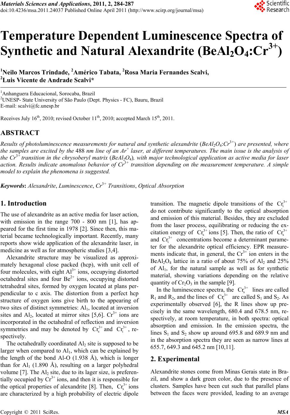 Materials Sciences and Applicatio ns, 2011, 2, 284-287 doi:10.4236/msa.2011.24037 Published Online April 2011 (http://www.scirp.org/journal/msa) Copyright © 2011 SciRes. MSA Temperature Dependent Luminescence Spectra of Synthetic and Natural Alexandrite (BeAl2O4:Cr3+) 1Neilo Marcos Trindade, 2Américo Tabata, 2Rosa Maria Fernandes Scalvi, 2Luis Vicente de Andrade Scalvi* 1Anhanguera Educacional, Sorocaba, Brazil 2UNESP- State University of São Paulo (D ept. Phy sics - FC), Bauru, Brazil E-mail: [email protected] Receives July 16th, 2010; revised October 11th, 2010; accepted March 15th, 2011. ABSTRACT Results of photoluminescen ce measurements for natural and synth etic alexandrite (BeAl2O4:Cr3+) are pre sented, wher e the samples are excited by the 488 nm line of an Ar+ laser, at different temperatures. The main issu e is the analysis of the Cr3+ transition in the chrysoberyl matrix (BeAl2O4), with major technological application as active media for laser action. Results indicate anomalous behavior of Cr3+ transition depending on the measurement temperature. A simple model to explain the phenomena is suggested. Keywords: Alexandrite, Luminescence, Cr3+ Transitions, Optical Absorption 1. Introduction The use of alexandrite as an active media for laser action, with emission in the range 700 - 800 nm [1], has ap- peared for the first time in 1978 [2]. Since then, this ma- terial became technologically important. Recently, many reports show wide application of the alexandrite laser, in medicine as well as for atmospheric studies [3,4]. Alexandrite structure may be visualized as approxi- mately hexagonal close packed (hcp), with unit cell of four molecules, with eight Al3+ ions, occupying distorted octahedral sites and four Be2+ ions, occupying distorted tetrahedral sites, formed by oxygen located at plans per- pendicular to c axis. The distortion from a perfect hcp structure of oxygen ions give birth to the appearing of two sites of distinct symmetries: Al1, located at inversion sites and Al2, located at mirror sites [5,6]. Cr3+ ions are incorporated in the octahedral of reflection and inversion symmetries and may be denoted by and , re- spectively. 3+ s Cr 3+ i Cr The octahedrally coordinated Al2 site is supposed to be larger when compared to Al1, which can be explained by the length of the bond Al-O (1.938 Å), which is longer than for Al1 (1.890 Å), resulting on a larger polyhedral volume [7]. The Al2 site, due to its lager size, is preferen- tially occupied by Cr3+ ions, and then it is responsible for the optical properties of alexandrite [8]. Then, sions are characterized by a high probability of electric dipole transition. The magnetic dipole transitions of the 3+ Cr 3+ Cr 3+ Cr i do not contribute significantly to the optical absorption and emission of this material. Besides, they are excluded from the laser process, equilibrating or reducing the ex- citation energy of sions [5]. Then, the ra tio of s and i concentrations become a determinant parame- ter for the alexandrite optical efficiency. EPR measure- ments indicate that, in general, the Cr3+ ion enters in the BeAl2O4 lattice in a ratio of about 75% of Al2 and 25% of Al1, for the natural sample as well as for synthetic material, showing variations depending on the relative quantity of Cr2O3 in the sample [9]. 3+ Cr 3+ Cr In the luminescence spectra, the s lines are called R1 and R2, and the lines of i are called S1 and S2. As experimentally observed [6], the R lines show up pre- cisely in the same wavelength, 680.4 and 678.5 nm, re- spectively, at room temperature, in both spectra: optical absorption and emission. In the emission spectra, the lines S1 and S2 show up around 695.8 and 689.9 nm and in the absorption spectra they are seen as narrow lines at 655.7, 649.3 and 645.2 nm [10,11]. 3+ Cr 3+ Cr 2. Experimental Alexandrite stones come from Minas Gerais state in Bra- zil, and show a dark green color, due to the presence of clusters. Samples have been cut such that parallel plans between the faces were provided, leading to an average 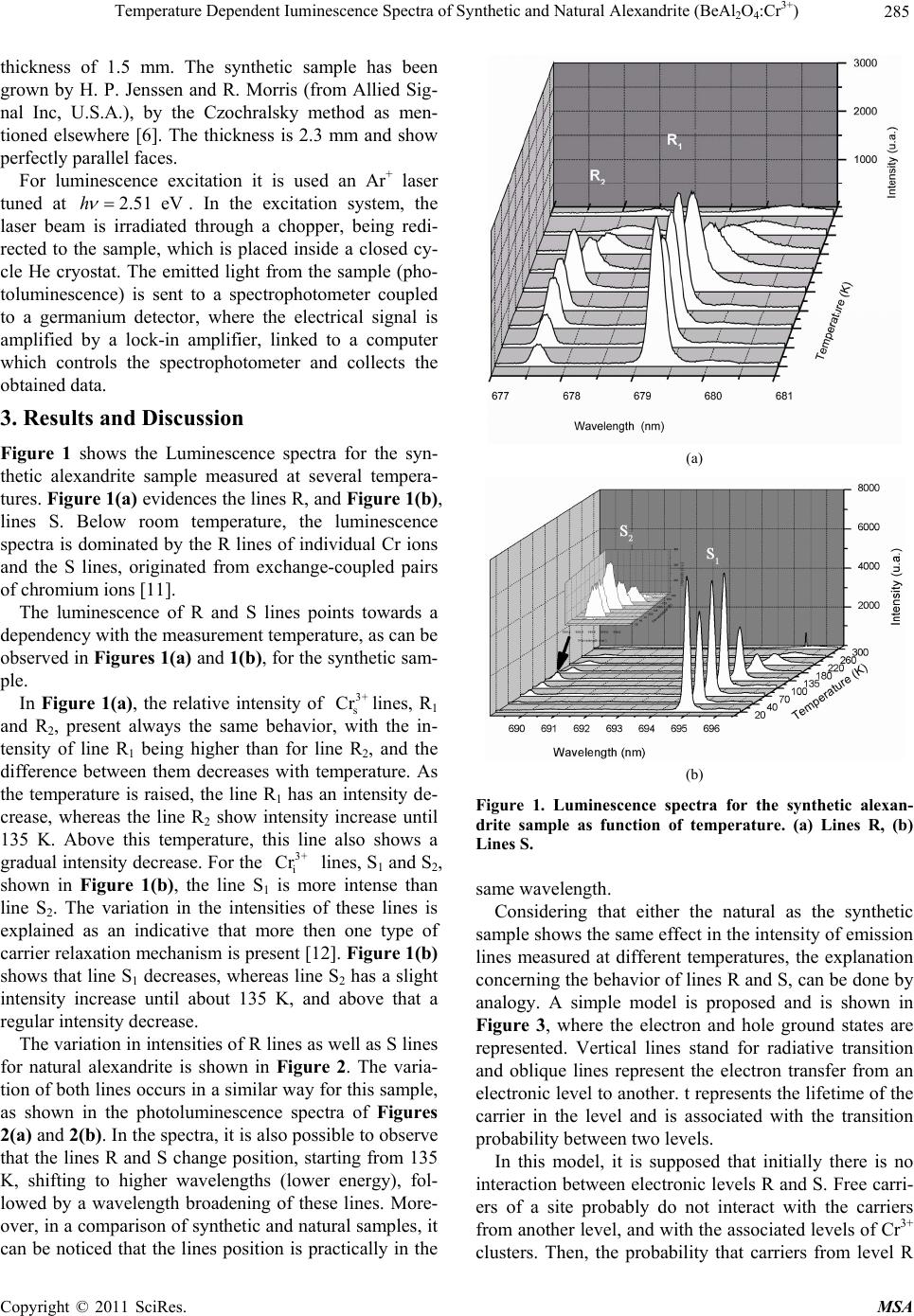 Temperature Dependent Iuminescence Spectra of Synthetic and Natural Alexandrite (BeAlO:Cr3+) 285 2 4 thickness of 1.5 mm. The synthetic sample has been grown by H. P. Jenssen and R. Morris (from Allied Sig- nal Inc, U.S.A.), by the Czochralsky method as men- tioned elsewhere [6]. The thickness is 2.3 mm and show perfectly parallel faces. For luminescence excitation it is used an Ar+ laser tuned at 2.51 eVh . In the excitation system, the laser beam is irradiated through a chopper, being redi- rected to the sample, which is placed inside a closed cy- cle He cryostat. The emitted light from the sample (pho- toluminescence) is sent to a spectrophotometer coupled to a germanium detector, where the electrical signal is amplified by a lock-in amplifier, linked to a computer which controls the spectrophotometer and collects the obtained data. 3. Results and Discussion Figure 1 shows the Luminescence spectra for the syn- thetic alexandrite sample measured at several tempera- tures. Fig ure 1(a) evidences the lines R, and Figure 1(b), lines S. Below room temperature, the luminescence spectra is dominated by the R lines of individual Cr ions and the S lines, originated from exchange-coupled pairs of chromium ions [11]. The luminescence of R and S lines points towards a dependency with the measurement temperature, as can be observed in Figures 1(a) and 1(b), for the synthetic sam- ple. In Figure 1(a), the relative intensity of lines, R1 and R2, present always the same behavior, with the in- tensity of line R1 being higher than for line R2, and the difference between them decreases with temperature. As the temperature is raised, the line R1 has an intensity de- crease, whereas the line R2 show intensity increase until 135 K. Above this temperature, this line also shows a gradual intensity decrease. For the lines, S1 and S2, shown in Figure 1(b), the line S1 is more intense than line S2. The variation in the intensities of these lines is explained as an indicative that more then one type of carrier relaxation mechanism is present [12]. Figure 1(b) shows that line S1 decreases, whereas line S2 has a slight intensity increase until about 135 K, and above that a regular intensity decrease. 3+ s Cr 3+ i Cr The variation in intensities of R lines as well as S lines for natural alexandrite is shown in Figure 2. The varia- tion of both lines occurs in a similar way for this sample, as shown in the photoluminescence spectra of Figures 2(a) and 2(b). In the spectra, it is also possible to observe that the lines R and S change position, starting from 135 K, shifting to higher wavelengths (lower energy), fol- lowed by a wavelength broadening of these lines. More- over, in a comparison of synthetic and natural samples, it can be noticed that the lines position is practically in the (a) (b) Figure 1. Luminescence spectra for the synthetic alexan- drite sample as function of temperature. (a) Lines R, (b) Lines S. same wavelength. Considering that either the natural as the synthetic sample shows the same effect in the intensity of emission lines measured at different temperatures, the explanation concerning the behavior of lines R and S, can be done by analogy. A simple model is proposed and is shown in Figure 3, where the electron and hole ground states are represented. Vertical lines stand for radiative transition and oblique lines represent the electron transfer from an electronic level to another. t represents the lifetime of the carrier in the level and is associated with the transition probability between two levels. In this model, it is supposed that initially there is no interaction between electronic levels R and S. Free carri- ers of a site probably do not interact with the carriers from another level, and with the associated levels of Cr3+ clusters. Then, the probability that carriers from level R C opyright © 2011 SciRes. MSA 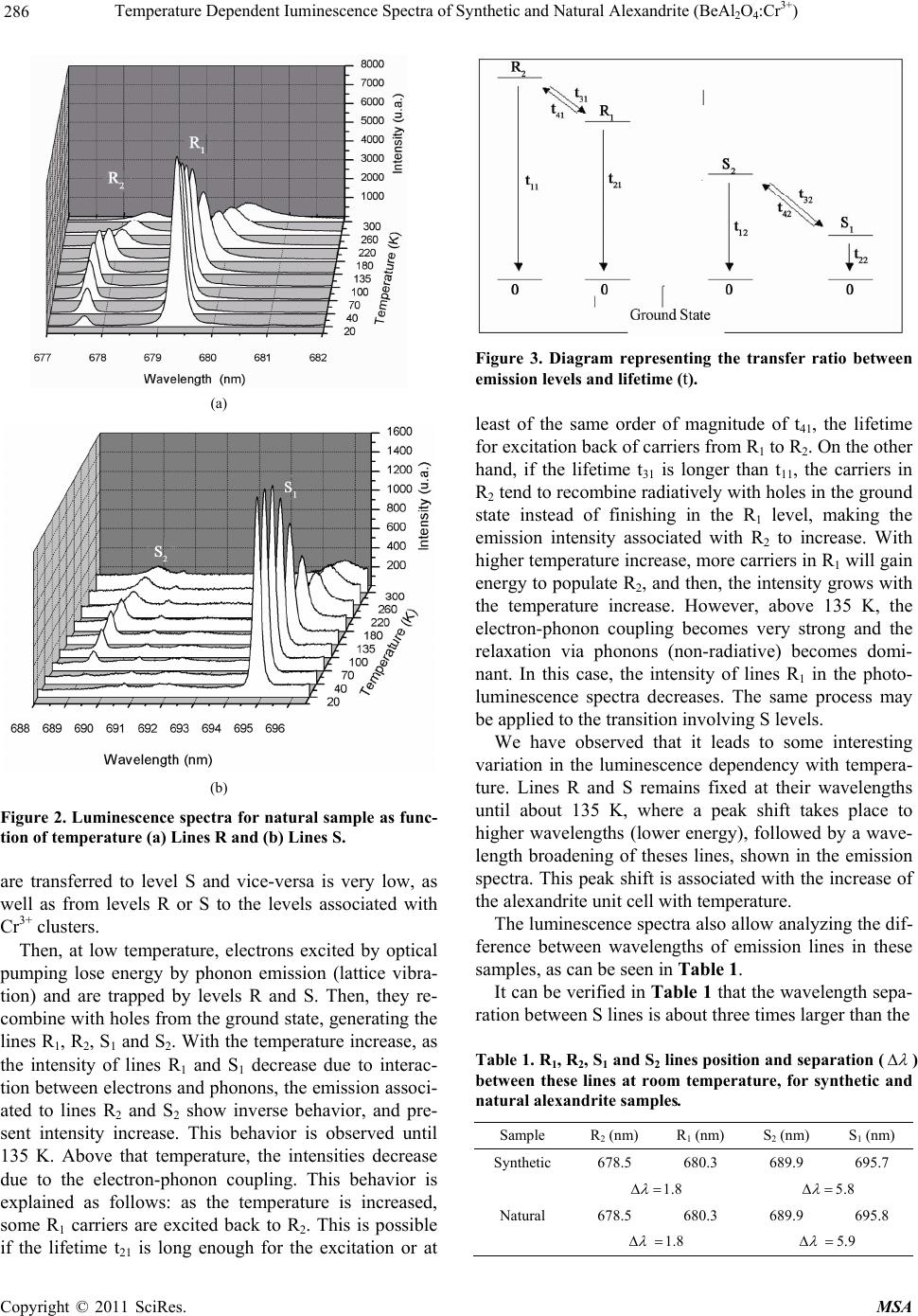 Temperature Dependent Iuminescence Spectra of Synthetic and Natural Alexandrite (BeAlO:Cr3+) 286 2 4 (a) (b) Figure 2. Luminescence spectra for natural sample as func- tion of temperature (a) Lines R and (b) Lines S. are transferred to level S and vice-versa is very low, as well as from levels R or S to the levels associated with Cr3+ clusters. Then, at low temperature, electrons excited by optical pumping lose energy by phonon emission (lattice vibra- tion) and are trapped by levels R and S. Then, they re- combine with holes from the ground state, generating the lines R1, R2, S1 and S2. With the temperature increase, as the intensity of lines R1 and S1 decrease due to interac- tion between electrons and phonons, the emission associ- ated to lines R2 and S2 show inverse behavior, and pre- sent intensity increase. This behavior is observed until 135 K. Above that temperature, the intensities decrease due to the electron-phonon coupling. This behavior is explained as follows: as the temperature is increased, some R1 carriers are excited back to R2. This is possible if the lifetime t21 is long enough for the excitation or at Figure 3. Diagram representing the transfer ratio between emission levels and lifetime (t). least of the same order of magnitude of t41, the lifetime for excitation back of carriers from R1 to R2. On the other hand, if the lifetime t31 is longer than t11, the carriers in R2 tend to recombine radiatively with holes in the ground state instead of finishing in the R1 level, making the emission intensity associated with R2 to increase. With higher temperature increase, more carriers in R1 will gain energy to populate R2, and then, the intensity grows with the temperature increase. However, above 135 K, the electron-phonon coupling becomes very strong and the relaxation via phonons (non-radiative) becomes domi- nant. In this case, the intensity of lines R1 in the photo- luminescence spectra decreases. The same process may be applied to the tran sition involving S levels. We have observed that it leads to some interesting variation in the luminescence dependency with tempera- ture. Lines R and S remains fixed at their wavelengths until about 135 K, where a peak shift takes place to higher wavelengths (lower energy), followed by a wave- length broadening of theses lines, shown in the emission spectra. This peak shift is as sociated with the increase of the alexandrite unit cell with temperature. The luminescence spectra also allow analyzing the dif- ference between wavelengths of emission lines in these samples, as can be seen in Table 1. It can be verified in Table 1 that the wavelength sepa- ration between S lines is about three times larger than the Table 1. R1, R2, S1 and S2 lines position and separation ( ) between these lines at room temperature, for synthetic and natural alexandrite samples. Sample R2 (nm) R1 (nm) S2 (nm) S1 (nm) Synthetic 678.5 680.3 689.9 695.7 1.8 5.8 Natural 678.5 680.3 689.9 695.8 1.8 5.9 C opyright © 2011 SciRes. MSA  Temperature Dependent Iuminescence Spectra of Synthetic and Natural Alexandrite (BeAl2O4:Cr3+) Copyright © 2011 SciRes. MSA 287 separation between R lines. This behavior indicates a stronger low symmetry component of the crystalline field for ions located in a inversion center [13]. 4. Conclusions Photoluminescence measurements carried out at several temperatures for alexandrite samples have shown up as very relevant to the analysis of Cr3+ transition in this ma- terial, because it is related to its potentiality to laser ac- tivity. The temperature dependency of the emission spec- tra exhibits an anomalous behavior for natural samples as well as for the synthetic one, compared to most materials, for instance, semiconductors. Then, a simple model is proposed, based on electronic transitions of levels R and S, characteristic of Cr3+ transitions. 5. Acknowledgements This work was supported by CAPES, CNPq, FAPESP and FUNDUNESP. The authors are grateful to Prof. Tomaz Catunda for the synthetic sample. REFERENCES [1] P. Fabeni, G. P. Pazzi and L. Salvini, “Impurity Centers for Tunable Lasers in the Ultraviolet and Visible Re- gions,” Journal of Physics and Chemistry of Solids, Vol. 52, No. 1, 1991, pp. 299-317. doi:10.1016/0022-3697(91)90069-C [2] G. V. Bukin, V. N. Matrosov, V. P. Orekhova, Yu. L. Remigailo, B. K. Sevastyanov, E. G. Syomin, V. P. Solnt- sev and E. G. Tsvetkov, “Growth of Alexandrite Crystals and Investigation of Their Properties,” Journal of Crystal Growth, Vol. 52, No. 2, April 1981, pp. 537-541. doi:10.1016/0022-0248(81)90335-3 [3] L. Li, T. Kono, W. F. Groff, H. H. Chan, Y. Kitazawa and M. Nozaki, “Comparison Study of a Long-Pulse Pulsed Dye Laser and a Long-Pulse Pulsed Alexandrite Laser in the Treatment of Port Wine Stains,” Journal of Cosmetic and Laser Therapy, Vol. 10, No. 1, March 2008, pp. 12-15. doi:10.1080/14764170701817023 [4] S. C. Collins, T. D. Wilkerson, V. B. Wickwar, D. Rees, J. C. Walling and D. F. Heller, “The Alexandrite Ring Laser: A Spectrally Narrow Lidar Light Source for Atmospheric Fluorescence and Absorption Observations,” In: A. Ans- mann, R. Neuber, P. Rairoux and U. Wandinger, Eds., Advances in Atmospheric Remote Sensing with Lidar, Springer Verlag, Berlin, 1997, pp. 577-580. [5] B. K. Sevast’yanov, “Excited-State Absorption Spectros- copy of Crystal Doped with Cr3+, Ti3+, and Nd3+ Ions, Review,” Crystallography Reports, Vol. 48, No. 6, 2003, pp. 989-1011. [6] R. M. F. Scalvi, M. S. Li and L. V. A. Scalvi, “Annealing Effects on Optical Properties of Natural Alexandrite,” Journal of Physics Condensed Matter, Vol. 15, No. 43, November 2003, pp. 7437-7443. doi:10.1088/0953-8984/15/43/025 [7] S. U. Weber, M. Grodzicki, W. Lottermoser, G. J. Redhammer, G. Tippelt, J. Ponahlo and G Amthauer, “57Fe Mossbauer Spectroscopy, X-Ray Single-Crystal Diffractometry, and Electronic Structure Calculations on Natural Alexandrite,” Physics and Chemistry of Minerals, Vol. 34, No. 7, 2007, pp. 507-515. doi:10.1007/s00269-007-0166-6 [8] G. V. Bukin, A. V. Eliseev, V. N. Matrosov, V. P. Solnt- sev, E. I. Kharchenko and E. G. Tsvetjov, “The Growth and Examination of Optical Properties of Gem Alexan- drite,” Proceedings of the XI General Meeting of IMA, Novosibirsk, 1980, pp. 317-328. [9] H. Rager, A. Bahshandh-Khiri and K. N. Schmetzer, “In- vestigation of the Intracrystalline Cr3+ Distribution in Natural and Synthetic Alexandrites,” Neues Jahrbuch für Mineralogie Monatshefte, Vol. 12, 1998, pp. 545-557. [10] R. C. Powell, L. Xi, X. Gang and G. J. Quarles, “Spec- troscopic Properties of Alexandrite Crystals,” Physical Review B, Vol. 32, No. 5, September 1985, pp. 2788- 2797. doi:10.1103/PhysRevB.32.2788 [11] S. P. Jamilson and G. F. Imbusch, “Temperature De- pendence of the Luminescence from Heavily Doped ruby,” Journal of Luminescence, Vol. 75, No. 2, Septem- ber 1997, pp. 143-147. [12] K. L. Schepler, “Fluorescence of Inversion Site Cr3+ Ions in Alexandrit e,” Journal of Applied Physics, Vol. 56, No. 5, September 1984, pp. 1314-1318. doi:10.1063/1.334119 [13] B. Suchocki , G. D. Gilliland, R. C. Powell and J. M. Bowen, “Spectroscopic Properties of Alexandrite Crystals II.,” Journal of Luminescenc, Vol. 37, No. 1, April 1987, pp. 29-37. doi:10.1016/0022-2313(87)90179-7 |