

Paper Menu >>
Journal Menu >>
 Surgical Science, 2011, 2, 66-68 doi:10.4236/ss.2011.22014 Published Online April 2011 (http://www.SciRP.org/journal/ss) Copyright © 2011 SciRes. SS Mixed Epithelial and Stromal Tumor of the Kidney: A Case Report Ebru Demiralay1, Cem Çomunoğlu1, Handan Özdemir1, Ramazan Yavuz Akman2 1Departmen t o f Pathology, Faculty of Medicine, Baskent University, Ankara, Turkey 2Department of Ur ol o gy, Faculty of Medicine, Baskent Universty, Ankara, Turkey E-mail: [email protected] Received December 2, 2010; revised January 25, 2011; accepted Februar y 25, 2011 Abstract Mixed epithelial and stromal tumor of the kidney (MESTK) is a recently described rare neoplasm. Malignant transformation, recurrence and metastasis are rare, therefore histopathological distinction from other renal neoplasms, especially from renal cell carcinoma is important. Histologically the tumor is composed of bi- phasic components including cysts and tubules embedded in the spindle cell stroma. We report a case of a MESTK in 60-year-old postmenopousal woman who presented with an incidental solid renal mass but no urinary complaint. Keywords: Kidney, Mixed Epithelial and Stromal Tumor 1. Introduction The mixed epithelial and stromal tumor of the kidney is a rare and newly defined entity which was included in the WHO 2004 renal tumor classification [1-7]. To the best of our knowledge, less than 100 MESTK cases have been reported. A similar lesion was previously referred to as cystic hamartoma of the renal pelvis, adult type of mesoblastic nephroma or cystic nephroma or mature nephroblastic tumor and cystic partially differentiated nephroblasto ma [1,4,5,7-10]. Differential diagnosis includes congenital mesoblastic nephroma, cystic nephroma and multilocular cystic ren al cell carcinoma [1,4,7,10]. Recent molecular studies sug- gest that, MESTK has no relationship to mesoblastic nephroma [8]. Microscopically, the tumor is composed of biphasic components including cysts and tubules embedded in the spindle cell stroma. The epithelial elements, composed of clusters of tubules with variable lining, are found scat- tered within the spindle cell stroma [2,4-6,8-10]. Cellular foci reminiscent of ovarian stroma or solitary fibrous tumor are also present. Immunohistochemically, desmin, smooth muscle actin and vimentin are often diffusely and strongly positive in the spindle cells of the tumors, whereas HMB-45 and CD 34 stains are negative [4,8,10]. Estrogen and progester- one receptors are frequently positive in the nuclei of the spindle cells [1,2,4,8,9]. Epithelial elements show usu- ally positive immunoreactivity with EMA and cy- tokeratin [1,10]. Although recently a few cases of malignant MESTK have been reported in the literature, malignant transfor- mation, recurrence and metastasis are rare. The aim of this study was to report a case of a MESTK in 60-year-old postmenopousal woman who presented with an incidental solid renal mass but without any uri- nary complaints. 2. Case Report A 60-year-old postmenopousal woman presented with cough for 15 days. She had a history of type 2 diabetes and hyperlipidemia. A computed tomography scan of thorax showed, as an incidental finding, a solid mass in right kidney. Contrast enhanced computed tomography scan of abdomen showed a uniformly lobulated solid mass with minimal contrast enhancement, situated in the upper medial aspect of the right kidney, measuring 3.5 × 3.5 × 3 cm. Neither radiological distinction of malig- nancy nor exclusion of renal cell carcinoma could be clearly obtained on magnetic resonance imaging. There was no evidence of distant metastasis. Physical examina- tion was unremarkable. She had high glucose level in her routine blood investigations. Urinary cytological exami- nation was normal. Radical nephrectomy was performed. 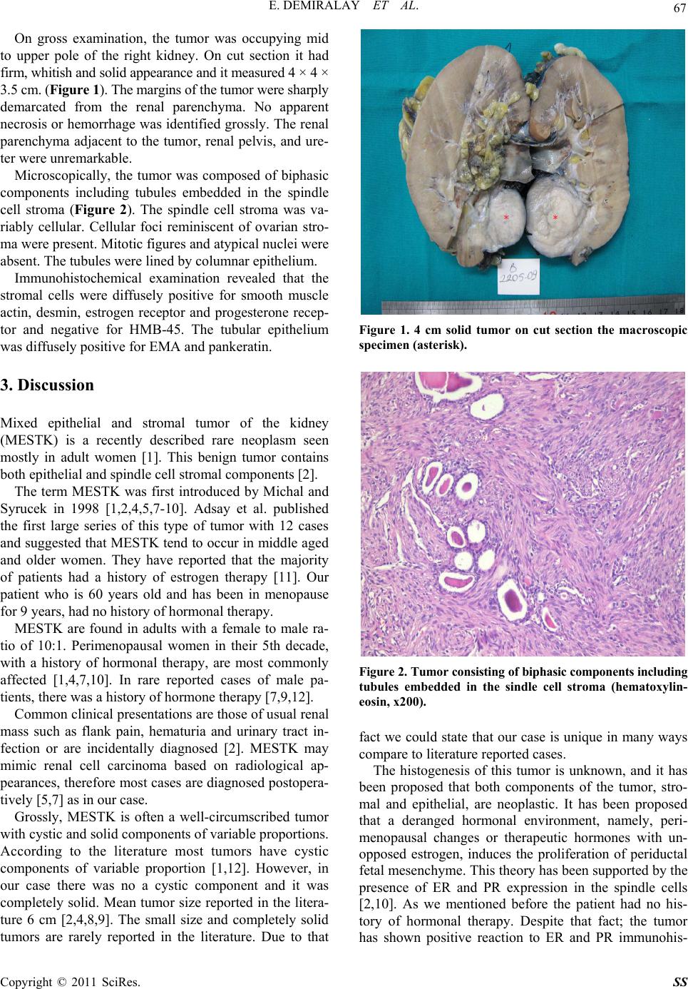 E. DEMIRALAY ET AL. 67 On gross examination, the tumor was occupying mid to upper pole of the right kidney. On cut section it had firm, whitish and solid appearance and it measured 4 × 4 × 3.5 cm. (Figure 1). The margins of the tumor were sharply demarcated from the renal parenchyma. No apparent necrosis or hemorrhage was identified grossly. The renal parenchyma adjacent to the tumor, renal pelvis, and ure- ter were unremarkable. Microscopically, the tumor was composed of biphasic components including tubules embedded in the spindle cell stroma (Figure 2). The spindle cell stroma was va- riably cellular. Cellular foci reminiscent of ovarian stro- ma were present. Mitotic figures and atypical nuc lei were absent. The tubules were lined by co lumnar epithelium. Immunohistochemical examination revealed that the stromal cells were diffusely positive for smooth muscle actin, desmin, estrogen receptor and progesterone recep- tor and negative for HMB-45. The tubular epithelium was diffusely positive for EMA and pankeratin. 3. Discussion Mixed epithelial and stromal tumor of the kidney (MESTK) is a recently described rare neoplasm seen mostly in adult women [1]. This benign tumor contains both epithelial and spindle cell stromal components [2]. The term MESTK was first introduced by Michal and Syrucek in 1998 [1,2,4,5,7-10]. Adsay et al. published the first large series of this type of tumor with 12 cases and suggested that MESTK tend to occur in middle aged and older women. They have reported that the majority of patients had a history of estrogen therapy [11]. Our patient who is 60 years old and has been in menopause for 9 years, had no history of hor monal the rapy. MESTK are found in adults with a female to male ra- tio of 10:1. Perimenopausal women in their 5th decade, with a history of hormonal therapy, are most commonly affected [1,4,7,10]. In rare reported cases of male pa- tients, there was a history of hormone therapy [7,9,12]. Common clinical presentations are those of usual renal mass such as flank pain, hematuria and urinary tract in- fection or are incidentally diagnosed [2]. MESTK may mimic renal cell carcinoma based on radiological ap- pearances, therefore most cases are diagnosed postopera- tively [5,7] as in our case. Grossly, MESTK is often a well-circumscribed tumor with cystic and solid components of variable proportio ns. According to the literature most tumors have cystic components of variable proportion [1,12]. However, in our case there was no a cystic component and it was completely solid. Mean tumor size reported in the litera- ture 6 cm [2,4,8,9]. The small size and completely solid tumors are rarely reported in the literature. Due to that Figure 1. 4 cm solid tumor on cut section the macroscopic specimen (asterisk). Figure 2. Tumor consisting of biphasic components including tubules embedded in the sindle cell stroma (hematoxylin- eosin, x200). fact we could state that our case is unique in many ways compare to literature reported cases. The histogenesis of this tumor is unknown, and it has been proposed that both components of the tumor, stro- mal and epithelial, are neoplastic. It has been proposed that a deranged hormonal environment, namely, peri- menopausal changes or therapeutic hormones with un- opposed estrogen, induces the proliferation of periductal fetal mesenchyme. This theory has been supported by the presence of ER and PR expression in the spindle cells [2,10]. As we mentioned before the patient had no his- tory of hormonal therapy. Despite that fact; the tumor has shown positive reaction to ER and PR immunohis- Copyright © 2011 SciRes. SS 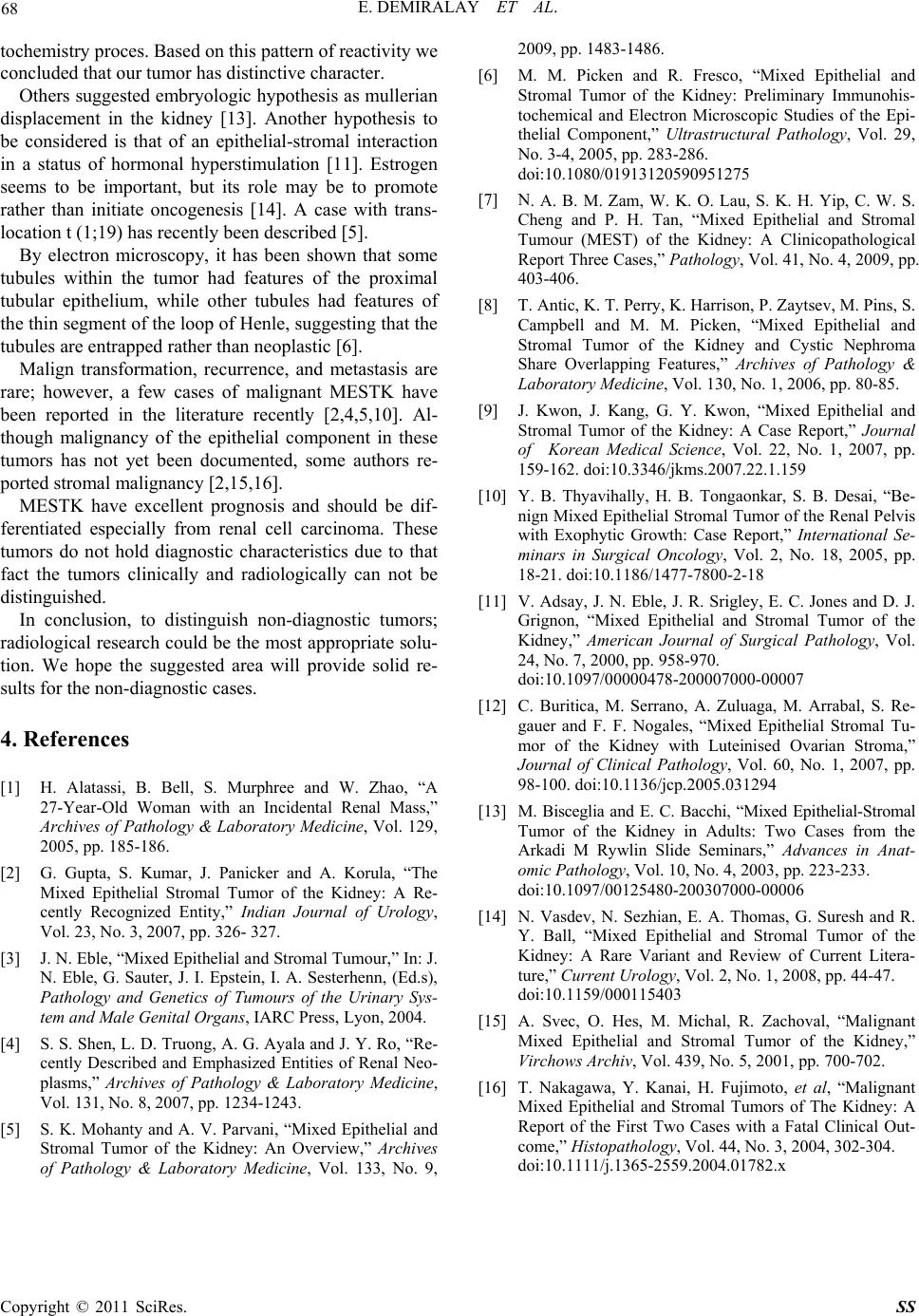 E. DEMIRALAY ET AL. Copyright © 2011 SciRes. SS 68 tochemistry proces. Based on this pattern of reactivity we concluded that our tumor has distinctive character. Others suggested embryologic hypothesis as mullerian displacement in the kidney [13]. Another hypothesis to be considered is that of an epithelial-stromal interaction in a status of hormonal hyperstimulation [11]. Estrogen seems to be important, but its role may be to promote rather than initiate oncogenesis [14]. A case with trans- location t (1;19) has recently been described [5]. By electron microscopy, it has been shown that some tubules within the tumor had features of the proximal tubular epithelium, while other tubules had features of the thin segment of the loop of Henle, suggesting that the tubules are entrapped rather than neoplastic [6]. Malign transformation, recurrence, and metastasis are rare; however, a few cases of malignant MESTK have been reported in the literature recently [2,4,5,10]. Al- though malignancy of the epithelial component in these tumors has not yet been documented, some authors re- ported stromal malignancy [2,15,16]. MESTK have excellent prognosis and should be dif- ferentiated especially from renal cell carcinoma. These tumors do not hold diagnostic characteristics due to that fact the tumors clinically and radiologically can not be distinguished. In conclusion, to distinguish non-diagnostic tumors; radiological research could be the most appropriate solu- tion. We hope the suggested area will provide solid re- sults for the non-diagnostic cases. 4. References [1] H. Alatassi, B. Bell, S. Murphree and W. Zhao, “A 27-Year-Old Woman with an Incidental Renal Mass,” Archives of Pathology & Laboratory Medicine, Vol. 129, 2005, pp. 185-186. [2] G. Gupta, S. Kumar, J. Panicker and A. Korula, “The Mixed Epithelial Stromal Tumor of the Kidney: A Re- cently Recognized Entity,” Indian Journal of Urology, Vol. 23, No. 3, 2007, pp. 326- 327. [3] J. N. Eble, “Mixed Epithel ial and St romal Tu mour,” In: J. N. Eble, G. Sauter, J. I. Epstein, I. A. Sesterhenn, (Ed.s), Pathology and Genetics of Tumours of the Urinary Sys- tem and Male Genital Organs, IARC Press, Lyon, 2004. [4] S. S. Shen, L. D. Truong, A. G. Ayala and J. Y. Ro, “Re- cently Described and Emphasized Entities of Renal Neo- plasms,” Archives of Pathology & Laboratory Medicine, Vol. 131, No. 8, 2007, pp. 1234-1243. [5] S. K. Mohanty and A. V. Parvani, “Mixed Epithelial and Stromal Tumor of the Kidney: An Overview,” Archives of Pathology & Laboratory Medicine, Vol. 133, No. 9, 2009, pp. 1483-1486. [6] M. M. Picken and R. Fresco, “Mixed Epithelial and Stromal Tumor of the Kidney: Preliminary Immunohis- tochemical and Electron Microscopic Studies of the Epi- thelial Component,” Ultrastructural Pathology, Vol. 29, No. 3-4, 2005, pp. 283-286. doi:10.1080/01913120590951275 [7] N. A. B. M. Zam, W. K. O. Lau, S. K. H. Yip, C. W. S. Cheng and P. H. Tan, “Mixed Epithelial and Stromal Tumour (MEST) of the Kidney: A Clinicopathological Report Three Cases,” Pathology, Vol. 41, No. 4, 2009, pp. 403-406. [8] T. Antic, K. T. Perry, K. Harrison, P. Zaytsev, M. Pins, S. Campbell and M. M. Picken, “Mixed Epithelial and Stromal Tumor of the Kidney and Cystic Nephroma Share Overlapping Features,” Archives of Pathology & Laboratory Medicine, Vol. 130, No. 1, 2006, pp. 80-85. [9] J. Kwon, J. Kang, G. Y. Kwon, “Mixed Epithelial and Stromal Tumor of the Kidney: A Case Report,” Journal of Korean Medical Science, Vol. 22, No. 1, 2007, pp. 159-162. doi:10.3346/jkms.2007.22.1.159 [10] Y. B. Thyavihally, H. B. Tongaonkar, S. B. Desai, “Be- nign Mixed Epithelial Stromal Tumor of the Renal Pelvis with Exophytic Growth: Case Report,” International Se- minars in Surgical Oncology, Vol. 2, No. 18, 2005, pp. 18-21. doi:10.1186/1477-7800-2-18 [11] V. Adsay, J. N. Eble, J. R. Srigley, E. C. Jones and D. J. Grignon, “Mixed Epithelial and Stromal Tumor of the Kidney,” American Journal of Surgical Pathology, Vol. 24, No. 7, 2000, pp. 958-970. doi:10.1097/00000478-200007000-00007 [12] C. Buritica, M. Serrano, A. Zuluaga, M. Arrabal, S. Re- gauer and F. F. Nogales, “Mixed Epithelial Stromal Tu- mor of the Kidney with Luteinised Ovarian Stroma,” Journal of Clinical Pathology, Vol. 60, No. 1, 2007, pp. 98-100. doi:10.1136/jcp.2005.031294 [13] M. Bisceglia and E. C. Bacchi, “Mixed Epithelial-Stromal Tumor of the Kidney in Adults: Two Cases from the Arkadi M Rywlin Slide Seminars,” Advances in Anat- omic Pathology, Vol. 10, No. 4, 2003, pp. 223-233. doi:10.1097/00125480-200307000-00006 [14] N. Vasdev, N. Sezhian, E. A. Thomas, G. Suresh and R. Y. Ball, “Mixed Epithelial and Stromal Tumor of the Kidney: A Rare Variant and Review of Current Litera- ture,” Current Urology, Vol. 2, No. 1, 2008, pp. 44-47. doi:10.1159/000115403 [15] A. Svec, O. Hes, M. Michal, R. Zachoval, “Malignant Mixed Epithelial and Stromal Tumor of the Kidney,” Virchows Archiv, Vol. 439, No. 5, 2001, pp. 700-702. [16] T. Nakagawa, Y. Kanai, H. Fujimoto, et al, “Malignant Mixed Epithelial and Stromal Tumors of The Kidney: A Report of the First Two Cases with a Fatal Clinical Out- come,” Histopathology, Vol. 44, No. 3, 2004, 302-304. doi:10.1111/j.1365-2559.2004.01782.x |