

Paper Menu >>
Journal Menu >>
 Surgical Science, 2011, 2, 57-61 doi:10.4236/ss.2011.22012 Published Online April 2011 (http://www.SciRP.org/journal/ss) Copyright © 2011 SciRes. SS Sentinel Lymph Node Biopsy as Guidance for Lateral Neck Dissection in Patients with Papillary Thyroid Carcinoma Yoshifumi Ikeda Department of Surgery, Teiky o Uni versi t y School of Medi cine, 2-11-1 Kaga, Itabashi-ku, Tokyo 173-8605, Japan E-mail: [email protected] Received January 11, 2011; revised January 20, 2011; accepted Ja n u ary 27, 2011 Abstract Introduction: The surgical management of lateral lymph nodes in differentiated thyroid carcinoma is contro- versies. Therefore, we analyzed whether sentinel lymph nodes (SLN) biopsy of the first draining nodes in the jugulo-carotid chain is an accurate technique to select patients with true-positive but nonpalpable lymph nodes for selective lateral node dissection. Materials and Methods: From January 2009 to December 2009, 12 patients with solitary papillary carcinoma measuring 2 cm by ultrasonography were included in this study. After the thyroid gland was exposed to avoid injuring the lateral thyroid lymphatic connection, approxi- mately 0.2 ml of 5mg/ml indocyanine green was injected into the parenchyma of upper and lower thyroid gland. Some stained lymph nodes in the jugulo-carotid chain could be identified following the stained lym- phatic duct and dissected as the SLN. After that, thyroidectomy with modified neck dissection was per- formed. Results: The mean tumor size was 22.1 ± 4.6 mm. Identification and biopsy of stained SLN in the ipsilateral jugulo-carotid chain was successful in all 12 cases. In 6 cases, histopathological analysis of SLNs revealed metastases of the papillary thyroid carcinoma. Among them, 2 cases had additional metastatic lymph nodes in the ipsilateral compartment. Of the 6 patients who had negative lymph node metastasis (LNM) in SLNs, all patients had negative LNM in the ipsilateral compartment. Conclusions: The method may be helpful in the detection of true-positive but nonpalpable lymph nodes and may support a decision to perform a selective lateral node dissection in patients with papillary thyroid carcinoma. Keywords: Sentinel Lymph Node, Thyroid Carcinoma, Lateral Neck Dissection 1. Introduction Well differentiated papillary thyroid carcinoma has a high propensity to spread to regional lymph nodes and lymph node metastases are frequently occurred in pa- tients with clinically negative lymph nodes [1]. Since lymph node metastasis has not been considered progno s- tic poor survival, prophylactic lymph node dissection is controversial [2]. There is wide consensus on an anato- mosurgical subdivision of potentially involved lymph nodes in four compartments; central, ipsilateral, conta- lateral, and mediastinal [4]. The central and ipsilateral compartments are two representative compartments to which papillary carcinoma can metastasize. The central compartment can be dissected via the same surgical inci- sion used for thyroidectomy. Furthermore, a second sur- gery for recurrence to the central nodes increases the risk of complications such as recurrent nerve paralysis and parathyroid dysfunction [5,6]. Therefore, routine central node dissection is recommended at the initial surgery regardless of whether the dissection is therapeutic or prophylactic. In contrast, the lateral compartment dissec- tion, regarded as the second region to which papillary carcinoma metastasizes, is less accessible because wound extension and a wide range of tissue peeling are neces- sary for lateral node dissection, and postoperative com- plaints of patients such as pain, limitation of neck movement, edema, and sensory abnormality increase. Therefore, lateral node dissection should be performed for patients with papillary carcinoma demonstrating me- tastasis in the lateral compartment. Although preopera- tive radiological investigations such as ultrasonography (US), enhanced computed tomography (CT), and mag- netic resonance imaging (MRI) were improved, its diag- nostic accuracy is far from perfect. The concept of sen tinel lymp h nodes (SLN) as the first  58 Y. IKEDA node(s) to receive drainage from a tumor was introduced by Ramon Cabanas [7] and later well established in skin melanoma [8] and breast cancer. [9,10] Controversies in the surgical management of lymph nodes in differenti- ated thyroid carcinoma, have led some authors to pro- pose that SLN should be sampled in patients with thyroid carcinoma to support the decision whether to perform lymph node dissection or not. The technique is feasible and safe and may be used to select patients with true positive lymph nodes fo r selective lymph nod e dissection. [11-17] In previous studies, the stained lymphatic channels in the central compartment were traced and followed up to the first blue-stained lymph node, which was defined as the SLN. However, central and lateral compartment each has its own lymphatic vessels. We analyzed whether SLN biopsy of the first draining node(s) in the jugulo- carotid chain is an accurate technique to select patients with true-positive but nonpalpable lymph nodes for se- lective lateral node dissection. 2. Materials and Methods 2.1. Patients From January 2009 to December 2009, 12 patients with well differentiated papillary thyroid carcinoma under- went SLN biopsy in the lower jugulo-carotid chain. Pre- operatively, all patients had clinical ex amination for sus- picious thyroid tumors, US of the neck, fine needle aspi- ration biopsy, CT of the neck, chest X-ray, and blood tests of thyroid hormones, thyrotrophin (TSH), serum thyroglobulin, and anti-th yroglobu lin antibody lev els. No patient had a history of neck surgery for head and neck cancers. Patients with solitary papillary carcinoma mea- suring 2 cm by US without massive extrathyroid exten- sion (corresponding T4 or pT4), clinically apparent lymph node in lateral component or distant metastasis at diagnosis were included in this study. The study was approved by the Institutional Review Board of our insti- tution, and written informed consent sheet was obtained from each patient. 2.2. Surgical Procedure and Detection of SLN Under general anesthesia, the patient is placed in a su- pine position with the neck extended. A transversal skin incision was made on the anterior border of the sterno- cleidomastoid muscle of the tumor side, 3 cm above the sternal notch. The superior and inferior skin flaps devel- oped and the strap muscles were separated from the sternocleidomastoid muscle. The sternohyoid muscle was separated from the sternothyroid muscle. After di- viding the sternohyoid and sternothyroid muscle, the sternohyoid muscle was retracted medially and the ster- nothyroid muscle was retracted laterally. Then the thy- roid gland could be exposed to avoid injuring the lateral thyroid lymphatic connection. By using a 27-gauge nee- dle, approximately 0.2 ml of 5mg/ml indocyanine green was injected into the parenchyma of upper and lower thyroid gland. Care was taken not to stain any surround- ing tissue with the indocyanine green. After 5 minutes, connective tissue between the sternothyroid and sterno- cleidomastoid muscles was sharply divided and the ca- rotid artery and the internal jugular vein were exposed. During this procedure, we could usually find a stained lymphatic duct cross the carotid artery. Some stained lymph nodes in the jugulo-carotid chain could be identi- fied following the stained lymphatic duct. The first stained lymph node was defined as the SLN. After that, the lateral neck dissection was continued above and be- yond the omohyoid muscle, around the internal jugular vein, extending from the carotid artery to the trapezoid muscle and total thyroid lobectomy and dissection of the central neck compartment were performed (Figure 1). All dissected nodes were examined by conventional (hema- toxylin eosin) histopathological examination. Postopera- tively, thyroid-stimulating hormone suppression thyrox- ine therapy was administered to all patients. All patients were followed regularly by clinical and US examination, whole-body radioiodine scanning, and serum thyroglobu- lin measurements. 2.3. Data Analysis Pathologic specimens were st ai ned wi t h hematoxyl i n and 30 40 Figure 1. Macroscopic appearance of resected specimen (lymph node was separated from each compartment): (red circle) central compartment, (blue circle) lateral compart- ment, (black circle) sentinel lymph nodes in lateral com- partment. Copyright © 2011 SciRes. SS 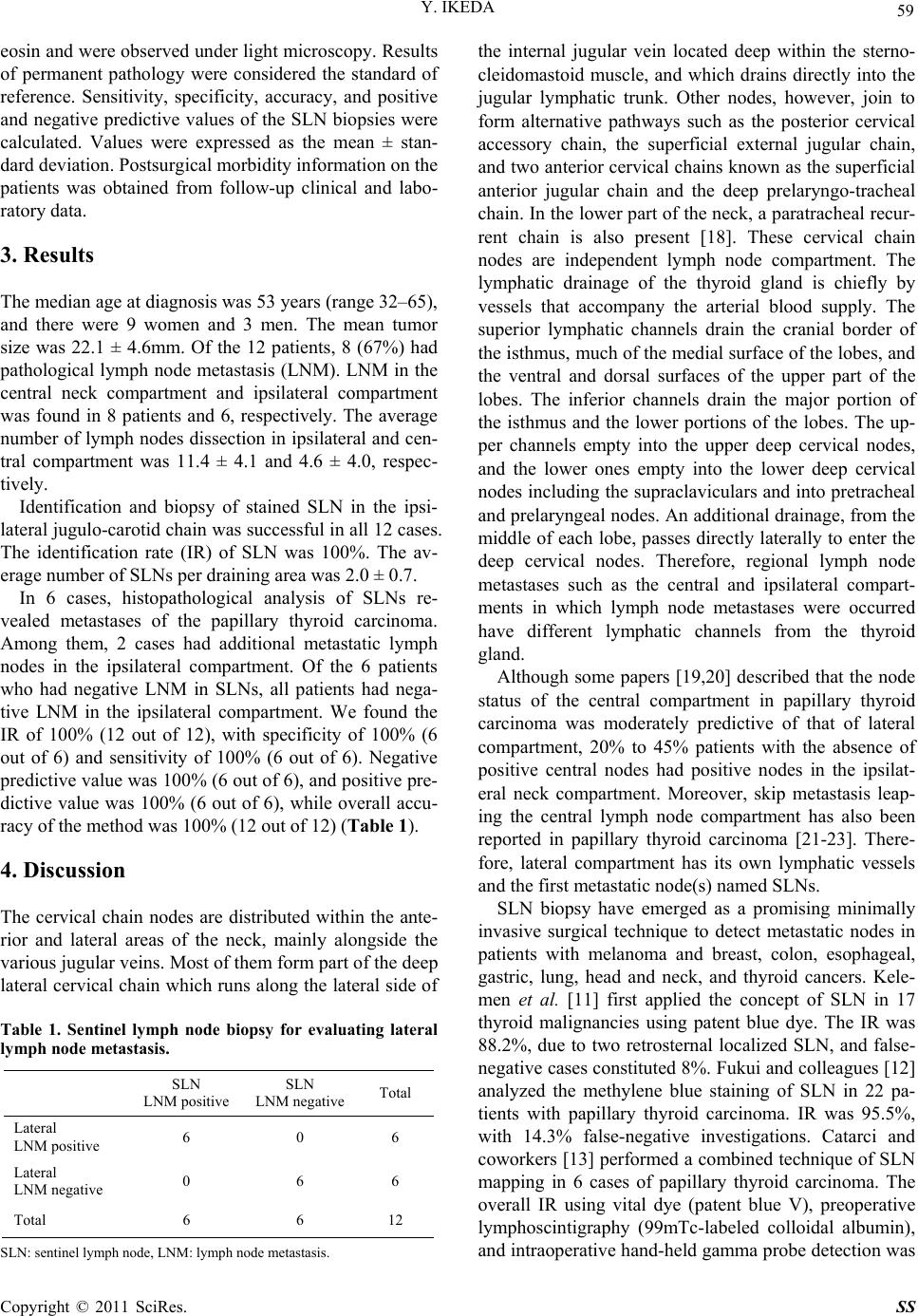 Y. IKEDA 59 eosin and were observed under light microscopy. Results of permanent pathology were considered the standard of reference. Sensitivity, specificity, accuracy, and positive and negative predictive values of the SLN biopsies were calculated. Values were expressed as the mean ± stan- dard deviation. Postsurgical morbidity information on the patients was obtained from follow-up clinical and labo- ratory data. 3. Results The median age at diagnosis was 53 years (range 32–65), and there were 9 women and 3 men. The mean tumor size was 22.1 ± 4.6mm. Of the 12 patients, 8 (67%) had pathological lymph node metastasis (LNM). LNM in the central neck compartment and ipsilateral compartment was found in 8 patients and 6, respectively. The average number of lymph nodes dissection in ipsilateral and cen- tral compartment was 11.4 ± 4.1 and 4.6 ± 4.0, respec- tively. Identification and biopsy of stained SLN in the ipsi- lateral jugulo-carotid chain was successful in all 12 cases. The identification rate (IR) of SLN was 100%. The av- erage number of SLNs per draining area was 2.0 ± 0.7. In 6 cases, histopathological analysis of SLNs re- vealed metastases of the papillary thyroid carcinoma. Among them, 2 cases had additional metastatic lymph nodes in the ipsilateral compartment. Of the 6 patients who had negative LNM in SLNs, all patients had nega- tive LNM in the ipsilateral compartment. We found the IR of 100% (12 out of 12), with specificity of 100% (6 out of 6) and sensitivity of 100% (6 out of 6). Negative predictive va lue was 100% (6 out of 6), and pos itive pre- dictive value was 100% (6 out of 6), while overall accu- racy of the method was 100% (12 out of 12) (Table 1). 4. Discussion The cervical chain nodes are distributed within the ante- rior and lateral areas of the neck, mainly alongside the various jugular veins. Most of them form part of the deep lateral cervical chain which runs along the lateral side of Table 1. Sentinel lymph node biopsy for evaluating lateral lymph node metastasis. SLN LNM positiveSLN LNM negative Total Lateral LNM positive 6 0 6 Lateral LNM negative 0 6 6 Total 6 6 12 SLN: sentinel lymph no d e, LNM: lymph node me t astasis. the internal jugular vein located deep within the sterno- cleidomastoid muscle, and which drains directly into the jugular lymphatic trunk. Other nodes, however, join to form alternative pathways such as the posterior cervical accessory chain, the superficial external jugular chain, and two anterior cervical chains known as the superficial anterior jugular chain and the deep prelaryngo-tracheal chain. In the lower part of the neck, a paratracheal recur- rent chain is also present [18]. These cervical chain nodes are independent lymph node compartment. The lymphatic drainage of the thyroid gland is chiefly by vessels that accompany the arterial blood supply. The superior lymphatic channels drain the cranial border of the isthmus, much of the medial surface of the lobes, and the ventral and dorsal surfaces of the upper part of the lobes. The inferior channels drain the major portion of the isthmus and the lower portions of the lobes. The up- per channels empty into the upper deep cervical nodes, and the lower ones empty into the lower deep cervical nodes including the supraclaviculars and into pretracheal and prelaryngeal n odes. An ad ditional drainage, from the middle of each lobe, passes directly laterally to enter the deep cervical nodes. Therefore, regional lymph node metastases such as the central and ipsilateral compart- ments in which lymph node metastases were occurred have different lymphatic channels from the thyroid gland. Although some papers [19,20] described that the node status of the central compartment in papillary thyroid carcinoma was moderately predictive of that of lateral compartment, 20% to 45% patients with the absence of positive central nodes had positive nodes in the ipsilat- eral neck compartment. Moreover, skip metastasis leap- ing the central lymph node compartment has also been reported in papillary thyroid carcinoma [21-23]. There- fore, lateral compartment has its own lymphatic vessels and the first metastatic node(s) named SLNs. SLN biopsy have emerged as a promising minimally invasive surgical technique to detect metastatic nodes in patients with melanoma and breast, colon, esophageal, gastric, lung, head and neck, and thyroid cancers. Kele- men et al. [11] first applied the concept of SLN in 17 thyroid malignancies using patent blue dye. The IR was 88.2%, due to two retrosternal localized SLN, and false- negative cases constitu ted 8%. Fukui and colleagu es [12] analyzed the methylene blue staining of SLN in 22 pa- tients with papillary thyroid carcinoma. IR was 95.5%, with 14.3% false-negative investigations. Catarci and coworkers [13] performed a combined technique of SLN mapping in 6 cases of papillary thyroid carcinoma. The overall IR using vital dye (patent blue V), preoperative lymphoscintigraphy (99mTc-labeled colloidal albumin), and intraoperative hand-held gamma probe detection was Copyright © 2011 SciRes. SS  60 Y. IKEDA 100%, while IR was 66%, 50%, and 83% for each of th e methods respectively. In a recent study, Roh et al. [24] showed that SLNs were identified in 46 of 50 patients (92%). Of these, 14 SLNs were positive and 32 SLNs were negative on intraoperative frozen sections. One patient had a positive SLN in the jug ular region and sub- sequently underwent modified radical neck dissection. Final pathologic examination revealed that 18 patients (36%), including 4 who had negative SLNs, had central lymph node metastasis. Thus, the sensitivity, specificity, accuracy, and positive and negative predictive values of SLN biopsy were 77.8%, 100%, 92%, 100%, and 88.9%, respectively. Temporary and permanent hypocalcemia developed in 19 patients and 1 patient, respectively. There were no di rect c omplications of SL N s ampling. Overall, the previous studies have shown that stained SLNs were found in the central or lateral neck compart- ments, or both, and SLN biopsy has the potential to iden- tify which patients might be most likely to benefit from central neck dissection at the time of initial surgery [2 4]. Since central neck dissection did not increase permanent morbidity and revealed a significant rate of non-clinically evident node metastases, central neck dissection should be routinely combined with thyroidectomy in papillary thyroid carcinoma [25]. Taking into consideration the central and ipsilateral compartments have different lymphatic channels from the thyroid gland and its own SLN and central neck dis- section was routinely performed in our hospital, our study was designed to trace the stained nodes in the ju- gulo-carotid chain to identify which patients might be most likely to ben efit from lateral neck dissection in cas- es of papillary thyroid carcinoma. In our study we used total of 0.4 ml of 5 mg/ml indo- cyanine green injected into the parenchyma of upper and lower thyroid gland for intraoperative lymphatic map- ping. After 5 minutes, the whole thyroid lobe in which indocyanine green injected dyed well. Although the ma- jority of lymphatic channels an d lymph nodes were dyed blue in central compartment, several lymphatic channels were identified in lateral compartment. The SLNs in lat- eral compartment were identified following a stained lymphatic channel. Although the sample size was small in our study, IR was 100 %, with specificity of 100% and sensitivity of 100%. Negativ e pred ictive value was 100 %, and positive predictive value was 100%. Moreover, 4 in 6 cases with metastasis to the lateral compartment, the SLN is the only positive nod e. Our report suggest that SLN biopsy of lateral com- partment in patients who have papillary thyroid carci- noma without suspected lymphadenopathy can identify occult metastases early and can avoid the morbidity as- sociated with routine and unnecessary lymph node dis- section. However, additional studies are needed that in- clude diagnostic sensitivity in frozen section, larger pa- tient samples, a control group and longer follow-up. 5. Conclusions Our results imply that SLN biopsy in the lower jugulo- carotid chain using indocyanine green mapping is a fea- sible, safe, and accurate method of estimating lymph node status in the lateral neck compartment. The method may be helpful in the detection of true-positive but non- palpable lymph nodes and may support a decision to perform a selective lateral node dissection in patients with papillary thyroid carcinoma. 6. References [1] M. Noguchi, S. Kinami and K. Kinoshita, et al, “Risk of Bilateral Cervical Lymph Node Metastases in Papillary Thyr oid Cancer,” Journal of Surgical Oncology, Vol. 52, No. 3, 1993, pp. 155-159. doi:10.1002/jso.2930520307 [2] P. A. Singer, D. S. Cooper and G. H. Daniels, et al, “Treatment Guidelines for Patients with Thyroid Nodules and Well-Differentiated Thyroid Cancer,” Archives of Internal Medicine, Vol. 156, No. 19, 1996, pp. 2165-2172. doi:10.1001/archinte.156.19.2165 [3] D. Simon, P. E. Goretzki and J. Witte, et al, “Incidence of Regional Recurrence Guiding Radicality in Differentiated Thyroid Carcinoma,” World Journal of Surgery, 1996, Vol. 20, No. 7, pp. 860-866. doi:10.1007/s002689900131 [4] G. F. Scheumann, O. Gimm and G. Wegener, et al, “Prognostic Significance and Surgical Management of Locoregional Lymph Node Metastases in Papillary Thy- roid Cancer,” World Journal of Surgery, Vol. 18, No. 4, 1994, pp. 559-67. doi:10.1007/BF00353765 [5] H. Martenson and J. Terins, “Recurrent Laryngeal Nerve Palsy in Thyroid Gland Surgery Related to Operations and Nerves at Risk,” Archives of Surgery, Vol. 120, No. 4, 1985, pp. 475-482. [6] G. F. Scheumann, H. Seeliger and T. F. Musholt, et al, “Completion Thyroidectomy in 131 Patients with Differ- entiated Thyroid Carcinoma,” The European journal of surgery, Vol. 162, No. 9, 1996, pp. 677-684. [7] R. M. Cabanas, “An Approach for the Treatment of Penile Carcinoma,” Cancer, Vol. 39, No. 2, 1977, pp. 456-466. doi:10.1002/1097-0142(197702)39:2<456::AID-CNCR2 820390214>3.0.CO;2-I [8] D. L. Morton, D-R Wen and J. H. Wong, et al, “Techni- cal Details of Intraoperative Lymphatic Mapping for Early Stage Melanoma,” Archives of Surgery, Vol. 127, No. 4, 1992, pp. 392-399. [9] A. E. Giuliano, D. M. Kirgan and J. M. Guenther, et al, “Lymphatic Mapping and Sentinel Lymphadenectomy for Breast Cancer,” Annals of Surgery, Vol. 220, No. 3, 1994, pp. 391-401. doi:10.1097/00000658-199409000-00015 Copyright © 2011 SciRes. SS  Y. IKEDA Copyright © 2011 SciRes. SS 61 [10] D. N. Krag, D. L. Weaver and J. C. Alex, et al, “Surgical Resection and Radio Localisation of the Sentinel Node in Breast Cancer Using Gamma Probe,” Surgical Oncology, Vol. 2, No. 6, 1993, pp. 335-340. doi:10.1016/0960-7404(93)90064-6 [11] P. R. Kelemen, A. J. Van Herle and A. E. Giuliano, “Sen- tinel Lymphadenectomy in Thyroid Malignant Neo- plasms,” Archives of Surgery, Vol. 133, No. 3, 1998, pp. 288-292. [12] Y. Fukui, T. Yamakawa and T. Taniki, et al, “Sentinel Lymph Node Biopsy in Patients with Papillary Thyroid Carcinoma,” Cancer, Vol. 92, No. 11, 2001, pp. 2868- 2874. doi:10.1002/1097-0142(20011201)92:11<2868::AID-CN CR10129>3.0.CO;2-I [13] J. Arch-Ferrer, D. Velazquez and R. Fajardo, et al, “Ac- curacy of Sentinel Lymph Node in Papillary Thyroid Carcinoma,” Surgery, Vol. 130, No. 6, 2000, pp. 907- 913. [14] M. Catarci, F. Zaraca and R. Angeloni, et al, “Preopera- tive Lymphoscintigraphy and Sentinel Lymph Node Bi- opsy in Papillary Thyroid Carcinoma. A Pilot Study,” Journal of Surgical Oncology, Vol. 77, No. 1, 2001, pp. 21-24. doi:10.1002/jso.1058 [15] H. Takami, K. Sasaki, Y. Ikeda, et al, “Detection of Sen- tinel Lymph Nodes in Patients with Papillary Thyroid Cancer,” Asian Journal of Surgery, Vol. 26, No. 3, 2003, pp. 145-148. doi:10.1016/S1015-9584(09)60373-0 [16] K. Tsugawa, I. Ohnishi, M. Nakamura, et al, “Intraopera- tive Lymphatic Mapping and Sentinel Lymph Node Bi- opsy in Patients with Papillary Carcinoma of the Thyroid Gland,” Biomed Pharmacother, Vol. 56, No. 1, 2002, pp. 100-103. doi:10.1016/S0753-3322(02)00276-7 [17] E. Dixon, J. G. McKinnon, J. L. Pasieka, “Feasibility of Sentinel Lymph Node Biopsy and Lymphatic Mapping in Nodular Thyroid Neoplasms,” World Journal of Surgery, Vol. 24, No. 11, 2000, pp. 1396- 1401. doi:10.1007/s002680010231 [18] B. Lengelé, M. Hamoir, P. Scalliet, V. Grégoire, “Ana- tomical Bases for the Radiological Delineation of Lymph Node Areas. Major Collecting Trunks, Head and Neck,” Radiotherapy & Oncology, Vol. 85, No. 1, 2007, pp. 146- 55. [19] G. Z. Xiao and L. Gao, “Central Lymph Node Metastasis: Is It a Reliable Indicator of Lateral Node Involvement in Papillary Thyroid Carcinoma?” World Journal of Surgery, Vol. 34, No. 2, 2010, 237-41. doi:10.1007/s00268-009-0347-1 [20] A. Machens, S. Hauptmann and H. Dralle, “Lymph Node Dissection in the Lateral Neck for Completion in Central Node-Positive Papillary Thyroid Cancer,” Surgery, Vol. 145, No. 2, 2009, pp. 176-81. doi:10.1016/j.surg.2008.09.003 [21] O. Gimm, F. W. Rath and H. Dralle, “Pattern of Lymph Node Metastases in Papillary Thyroid Carcinoma,” British Journal of Surgery, Vol. 85, No. 2, 1998, pp. 252- 254. doi:10.1046/j.1365-2168.1998.00510.x [22] Y. Ito, T. Jikuzono, T. Higashiyama, et al, “Clinical Sig- nificance of Lymph Node Metastasis of Thyroid Papillary Carcinoma Located in One Lobe,” World Journal of Surgery, Vol. 30, No. 10, 2006, pp. 1821-1828. doi:10.1007/s00268-006-0211-5 [23] A. Machens, H. J. Holzhausen, H. Dralle, “Skip Metasta- ses in Thyroid Cancer Leaping the Central Lymph Node Compartment,” Archives of Surgery, Vol. 139, No. 1, 2004, pp. 43-45. [24] J. L. Roh and C. I. Park, “Sentinel Lymph Node Biopsy as Guidance for Central Neck Dissection in Patients with Papillary Thyroid Carcinoma,” Cancer, Vol. 113, No. 7, 2008, pp. 1527-1531. doi:10.1002/cncr.23779 [25] N. Palestini, A. Borasi and L. Cestino, et al, “Is Central Neck Dissection a Safe Procedure in the Treatment of Papillary Thyroid Cancer? Our Experience,” Langen- beck’s Archives of Surgery, Vol. 393, No. 5, 2008, 693- 698. doi:10.1007/s00423-008-0360-0 |