

Paper Menu >>
Journal Menu >>
 Surgical Science, 2011, 2, 39-44 doi:10.4236/ss.2011.22009 Published Online April 2011 (http://www.SciRP.org/journal/ss) Copyright © 2011 SciRes. SS The Presence but not the Location of an Appendicolith Affects the Success of Interval Appendectomy in Children with Ruptured Appendicitis Iyore A. James, Stephen Druhan, Donna A. Caniano, Gail E. Besner Nationwide Children’s Hospital and The Ohio State University College of Medicine Columbus, OH 43205, USA E-mail: [email protected] Received July 20, 2010; revised February 25, 2011; accepted February 28, 2011 Abstract Purpose: To determine whether the presence and/or location of an appendicolith can predict failure of initial non-operative management in children with ruptured appendicitis. Methods: A retrospective chart review of pediatric patients presenting with ruptured appendicitis was performed. Patients in whom the intent to treat was with initial non-operative management and interval appendectomy (IA) were selected (n = 117). One patient was excluded due to the presence of both intraluminal and extraluminal appendicoliths. Children who failed initial non-operative management were assigned to the “failure” group (n = 22). Children that im- proved and underwent elective IA were assigned to the “success” group (n = 94). Age, gender, duration of symptoms, presence of an appendicolith, appendicolith location (intraluminal/extraluminal), presence of a drainable abscess, and complications were reviewed. Results: There was an overall 18.8% failure rate for IA. Patients with an appendicolith had a 41.7% failure rate, and patients without an appendicolith had a 13% failure rate (p = 0.003). Patients with intraluminal or extraluminal appendicoliths each had a 41.7% failure rate. The presence or absence of a drainable intra-abdominal abscess did not affect the failure rate. Children in the failure group presented to the hospital earlier (6.57 ± 2.59 vs. 10.02 ± 7.21 days; p = 0.030). Conclu- sions: The presence of an appendicolith increases the likelihood of failure of initial non-operative manage- ment of ruptured appendicitis in children; however, the location of the appendicolith is not a predictor of failure. Keywords: Ruptured Appendicitis, Failure Non-Operative Management, Appendicolith 1. Introduction The management of ruptured appendicitis in a child that presents with a relatively indolent course remains a sub- ject of debate [1]. There is a growing consensus for treating these patients non-operatively with intravenous antibiotics and percutaneous drainage of abscess (if amenable), followed by an interval appendectomy in 6-8 weeks [2-5]. However, the routine performance of inter- val appendectomy remains controversial [6-8]. Failure rates of initial non-operative approaches for ruptured appendicitis range from 6%-26% [9-13]. Sev- eral studies on predictors of failure of non-operative management have yielded inconsistent results [10-14]. Although some authors report that the presence of an appendicolith is predictive of failure [10,11] others did not demonstrate this finding [13,14]. The discrepancy in these reports may be related to the location of the appen- dicolith, however, there have been no studies of this re- ported in the literature. The aim of the current study was to determine whether the location of an appendicolith could predict failure of non-operative management in children with ruptured appendicitis. 2. Methods The study was approved by the Nationwide Children’s Hospital Institutional Review Board (Protocol #IRB08- 00317). A search of all pediatric patients presenting to our institution with ruptured appendicitis between Janu- ary 1994 and March 2009 was performed on the Nation- wide Children’s Internal Decision Support Data System. 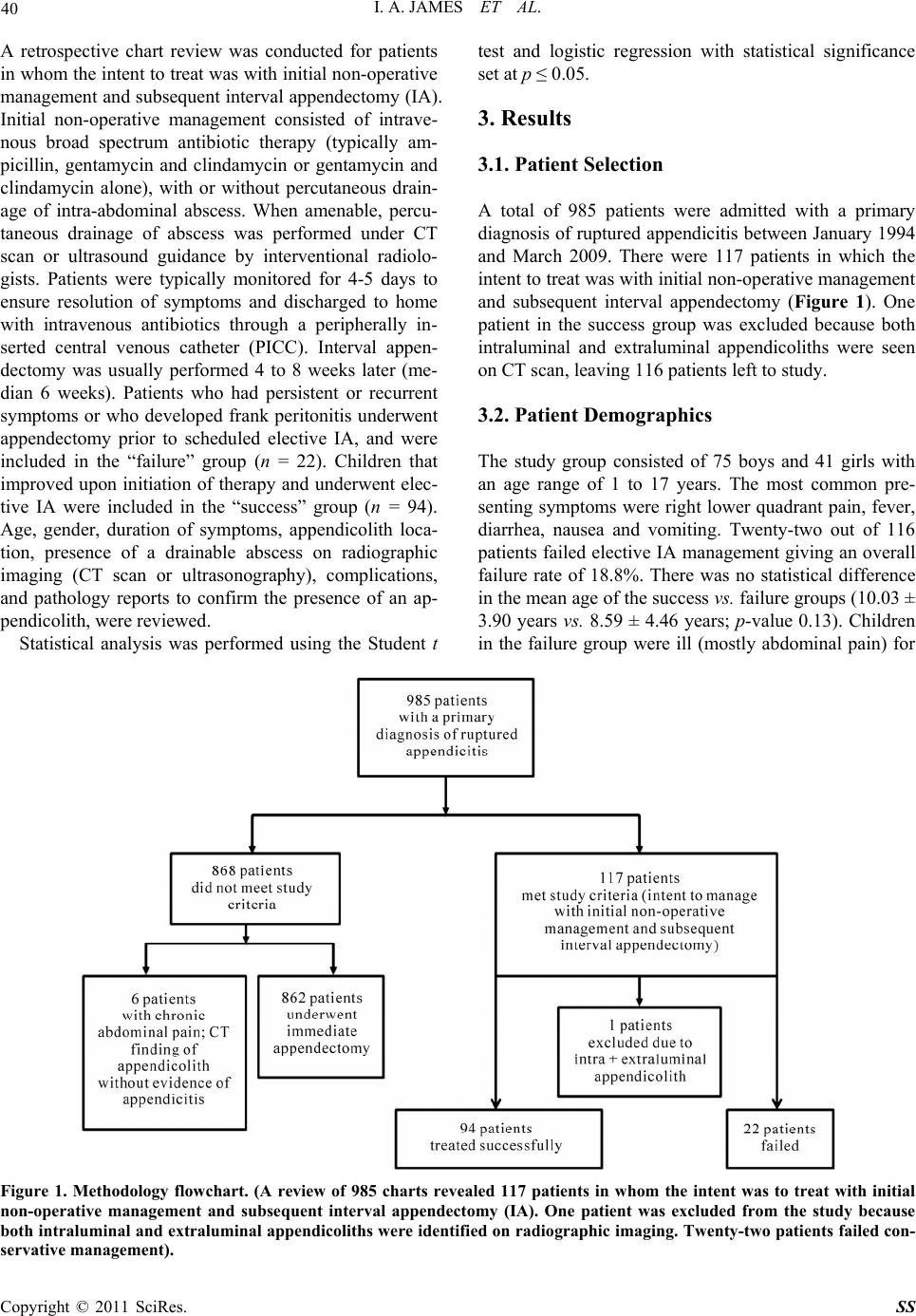 40 I. A. JAMES ET AL. A retrospective chart review was conducted for patients in whom the intent to treat was with initial non-operative management and subsequent interval appendectomy (IA). Initial non-operative management consisted of intrave- nous broad spectrum antibiotic therapy (typically am- picillin, gentamycin and clindamycin or gentamycin and clindamycin alone), with or without percutaneous drain- age of intra-abdominal abscess. When amenable, percu- taneous drainage of abscess was performed under CT scan or ultrasound guidance by interventional radiolo- gists. Patients were typically monitored for 4-5 days to ensure resolution of symptoms and discharged to home with intravenous antibiotics through a peripherally in- serted central venous catheter (PICC). Interval appen- dectomy was usually performed 4 to 8 weeks later (me- dian 6 weeks). Patients who had persistent or recurrent symptoms or who developed frank peritonitis underwent appendectomy prior to scheduled elective IA, and were included in the “failure” group (n = 22). Children that improved upon initiation of therapy and underwent elec- tive IA were included in the “success” group (n = 94). Age, gender, duration of symptoms, appendicolith loca- tion, presence of a drainable abscess on radiographic imaging (CT scan or ultrasonography), complications, and pathology reports to confirm the presence of an ap- pendicolith, were reviewed. Statistical analysis was performed using the Student t test and logistic regression with statistical significance set at p ≤ 0.05. 3. Results 3.1. Patient Selection A total of 985 patients were admitted with a primary diagnosis of ruptured appendicitis between January 1994 and March 2009. There were 117 patients in which the intent to treat was with initial non-operative management and subsequent interval appendectomy (Figure 1). One patient in the success group was excluded because both intraluminal and extraluminal appendicoliths were seen on CT scan, leaving 116 patients left to study. 3.2. Patient Demographics The study group consisted of 75 boys and 41 girls with an age range of 1 to 17 years. The most common pre- senting symptoms were right lower quadrant pain, fever, diarrhea, nausea and vomiting. Twenty-two out of 116 patients failed elective IA management giving an overall failure rate of 18.8%. There was no statistical difference in the mean age of the success vs. failure groups (10.03 ± 3.90 years vs. 8.59 ± 4.46 years; p-value 0.13). Children in the failure group were ill (mostly abdominal pain) for Figure 1. Methodology flowchart. (A review of 985 charts revealed 117 patients in whom the intent was to treat with initial non-operative management and subsequent interval appendectomy (IA). One patient was excluded from the study because both intraluminal and extraluminal appendicoliths were identified on radiographic imaging. Twenty-two patients failed con- ervative management). s Copyright © 2011 SciRes. SS 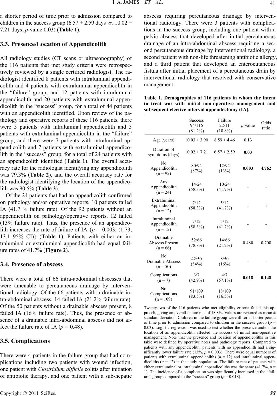 I. A. JAMES ET AL. Copyright © 2011 SciRes. SS 41 a shorter period of time prior to admission compared to children in the success group (6.57 ± 2.59 days vs. 10.02 ± 7.21 days; p-value 0.03) (Table 1). 3.3. Presence/Location of Appendicolith All radiology studies (CT scans or ultrasonography) of the 116 patients that met study criteria were retrospec- tively reviewed by a single certified radiologist. The ra- diologist identified 8 patients with intraluminal appendi- colith and 4 patients with extraluminal appendicolith in the “failure” group, and 12 patients with intraluminal appendicolith and 20 patients with extraluminal appen- dicolith in the “success” group, for a total of 44 patients with an appendicolith identified. Upon review of the pa- thology and operative reports of these 116 patients, there were 5 patients with intraluminal appendicolith and 5 patients with extraluminal appendicolith in the “failure” group, and there were 7 patients with intraluminal ap- pendicolith and 7 patients with extraluminal appendico- lith in the “success” group, for a total of 24 patients with an appendicolith identified (Table 1). The overall accu- racy rate for the radiologist identifying any appendicolith was 79.3% (Table 2), and the overall accuracy rate for the radiologist identifying the location of the appendico- lith was 90.5% (Table 3). Of the 24 patients that had an appendicolith confirmed on pathology and/or operative reports, 10 patients failed IA (41.7 % failure rate). Of the 92 patients without an appendicolith on pathology/operative reports, 12 failed (13% failure rate). Thus, the presence of an appendico- lith increases the rate of failure of IA {p = 0.003; (1.73, 13.1 95% CI)} (Table 1). Patients with either an in- traluminal or extraluminal appendicolith had equal fail- ure rates of 41.7% (Figure 2). 3.4. Presence of abscess There were a total of 66 intra-abdominal abscesses that were amenable to percutaneous drainage by interven- tional radiology. Of the 66 patients with a drainable in- tra-abdominal abscess, 14 failed IA (21.2% failure rate). Of the 50 patients without a drainable abscess present, 8 failed IA (16% failure rate). Thus, the presence or ab- sence of a drainable intra-abdominal abscess did not af- fect the failure rate of IA (p = 0.48). 3.5. Complications There were 4 patients in the failure group that had com- plications including two patients with wound infection, one patient with Clostridium difficile colitis after initiation of antibiotic therapy, and one patient with a sub-hepatic abscess requiring percutaneous drainage by interven- tional radiology. There were 3 patients with complica- tions in the success group, including one patient with a pelvic abscess that developed after initial percutaneous drainage of an intra-abdominal abscess requiring a sec- ond percutaneous drainage by interventional radiology, a second patient with non-life threatening antibiotic allergy, and a third patient that developed an enterocutaneous fistula after initial placement of a percutaneous drain by interventional radiology that resolved with conservative management. Table 1. Demographics of 116 patients in whom the intent to treat was with initial non-operative management and subsequent elective interval appendectomy (IA). Success 94/116 (81.2%) Failure 22/11 (18.8%) p-value Odds ratio Age (years) 10.03 ± 3.90 8.59 ± 4.46 0.13 Duration of symptoms (days)10.02 ± 7.21 6.57 ± 2.59 0.03 No Appendicolith (n = 92) 80/92 (87%) 12/92 (13%) 0.003 4.762 Any Appendicolith (n = 24) 14/24 (58.3%) 10/24 (41.7%) - - Extraluminal Appendicolith (n = 12) 7/12 (58.3%) 5/12 (41.7%) 1 1 Intraluminal Appendicolith (n = 12) 7/12 (58.3%) 5/12 (41.7%) - - Drainable Abscess Present (n = 66) 52/66 (78.8%) 14/66 (21.2%) 0.480 0.708 No Drainable Abscess (n = 50) 42/50 (84%) 8/50 (16%) - - Complications (n = 7) 3/7 (42.9%) 4/7 (57.1%) 0.018 0.148 No Complications (n = 109) 91/109 (83.5%) 18/109 (16.5%) - - Twenty-two of the 116 patients who met eligibility criteria failed this ap- proach, giving an overall failure rate of 18.8%. Values are reported as mean ± standard deviation. Children in the failure group were ill for a shorter period of time prior to admission compared to children in the success group (p = 0.03). Logistic regression was used to test whether the presence and/or the location of an appendicolith affected the success of initial non-operative management. Note that the presence and location of appendicoliths in this table were defined by operative notes and pathology reports. Compared to patients with any appendicolith, patients with no appendicolith had a sig- nificantly lower failure rate (13%, p = 0.003). There were equal numbers of patients with extraluminal appendicoliths (n = 12) and intraluminal appen- dicoliths (n = 12) in the study population. The failure rate of patients with either extraluminal or intraluminal appendicoliths was the same (41.7%, p = 1). The incidence of a complication was significantly increased in the “fail- ure” group compared to the “success” group (p = 0.018). 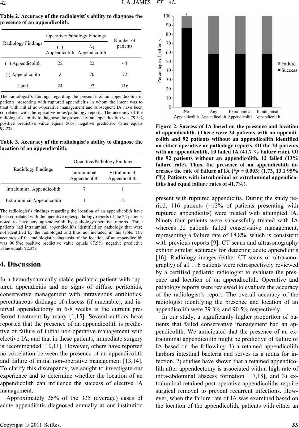 42 I. A. JAMES ET AL. Table 2. Accuracy of the radiologist’s ability to diagnose the presence of an appendicolith. Operative/Pathology Findings Radiology Findings (+) Appendicolith (-) Appendicolith Number of patients (+) Appendicolith 22 22 44 (-) Appendicolith 2 70 72 Total 24 92 116 The radiologist’s findings regarding the presence of an appendicolith in patients presenting with ruptured appendicitis in whom the intent was to treat with initial non-operative management and subsequent IA have been correlated with the operative notes/pathology reports. The accuracy of the radiologist’s ability to diagnose the presence of an appendicolith was 79.3%; positive predictive value equals 50%; negative predictive value equals 97.2%. Table 3. Accuracy of the radiologist’s ability to diagnose the location of an appendicolith. Operative/Pathology Findings Radiology Findings Intraluminal Appendicolith Extraluminal Appendicolith Intraluminal Appendicolith 7 1 Extraluminal Appendicolith 1 12 The radiologist’s findings regarding the location of an appendicolith have been correlated with the operative notes/pathology reports of the 24 patients noted to have any appendicolith by pathology/operative reports. Three patients had intraluminal appendicoliths identified on pathology that were not identified by the radiologist and thus not included in this table. The accuracy of the radiologist’s diagnosis of the location of an appendicolith was 90.5%; positive predictive value equals 87.5%; negative predictive value equals 92.3%. 4. Discussion In a hemodynamically stable pediatric patient with rup- tured appendicitis and no signs of diffuse peritonitis, conservative management with intravenous antibiotics, percutaneous drainage of abscess (if amenable), and in- terval appendectomy in 6-8 weeks is the current pre- ferred treatment by many [1,15]. Several authors have reported that the presence of an appendicolith is predic- tive of failure of initial non-operative management with elective IA, and that in these patients, immediate surgery is recommended [10,11]. However, others have reported no correlation between the presence of an appendicolith and failure of initial non-operative management [13,14]. To clarify this discrepancy, we sought to investigate our experience and to determine whether the location of an appendicolith can influence the success of elective IA management. Approximately 26% of the 325 (average) cases of acute appendicitis diagnosed annually at our institution Figure 2. Success of IA based on the presence and location of appendicolith. (There were 24 patients with an appendi- colith and 92 patients without an appendicolith identified on either operative or pathology reports. Of the 24 patients with an appendicolith, 10 failed IA (41.7 % failure rate). Of the 92 patients without an appendicolith, 12 failed (13% failure rate). Thus, the presence of an appendicolith in- creases the rate of failure of IA {*p = 0.003; (1.73, 13.1 95% CI)} Patients with intraluminal or extraluminal appendico- liths had equal failure rates of 41.7%). present with ruptured appendicitis. During the study pe- riod, 116 patients (~12% of patients presenting with ruptured appendicitis) were treated with attempted IA. Ninety-four patients were successfully treated with IA whereas 22 patients failed conservative management, representing a failure rate of 18.8%, which is consistent with previous reports [9]. CT scans and ultrasonography exhibit similar accuracy for detecting acute appendicitis [16]. Radiology images (either CT scans or ultrasono- graphy) of all 116 patients were retrospectively reviewed by a certified pediatric radiologist to evaluate the pres- ence and location of an appendicolith. Operative and pathology reports were reviewed to evaluate the accuracy of the radiologist’s report. The overall accuracy of the radiologist identifying the presence and location of an appendicolith were 79.3% and 90.5% respectively. In our study, a significantly higher proportion of pa- tients that failed conservative management had an ap- pendicolith. We anticipated that the presence of an ex- traluminal appendicolith might be predictive of failure of IA based on the following: 1) a retained appendicolith harbors intestinal bacteria and serves as a nidus for in- fection, 2) studies have shown that a retained appendico- lith after appendectomy is associated with a high rate of intra-abdominal abscess formation [17,18], and 3) ex- traluminal retained post-operative appendicoliths require surgical removal to prevent recurrent infections. How- ever, when the failure rate of IA was examined based on the location of the appendicolith, patients with either an Copyright © 2011 SciRes. SS  I. A. JAMES ET AL. 43 intraluminal or extraluminal appendicolith had a similar failure rate of 41.7%. Since only 20.7% (n = 24) of our study population presenting with perforated appendicitis and intent to treat with IA had an appendicolith docu- mented on pathology or operative reports, it is possible that the location of an appendicolith was not predictive of failure due to small sample size. The recorded inci- dence of appendicolith in children presenting with per- forated appendicitis has not been definitively established. In one study by Fraser et al., the incidence of appendico- lith based on histopathology reports was 26.3% in chil- dren with perforated appendicitis, and this was signifi- cantly higher compared to children that had either “un- complicated appendicitis,” “non-inflamed appendix,” or “incidental appendectomy” [19]. Nadler et al. reported that the need for abscess drainage doubles the likelihood of failure of initial non-operative management due to inadequate source control [13]. We did not demonstrate this finding in our study. Statistically significant characteristics of patients that failed conser- vative treatment were the presence of an appendicolith and duration of symptoms. The fact that significantly more patients in the failure group presented to the hospi- tal earlier in the course of their illness suggests that the severity of their symptoms may have prompted the care- giver to bring the child to the emergency department sooner. We have not verified this assumption in this study because of the lack of a reliable standardized ob- jective marker for assessing the severity of presenting symptoms. In a matched analysis comparing clinical characteristics of patients treated with initial non-operative management versus immediate appendectomy for perfo- rated appendicitis, Henry et al. observed that the only statistically significant difference between the two groups was duration of abdominal pain, where patients with pain less than 5 days in duration were more likely to undergo immediate appendectomy [2]. These investigators also demonstrated that patients who failed nonoperative management had a shorter duration of abdominal pain (average 4.4 days) compared to patients that were suc- cessfully treated with nonoperative management (aver- age 7.3 days). Their findings are consistent with our re- sults. Our study demonstrates that the presence of an appen- dicolith increases the likelihood of failure of initial non-operative management of ruptured appendicitis in children, but the location of the appendicolith is not a predictor of failure of initial non-operative management in these patients. Furthermore, it is apparent that the ra- diologist at times identifies appendicoliths that are not confirmed on operative/radiology reports. Thus, the presence of an appendicolith on admission CT scan or ultrasonography should not necessarily dissuade the practitioner from attempting initial non-operative man- agement with IA, whether the appendicolith is reported to be intraluminal or extraluminal. While established predictors for failure of non-operative management may be essential for standardizing care of ruptured appendici- tis in pediatric patients, careful clinical judgment contin- ues to be imperative in the management of these patients. 5. References [1] C. Chen, C. Botelho, A. Cooper, P. Hibberd and S. K. Parsons, “Current Practice Patterns in the Treatment of Perforated Appendicitis in Children,” Journal of the American College of Surgeons, Vol. 196, No. 2, February 2003, pp. 212-221. doi:10.1016/S1072-7515(02)01666-6 [2] M. C. W. Henry, G. Gollin, S. Islam, K. Sylvester, A. Walker, B. L. Silverman and R. Lawrence Moss, “Matched Analysis of Nonoperative Management Vs Immediate Appendectomy for Perforated Appendicitis,” Journal of Pediatric Surgery, Vol. 42, No. 1, January 2007, pp. 19-24. doi:10.1016/j.jpedsurg.2006.09.005 [3] T. R. Weber, M. A. Keller, R. J. Bower, G. Spinner and K. Vierling, “Is Delayed Operative Treatment Worth the Trouble with Perforated Appendicitis in Children?” The American Journal of Surgery, Vol. 186, No. 6, December 2003, pp. 685-689. doi:10.1016/j.amjsurg.2003.08.027 [4] D. W Vane and N. Fernandez, “Role of Interval Appen- dectomy in the Management of Complicated Appendicitis in Children,” World Journal of Surgery, Vol. 30, No. 1, January 2006, pp. 51-54. doi:10.1007/s00268-005-7946-2 [5] M. Samuel, G. Hosie, and K. Holmes, “Prospective Evaluation of Nonsurgical Versus Surgical Management of Appendiceal Mass,” Journal of Pediatric Surgery, Vol. 37, No. 6, June 2002, 882-886. doi:10.1053/jpsu.2002.32895 [6] D. Puapong, S. L. Lee, P. I. Haigh, A. Kaminski, I. L. Liu and H. Applebaum, “Routine Interval Appendectomy in Children Is Not Indicated.” Journal of Pediatric Surgery, Vol 42, No. 5, May 2007, pp. 1500-1503. doi:10.1016/j.jpedsurg.2007.04.011 [7] I. Karaca, Z. Altintoprak, A. Karkiner, G. Temir and E. Mir, “The Management of Appendiceal Mass in Children: Is Interval Appendectomy Necessary?” Surgery Today, Vol. 31, No. 8, August 2001, pp. 675-677. doi:10.1007/s005950170068 [8] S. H. Ein and B. Shandling B, “Is Interval Appendectomy Necessary after Rupture of an Appendiceal Mass?” Journal of Pediatric Surgery, Vol. 31, No. 6, June1996, pp. 849-850. doi:10.1016/S0022-3468(96)90151-7 [9] R. E. Andersson and M. G. Petzold, “Nonsurgical Treat- ment of Appendiceal Abscess or Phlegmon: A Systemic Review And Meta-Analysis,” Annals of Surgery, Vol. 246, No. 5, November 2007, pp. 741-s748. doi:10.1097/SLA.0b013e31811f3f9f [10] S. H. Ein, J. C. Langer and A. Daneman, “Nonoperative Management of Pediatric Ruptured Appendix with In- Copyright © 2011 SciRes. SS  I. A. JAMES ET AL. Copyright © 2011 SciRes. SS 44 flammatory Mass or Abscess: Presence of an Appendico- lith Predicts Recurrent Appendicitis,” Journal of Pediat- ric Surgery, Vol. 40, No. 10, October 2005, pp. 1612- 1615. doi:10.1016/j.jpedsurg.2005.06.001 [11] C. J. Aprahamian, D. C. Barnhart and S. E. Bledsoe, “Failure in The Nonoperative Management of Pediatric Ruptured Appendicitis: Predictors and Consequences,” Journal of Pediatric Surgery, Vol. 42, No. 6, June 2007, pp. 934-938. doi:10.1016/j.jpedsurg.2007.01.024. [12] J. Gillick, M. Velayudham and P. Puri, “Conservative Management of Appendix Mass in Children” British Journal of Surgery, Vol. 88, No. 11, November 2001, pp. 539-542. [13] E. P. Nadler, K. K. Reblock and K. G. Vaughan, “Pre- dictors of Outcome for Children with Perforated Appen- dicitis Initially Treated with Non-Operative Manage- ment,” Surgical Infections, Vol. 5, No. 4, Winter 2004, pp. 349-356. doi:10.1089/sur.2004.5.349 [14] T. Levin, C. Whyte, R. Borzykowski, B. Han, N. Blitman and B. Harris, “Nonoperative Management of Perforated Appendicitis in Children: Can CT Predict Outcome?” Pediatric Radiology, Vol. 37, No. 3, March 2007, pp. 251-255. doi:10.1007/s00247-006-0384-y [15] S. G. Muehlstedt, T. Q. Pham and D. J. Schmeling, “The Management of Pediatric Appendicitis: A Survey of North American Pediatric Surgeons,” Journal of Pediat- ric Surgery, Vol. 39, No. 6, June 2004, pp. 875- 859. doi:10.1016/j.jpedsurg.2004.02.035 [16] P. Poortman P, P. N. M. Lohle, C. M. C Schoemaker, H. J Oostvogel, H. J. Teepen, K. A. Zwinderman and J. F. Hamming, “Comparison of CT and Sonography in the Diagnosis of Acute Appendicitis: A Blinded Prospective Study,” American Journal of Roentgenology, Vol. 181, No. 5, November 2003, pp. 1355- 1359. [17] T. E. Monfore and F. J. Montegut, “The Case of the Missing Fecalith,” Archives of Surgery, Vol. 86, No. 4, April 1963, pp. 655- 658. [18] M. Mulder, “Retained Fecalith as Late Complication of Appendectomy,” Journal of American Medica Associa- tion, Vol. 225, No. 6, August 1973, pp. 639. [19] N. Fraser, C. Gannon and M. D.Stringer, “Appendicular Colic and the Non-Inflamed Appendix: Fact or Fiction?” European Journal of Pediatric Surgery, Vol. 14, No. 1, February 2004, pp. 21-24. |