

Paper Menu >>
Journal Menu >>
 Advances in Anthropology 2014. Vol.4, No.1, 1-6 Published Online February 2014 in SciRes (http://www.scirp.org/journal/aa) http://dx.doi.org/10.4236/aa.2014.41001 Sexual Dimorphism and Tooth Size Variation in the Permanent Dentition of the Uva Bintenna Veddas of Sri Lanka Thushari H e wapathira na 1, Roshan Peiris1, Deepthi Nanayakkara1, Malkanthi Chandrasekara2, Eugine Wikramanayake2 1Department of Basic Sciences, Faculty of Dental Sciences, University of Peradeniya, Peradeniya, Sri Lanka 2Department of Anatomy, Faculty of Medicine, University of Peradeniya, Peradeniya, Sri Lanka Email: [email protected] Received October 14th, 2013; revised November 16th, 2013; accepted December 13th, 2013 Copyright © 2014 Thushari Hewapathirana et al. This is an open access article distributed under the Creative Commons Attribution License, which permits unrestricted use, distribution, and reproduction in any medium, provided the original work is properly cited. In accordance of the Creative Commons Attribution License all Copyrights © 2014 are reserved for SCIRP and th e ow n er of th e in tellectual p rop erty Thush ari Hewap ath irana et al. All Copyright © 2014 are guarded by law and by SCIRP as a guardian. Forty-eight dental casts (37 males, 11 females) of Uva Bintenna Veddas were the population. Mesiodistal (MD) and buccolingual (BL) diameters were measured and analyzed for sexual dimorphism and size variation. In males, MD of maxillary canines and mandibular first molars were significantly greater at p < 0.01 and maxillary first molars and mandibular second molars at p < 0.05. Crown area was significantly greater at p < 0.01 for maxillary and mandibular first molars. Crown module was significantly greater at p < 0.01 for mandibular first molars and at p < 0.05 for maxillary first premolars, first molars and mandibu- lar second molars. Maxillary canines, maxillary first molars and mandibular canines were the most sex ually dimorphic. First tooth of each tooth class I1, P1, and M1 showed least variability in both arches and in both sexes. In males, both MD and BL showed high variability in maxillary lateral incisors and man- dibular second premolars. Keywords: Uva Bintenna Veddas; Sexual Dimorphism; Size Variation; Crown Diameters; Permanent Teeth Introduction The earliest account of the existence of, habitations, life style, language and religious ceremonies of the Veddas of Sri Lanka was by Knox (1681). However, the scientific study of their physical anthropology had to await the publications of Virchow and the Sarasins two centuries later in 1886 (Virchow, 1886; Sarasin & Sarasin, 1886). After a lapse of another fifty years Osman Hill, Professor of Anatomy, Ceylon Medical College initiated his studies on personally identified Veddas (Hill, 1932). The somatometry, craniometry and the variations of the dentition have been reported (Hill, 1945). Meanwhile, from 1937-1939, the anthropological and anth- roposcopic data for the Ethnological Survey of Ceylon was collected by H. R. de la Haule Marett an assistant in Ethnology, Colombo Museum. The Veddas were identified by their Wa- riges (clan names). Marett regarded the Veddas with the Sinha- lese and Tamils as the primary ethnic groups of Sri Lanka. Ma- rett had included variation in the dentition. After the untimely death of Marett, the data was analyzed and published as the Physical Anthropology of Ceylon (Stoudt, 1961). The Vedda country had been divided geographically into a northern Tam- mankaduwa and a southern Binntenne, with Binntenne further subdivided into Uva Binntenne in the west and Wellassa in the east. The Uva Binntenne Veddas were clustered in Kandegan- vila and Dambana (Hill, 1941). Following on the footsteps of Hill, a team of researchers from the University of Peradeniya commenced a comprehen- sive longitudinal biometric study of the genetics, somatometry, nutrition, oral health and dentition of the Uva Binntenne Ved- das of Kandeganvila from 1971-1973. As these Veddas had been colonized in 1958, the field studies could be organized at the level of identified households. For the first time in Vedda studies, three-generation pedigrees were compiled for the dif- ferent Wariges. Veddas of Vedda matings of at least three gen- erations identified from these pedigrees were the research pop- ulation. The results of the first phase have been published, the genetics (Ellepola & Wikramanayake, 1986; Hanihara et al., 1988), secular trends in physical anthropology (Wikrama- nayake & Wikramanayake, 1992), oral disease patterns and tooth wear (Pathmanathan & Wikramanayake, 1993), and body mass index (Wikramanayake et al., 1994). This research was reviewed later (Wikramanayake, 2002). With the declaration of the traditional hunting grounds of the Uva Binntenne Veddas as a National Park in 1982, the Kande- ganvila Veddas were relocated in Hennanigala South of the Mahaweli Irrigation System C. In the second phase of the study from 1993-1996, it was decided to study the Uva Binntenne Veddas in the two different locations, those of Kandeganvila in their relocated households in Hennanigala and those of Dam- bana in their traditional habitat. Figure 1 gives a map of the Vedda country with the three locations studied Kandeganvila (1971-1973) and Hennanigala and Dambana (1993-1996) un- derlined. At Hennanigala, somatometry, nutrition, oral health OPEN ACCESS 1 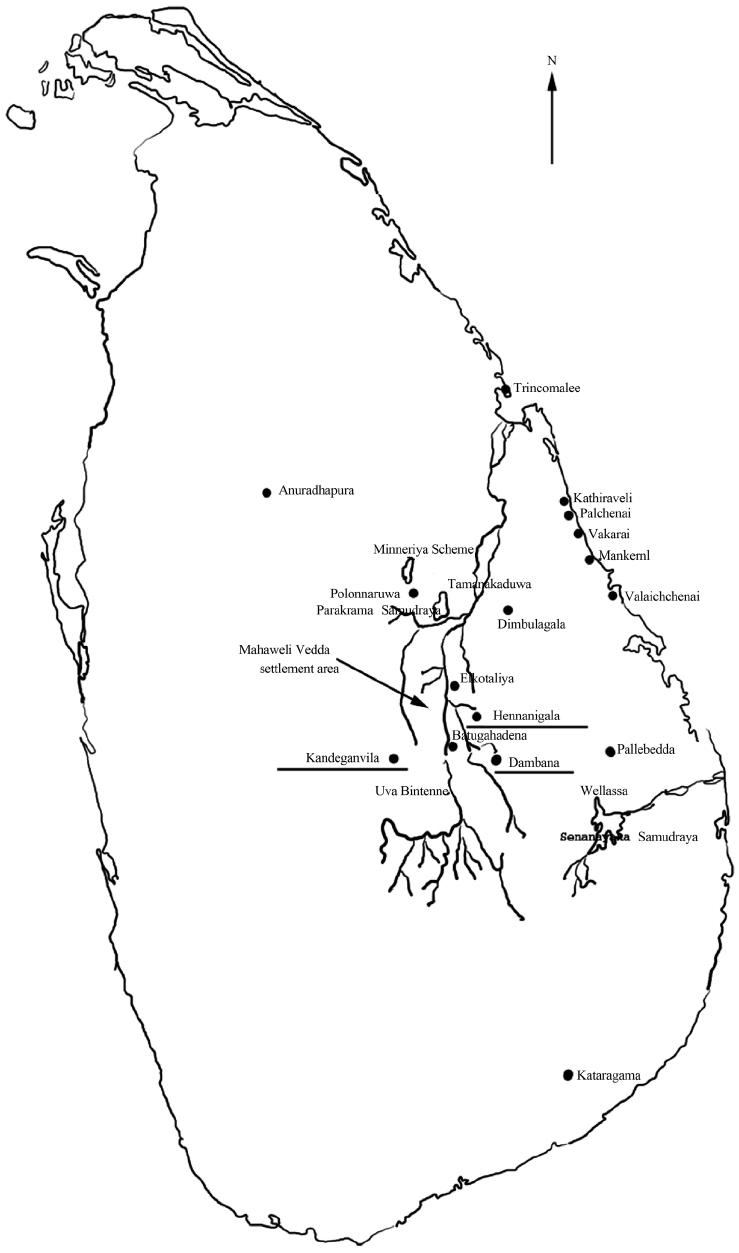 T. HEWAPATHIRANA ET AL. Figure 1. The location of Vedda settlements (three locations studied are underlined). OPEN ACCESS 2  T. HEWAPATHIRANA ET AL. and dentition were repeated. Additionally craniometry and blood pressure measurements were included. At Dambana, three-generation pedigrees of the different Wariges were com- piled. As in the first phase, Veddas identified from these pedi- grees as those from Vedda matings were the research popula- tion for the biometry both at Hennanigala and Dambana. Additionally, at Dambana alginate impressions of the denti- tion of Veddas were made in the field with informed consent. The present study based on these dental casts investigates the sexual dimorphism and tooth size variation of the permanent dentition of the Uva Bintenne Veddas of Dambana living in their traditional hunter-gatherer habitat. This is the first report of the dentition from in Sri Lanka. Materials and Methods The forty-eight dental casts, 37 from males (age range 17 - 70 years) and 11 from females were measured and analyzed. Measurements were done only if teeth were morphologically normal, fully erupted, and not noticeably affected by attrition or cavities. The selected teeth of both upper and lower arches of the left side were measured to the nearest 0.01 mm using a di- gital caliper. When a tooth on the left side could not be meas- ured because of absence, abnormality or heavy wear the cor- responding tooth on the right side was measured. The measure- ments of the mesiodistal (MD) and buccolingual (BL) crown diameters were made according to Fujita (1949). For the deter- mination of measurement errors, thirty casts were selected at random three months later. Measurements were redone on these casts and an analysis of error was done according to the method of Dahlberg (1940). Using the equation, Error = d2/2N (d = difference between two determinations, N = number of double determinations). The measurement errors ranged from 0.04 mm to 0.11 mm. As these were small values compared with the means, no effect on the statistical analysis was expected. Using the MD and BL diameters the following indices, crown index, crown area and crown module were also calculated for the posterior teeth. Crown index = MD/BL × 100, Crown area = MD × BL, Crown module = MD + BL/2. Results Table 1 gives the results of the MD diameters of the mandi- bular and maxillary permanent dentition and Table 2 gives the results of their BL diameter. Both maxillary and mandibular teeth of males were greater in MD and BL diameters than those of females except for the MD of the mandibular central incisor and BL of the mandibular lateral incisor. The differences were significant at p < 0.01 for MD of maxillary canines and mandi- bular first molar and at p < 0.05 for MD of maxillary first molar, maxillary canine, mandibular second molar, BL of maxillary canine, maxillary first and second molar, and the mandibular first molar. The coefficients of variation were taken as indica- tors of trends in the relative variability of tooth size. In each arch the first molar was the least variable tooth in both MD and BL diameters. There was no clearly defined pattern of relative variability in the MD and BL diameters in either sex. In the males, both MD and BL of the maxillary lateral incisor and mandibular second premolar showed high variability. In the females, variability was randomly distributed. Table 3 gives the crown index, Table 4 the crown area and Table 5 the crown module of the maxillary and mandibular premolars and molars (first and second). All the indices were greater in males. The differences were significant at p < 0.01 Table 1. Mesiodistal diameters for permanent teeth of mandibular and maxillary teeth of the study. Male Female Sex difference N Mean SD Coefficient of variation N Mean SD Coefficient of variation Maxillary I1 25 8.06 0.65 8.06 12 8.06 0.62 7.69 I2 27 6.61 0.54 8.16 11 6.37 0.39 6.12 C 29 7.50 0.52 6.93 13 6.95 0.46 6.61 ** P1 31 6.81 0.47 6.90 13 6.54 0.65 9.93 P2 31 6.42 0.56 8.72 13 6.11 0.55 9.00 M1 31 10.14 0.52 5.12 13 9.72 0.67 6.89 * M2 31 9.27 0.70 7.55 13 8.96 0.91 10.47 Mandibular I1 23 5.06 0.46 9.09 11 5.27 1.12 21.25 I2 24 5.73 0.43 7.50 10 5.38 0.69 12.82 C 30 6.58 0.30 4.55 12 6.28 0.61 9.71 * P1 31 6.71 0.48 7.15 13 6.59 0.32 4.85 P2 31 6.75 0.65 9.62 12 6.61 0.76 11.49 M1 29 10.93 0.58 5.30 12 10.09 0.75 7.43 ** M2 30 10.11 0.71 7.02 12 9.41 0.96 10.20 * Note: *p < 0.05; **p < 0.01. OPEN ACCESS 3  T. HEWAPATHIRANA ET AL. Table 2. Buccolingual diameters for permanent teeth of mandibular and maxillary teeth of the study. Male Female Sex difference N Mean SD Coefficient of variation N Mean SD Coefficient of variation Maxillary I1 25 7.10 0.66 9.29 12 6.91 0.78 11.28 I2 27 6.45 0.74 11.47 11 6.02 0.52 8.63 C 28 7.86 0.51 6.48 13 7.41 0.57 7.69 * P1 31 8.76 0.60 6.84 13 8.39 0.57 6.79 P2 31 8.53 0.75 8.79 13 8.45 0.89 10.53 M1 31 10.73 0.67 6.24 13 10.18 0.77 7.56 * M2 31 10.39 0.78 7.50 13 9.73 1.20 12.33 * Mandibular I1 25 5.98 0.53 8.86 12 6.02 0.89 14.78 I2 25 6.28 0.62 9.87 10 6.41 0.99 15.44 C 30 7.26 0.52 7.16 12 7.07 1.12 15.84 P1 31 7.49 0.53 7.07 13 7.16 1.00 13.96 P2 31 7.68 0.66 8.59 12 7.39 1.08 14.61 M1 29 10.14 0.49 4.83 12 9.63 0.87 9.03 * M2 30 9.68 0.63 6.50 12 9.31 1.04 11.17 Note: *p < 0.05; **p < 0.01. Table 3. Crown index of the permanent prem olars and molars of the study populat ion. Male Female N Mean Standard devi ation Coefficient of variation N Mean Standard deviation Coefficient of variation Maxillary P1 31 79.01 7.76 9.82 13 78.14 6.52 8.34 P2 31 75.82 5.65 7.45 13 72.65 7.15 9.84 M1 31 94.76 5.82 6.14 13 95.68 6.73 7.03 M2 31 89.81 8.93 9.94 13 93.20 14.19 15.22 Mandibular P1 31 89.40 7.86 8.79 13 92.30 11.31 12.25 P2 31 88.29 9.13 10.34 12 90.88 15.58 17.14 M1 29 107.53 6.02 5.59 12 105.36 8.43 8.00 M2 30 104.49 4.07 3.89 12 101.92 12.1 11.87 for crown area and crown module of mandibular first molar and at p < 0.05 for crown area and crown module of maxillary first premolar, maxillary first molar and mandibular second molar. In both males and females, crown index and crown module of the first premolar and the first molar were greater than that of the second premolar and second molar respectively. In males, in both dentitions the crown index and crown mod- ule of the first molar was significantly greater than that of the second in the maxilla at p < 0.01 and mandible at p < 0.05. Additionally the crown module of the maxillary first premolar was significantly greater than the second at p < 0.05. The crown area was greater in the first molar than that of the second molar in both dentitions with the maxilla at p < 0.01 and the mandible at p < 0.05. Variability in the indices was not analyzed statisti- cally in the females. Discussion The present study deals with 48 dental casts (37 male and 11 female) of adult contemporary Uva Bintenne Veddas of Sri Lanka. The subjects were identified from Vedda matings of at least three generations and had normal occlusion. The MD and BL crown diameters of both maxillary and mandibular teeth except the third molars were measured on the left side. Using the crown diameters the crown index, crown area and crown module of the premolars and first and second OPEN ACCESS 4  T. HEWAPATHIRANA ET AL. Table 4. Crown area of the permanent prem olars and molars of the s tudy population. Male Female N Mean Standard deviation Coefficient of variation N Mean Standard deviation Coefficient of variation Maxillary P1 31 60.72* 8.99 14.80 13 55.08 8.09 14.68 P2 31 55.33 8.54 15.43 13 51.98 9.02 17.35 M1 31 108.96* 10.72 9.83 13 99.48 12.80 12.86 M2 31 96.92 12.51 12.90 13 87.73 17.35 19.77 Mandibular P1 31 50.20 6.31 12.56 13 48.05 7.73 16.08 P2 31 52.05 7.91 15.19 12 49.14 10.32 21.00 M1 29 110.78** 9.34 8.43 12 97.66 13.72 14.04 M2 30 98.33* 13.08 13.30 12 88.08 14.92 16.93 Note: Statistica lly significant difference between ma le and fem ale is denot ed by *. *p < 0.05; **p < 0.01. Table 5. Crown module of the permanent premolars and molars of the study populat ion. Male Female N Mean Standard deviation Coefficient of variation N Mean Standard deviation Coefficient of variation Maxillary P1 31 7.83* 0.56 7.15 13 7.46 0.54 7.23 P2 31 7.49 0.59 7.87 13 7.28 0.64 8.79 M1 31 10.43* 0.50 4.79 13 9.96 0.65 6.52 M2 31 9.58 0.62 6.47 13 9.34 0.91 9.74 Mandibular P1 31 7.09 0.44 6.20 13 6.93 0.58 8.36 P2 31 7.21 0.55 7.62 12 7.00 0.76 10.85 M1 29 10.52** 0.44 4.18 12 9.86 0.71 7.20 M2 30 9.90* 0.65 6.56 12 9.36 0.83 8.86 Note: Statistica lly significant difference between ma le and fem ale is denot ed by *. *p < 0.05; **p < 0.01. molars were calculated. The results were analyzed for sexual dimorphism and size variation of the dentition. The MD and BL diameters of all teeth except the MD of the mandibular central incisor and BL of the mandibular lateral incisor were greater in the males. The differences were signifi- cant at p < 0.01 for the MD of maxillary canine and mandibular first molar and at p < 0.05 for the maxillary first molar and mandibular second molar. The BL of the maxillary canine, first and second molars and mandibular first molar were signifi- cantly great er at p < 0.05. The crown dimension indices were also greater in the males. However, the crown index was not significantly greater in ei- ther arch. The crown area was significantly greater at p < 0.01 for the maxillary and mandibular first molars and at p < 0.05 for the maxillary first premolar and mandibular second molar. The crown module was significantly greater at p < 0.01 for the mandibular first molar and at p < 0.05 for the maxillary premo- lar, and first molar and the mandibular second molar. When the coefficients of variation were taken to indicate rel- ative variability of tooth size no clearly defined pattern in MD and BL were observed in either sex. In the males both MD and BL of the maxillary lateral incisor and mandibular second pre- molar showed high variability. In the females, variability was randomly distributed. Crown index and crown module of the first molar in both dentitions was significantly greater than that of the second in the maxilla at p > 0.01 ad mandible at p < 0.05. The crown module of the maxillary first premolar was significantly greater than that of the second at p < 0.05 . With reference to sexual dimorphism the MD identifies the maxillary canine and mandibular first molar at the higher probability and the maxillary first molar and mandibular at the lower. The BL identifies the maxillary canine, first and second molars and the mandibular first molar at the lower probability. The crown indices are limited to the posterior teeth. Crwon index has not been useful in assessing sexual dimorphism. Crown area identifies the maxillary and mandibular first molars at the higher probability and the maxillary first premolar and mandibular second molar at the lower. Crown module identifies the mandibular first molar at the higher probability and the OPEN ACCESS 5  T. HEWAPATHIRANA ET AL. maxillary premolar and first molar and mandibular second mo- lar at the lower probability. With reference to relative variability of tooth size, a definite pattern was not observed in either sex in the MD & BL. The variability was randomly distributed in the females. In both sexes in both arches the first tooth of each tooth class (I1, P1, and M1) showed less variability with the first molar being the least variable. Crown area has not been useful in assessing tooth size varia- bility. Crown index identifies the first molar significantly greater in the maxilla at the higher probability and the maxillary first premolar and mandibular second molar at the lower proba- bility. Crown module identifies the mandibular first molar at the higher probability and the maxillary premolar and first mo- lar and mandibular second molar at the lower probability. The study reveals that MD, crown area and crown module are more useful in identifying sexual dimorphism. The maxil- lary teeth are more dimorphic and the maxillary canine and first molar and mandibular first molar most dimorphic. With reference to variability of tooth size in the MD and BL diameters the first tooth of each class (I1, P1, and M1) in both arches with the first molar being the least variable in both sexes. In the males both MD and BL of the maxillary lateral incisor and mandibular second premolar showed high variability. The variability in MD and BL were randomly distributed in the females. With reference to the crown indices in both arches in the males, the crown index and crown module of the first molar was significantly greater than that of the second in the maxilla at the higher probability in the maxilla and in the mandible at the lower. The crown module of the maxillary first premolar was significantly greater than that of the second at the lower probability. In this study, the MD and BL diameters have not been in- formative of relative variability of tooth size in the dentition. The crown area too was not informative. However in the males both crown index and crown module have identified tooth size variation between the first and second molars at the higher probability in the maxillary teeth. Crown module has also iden- tified variation between the maxillary first premolar and second at the lower probability. The first tooth of each tooth class I1, P1, and M1 in both arches in both sexes showed less variability with the first molar being the least variable. In the males both MD and BL showed high variability in the maxillary lateral incisor and mandibular second molar. Differences in the crown dimension indices crown index, crown area and crown module are expected to show variability in the shape, robustness and bulkiness of the teeth respectively (Townsend & Brown, 1979). In this study crown index was not informative on sexual di- morphism and crown area on relative variability while crown module was informative of both. Further studies are necessary to confirm the relevance of these indices in the dentition. Conclusion In conclusion, this study is the first on dental casts of any population group in Sri Lanka establishing the sexual dimor- phism and tooth size variation in the permanent dentition of the contemporary Uva Bintenne Veddas. REFERENCES Dahlberg, G. (1940). Statistical methods for medical and biological students. London: George Allen and Unwin, Ltd. Ellepola, S. B., & Wirka manayake, E. R. (1986). A genetic study of the Veddas and Sinhalese. Ceylon Journal of Medical Science, 29, 1-21. Fujita, T. (1949). On the standard of the measurement of teeth. The Journal of the Anthropological Soc iety of Nippon, 61, 27-32. http://dx.doi.org/10.1537/ase1911.61.27 Hanihara, S., Saitou, N., Hirai, M., Gojobori, T., Park, K. S., Misawa, S., Ellepola, S. B., Ishida, T., & Omoto, K. (1988). Mitochondrial DNA polymorphism among five Asian populations. American Jour- nal of Human Genetics, 43, 134-143. Hill, W. C. O. (1932). Anthropology of Veddas. Nature, 130, 891-892. http://dx.doi.org/10.1038/130891b0 Hill, W. C. O. (1941). The physical anthropology of the existing Ved- das in Ceylon Part I. Ceylon Journal of Science, 3, 37-38. Hill, W. C. O. (1945). Veddahs—A living relic of primitive man. Loris, 201-206. Knox, R. (1981). An historical relation of Ceylon (pp. 194-199). Dehiwela: Tisara Prakasakayo Ltd. Pathmanathan, R. S., and Wikramanayake, E. R. (1993). Oral disease patterns and tooth wear in the Veddas. Ceylon Journal of Medical Science, 36, 43-49. Sarasin, C. F., and Sarasin, P. B. (1886). Outline of tw o years research in Ceylon. Journal of the Royal Asiatic Society (Ceylon Branch), 9, 289-305. Stoudt, H. W. (1961). The physical anthropology of Ceylon. Ceyl on N a- tional Museum Ethnographic Series 2 (pp. 149-154). Sri Lanka: Co- lombo National Museum. Townsend, G. C., and Brown, T. (1979). Tooth size characteristics of Australian Aborigines (pp. 17-38). Ocaational papers in human biol- ogy. Australian Institute of Aboriginal Studies, Canberra. Virchow, R. (1886). The Vedda of Ceylon and their relation to neigh- boring tribes. Journal of the Royal Asiatic Society (Ceylon Branch), 9, 349-495. Wikramanayake, E. (2002). Twenty five years research on the Ved- das—A review. Journ al Royal Asiatic Society of Sri Lan ka, 46, 105- 114. Wikramanayak e, E. R., Wikramanaayke, T. W., & Chan drasekera, M. S. (1994). Body mass index of kandeganvila Veddas. Ceylon Journal of Medical Science, 37, 13-17. Wikramanayake, T. W., & Wikramanayake, E. R. (1992). Secular trends in the physical anthropology of the Veddas of Sri Lanka. Cey- lon Journal of Medical Science, 35, 23-28. OPEN ACCESS 6 |