

Paper Menu >>
Journal Menu >>
 Journal of Environmental Protection, 2014, 5, 35-41 Published Online January 2014 (http://www.scirp.org/journal/jep) http://dx.doi.org/10.4236/jep.2014.51005 Influences of Cyanobacterial Toxins Microcystins on the Seedling of Plants Thanh-Son Dao1*, Thai-Hang Le1, Thanh-Luu Pham2, Lan-Chi Do-Hong3, Phuoc-Dan Nguyen4 1Institute for Environment and Resources, Ho Chi Minh City, Vietnam; 2Graduate School of Life and Environmental Sciences, Uni- versit y of Tsukuba, Japan; 3Vietnam National University, Ho Chi Minh City, Vietnam; 4University of Technology, Ho Chi Minh City, Vietnam. Email: *[email protected] Received November 26th, 2013; revised December 21st, 2013; accepted J anuary 9th, 2014 Copyright © 2014 Thanh-Son Dao et al. This is an open access article dist ributed under the C reative Commons Attri bution Licens e, which permits unrestricted use, distribution, and reproduction in any medium, provided the original work is properly cited. In accor- dance of the Creative Commons Attribution License all Copyrights © 2014 ar e reserved for SCIRP an d the owner of the intel lectual property Thanh-Son Dao et al. All Copyright © 2014 are guarded by law and by SCIRP as a guardian. ABSTRACT Cyanobacterial blooms associated by their toxins have been increasing in frequency in fresh water bodies throug ho ut the world. Among the cy anobacteria l toxins, microcystins (MC) are the most common and cause se- vere adverse impacts on plants, aquatic organisms and human beings. In this study, the effects of MC (at the concentrations of 20 and 200 µg·L−1) from field water and crude extract of cyanobacterial scum (mainly Micro- cystis spp.) from the Dau Tieng Reservoir, Vietnam, on the seedlings of three plants, Brassica rapa-chinensi s, B. narinosa and Nasturtium officinale, were investigated for over a period of 7 days. The results showed that MC reduced the fresh w eight , roo t and shoo t lengt h of the exposed see dlings. In ad dition, abno rmalities o f leaf shape and color of B. rapa-chinensis under exposure to MC were observed. The results implied that MC w ere ta ken up and might be accu mulated i n the seedl ings po ssessing pot ential r isk to co nsumers as seedling s of t hese plants a re a common food source for Vietna mese. To the best of our knowledge, this is the first report on the effects of MC on B. rapa-chinensis, B. narinosa and N. off icina le . KEYWORDS Microcystins; Adverse Effects; Fresh Weight; Root and Shoot Length; Abnormalities 1. Introduction In water bodies, microcystins (MC) have been the most commonly reported cyanobacterial toxins compared to other cyanotoxins (e.g. cylindrospermopsin, nodularin, saxito xins, anatoxi n-a(s), anatoxin-a) [1]. Cyanobacterial toxins cause a range of adverse effects on aquatic plants, animals and human beings. In addition, investigations showed that the toxins can accumulate and adversely affect on plants at different aspects of enzymatic res- pons es, photosynthesis, seedlings and growth. Recently, many studies on bioaccumulation of cyano- bacterial toxins (e.g. MC) in plants have been conducted and reported. The uptake and metabolism of MC-LR in stems, rhizomes and leaves of aquatic macrophytes were reported with the MC-LR conce ntra tion s be ing hig hest i n leaves, followed by shoots and lowest in stems [2,3]. Other plants such as broccoli, mustard and duckweed are able to accumulate MC in their leaves and bark protein, and whole plant up to 8.7 ng MC g−1 fresh weight (FW) after exposure [4-6]. Furthermore, Microcystis aerugi- nosa containing MC-LR can also be retained by salad letture (Lactuca sativa) after spraying with irrigation water containing the cyanobacterium [7]. Macrophytes and seedlings have been showed to dis- play the growth inhibitio n wh en irrigated with water co n- taining toxic cyanobacterial extract [8]. MC inhibit the growth and development of rape (Brassica napus), rice (Oryza sativa) [9], mustard (Sinapis alba) [4] and Wolffia arrhiza [5]. Exposure to MC-LR resulted in a decrease in germination, roof and leaf length of spinach [10]. MC- LR, M C-RR and anatoxin-a also reduced the chlorophyll and carotenoid concentrations in plants [8,11,12] and *Corresponding author. OPEN ACCESS JEP Influences of Cyanobacterial Toxins Microcystins on the Seedling of Plants 36 caused malformation on mustard [4]. Pietsch et al. [13] and Wiegand et al. [14] showed that the photosynthesis of macrophytes, Vesicularia dubyana and Ceratophyllum demersum, was inhibited by cyano- bacterial crude extracts or purified cyanotoxins, MC, microcin SF608 and anatoxin-a. These authors also rec- orded the activity alteration of the enzyme glutathione S-transferase in the macrophytes after toxin exposures. The activities of antioxidant and biotransformation en- zymes (superoxide dismutase, peroxidase, catalase, glu- tathione S-transferase, glutathione peroxidase) from plants were also significantly changed by cyanobacterial crude extracts containing MC, pure MC-LR or anatoxin- a at the concentration from 0.5 - 10 µg·L−1 [10,12,14]. MC-LR induced oxidative stress responses in Lepidium sativum seedlings including lipid peroxidation, change of tocopherol concentrations and profile, and elevation of glutathione enz yme acti vitie s [15] . MC-RR decreased the glutathione level and increased superoxide dismutase and catalase activities in cells of Arabidopsis thaliana and tobacco (Nicotiana tabucum) [16,17]. Today, seedlings of many different plants are common food ingredients especially in Asia. In the field plants could be irrigated with water containing cyanobacteria and their toxins. Hence these plants could be affected by and accumulate cyanotoxins. To our knowledge, there has been no information on the effects of MC on the seedlings of Brassica rapa-chinensis, B. narinosa and Nasturtium off icinale . T he aim o f this stud y is to obser ve the detrimental effects of MC from field water and cya- nobacterial crude extract obtained from Dau Tieng Re- servoir, Vietnam, at the environmentally relevant con- centrations (20 and 200 µg·L−1) on t he see dling s of thr ee different plants B. rapa-chinen sis, B. narinosa and N. officinale over 7 days. 2. Materials and Methods The seeds of B. rapa-chinensis, B. narinosa and N. o ffi- cinale were purchased from a super market in Hochiminh City. Two samples: 1) cyanobacterial scum (mainly Mi- crocystis spp) collected in July 2011; and 2) (raw) field water sample during cyanobacterial scum (mainly Ana- baena flos-aquae) from Dau Tieng Reservoir collected in September 2012, were used for experiments. 2.1. Sample Preparation for Experiments and Microcystins Analysis Crude extract from cyanobacterial scum collected in July 2011 was prepared according to Pietsch et al. [13] with minor modification. Briefly, the dried biomass of scum on GF/C filters was homogenized, suspended into re- versed osmosis water, sonicated, frozen at –70˚C over night and thawed at room temperature. The freeze/thaw cycle was repeated five times. After the last thawing cycle, samples were centrifuged at 4500 rpm, 4˚C for 15 min. Supernatant was collected and kept at –70˚C prior to exp e riments on t he plant se edling s. Field wate r sa mple collected in September 2012 was filtere d via plankton net (25 µm mesh size) then centrifuged at 4500 rpm, 4˚C for 15 min and supernatant was collected and stored at –70˚C prior to exposure to seedlings. Sub-samples of field water and cyanobacterial crude extract were centrifuged at 14,000 rpm 4˚C for 15 min and supernatants were collected for MC analysis by high performance liquid chromatography (HPLC). HPLC (Shimadzu, Japan) equipped with a silica based reverse phase C18 column (Waters SunFire™, Ireland) was maintained at 40˚C. A 0.05 M phosphate buffer (pH 2.5) in methanol (50/50, v/v), at a flow rate of 0.58 mL min–1, was used as mobile phase. MC congeners were detected by the UV detection at 238 nm with a photodiode UV- visible array detector. The MC variants MC-RR, -YR and -LR purchased from Wako chemicals company (Osaka, Japan) were used as standards. 2.2. Exposures of Brassica rapa-chinensis, B. narinosa and Nasturtium officinale to Micro cy sti ns The seedling experiment used 40 seeds of each plant species (with 3 replicates). The seeds were placed on tissues paper and watered daily with 5 mL of field water (containing 20 (DT20) or 200 (DT200) MC µg·L−1) or cyanobacterial crude extract (containing 20 (Sc20) or 200 (Sc200) MC µg·L−1). A co ntro l was inc lude d whic h con- sisted of seeds watered with distilled water only. Expe- riments were run at 25˚C ± 1˚C, in the dark for the first 2 days. From the third day to the end of incubation (7 days), the seedlings were placed under light with an inten sity of around 1500 Lux and a light: dark cycle of 12 h:12 h. The parameters recorded were fresh weight (FW), shoot lengt h and roo t length of t he seedli ngs at 2, 4 and 7 da ys of incubation. The FW was determined using a balance (Sartorius BP 201S, Germany) and the lengt h was meas- ured with a ruler of 1 mm spacing. 2.3. Statistical Analysis Sigmaplot, version 12.0 was used for data analysis. One- way Analysis of Variance (A NOVA) and Tukey test Post Hoc were applied to deter mine the sta tistically significant differences of the FW, shoot and root length of seedlings after the data were checked for variance homogeneity (Levenes) and normality (Shapiro-Wilk’s test). 3. Results and Discussion 3.1. Microcystins Concentration in the Field Water and Cyanobacterial Crude Extr act The HPLC analysis showed that the cyanobacterial crude OPEN ACCESS JEP 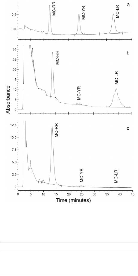 Influences of Cyanobacterial Toxins Microcystins on the Seedling of Plants 37 extract and field water contained MC-RR, MC-YR and MC-LR (Figure 1) at the total concentrations of 686.9 µg MC-LReq. g−1 dry weight and 1069.2 µg MC-LReq. L–1, respectively (Table 1). The MC concentration from cyanobacterial scum sample (collected in July 2011) in this st udy is in r ange o f pr e vio us r ec or d s fr om the f i eld i n Vietnam [18]. H owever , the M C co nc e ntr a tio n i n t he r aw water sample from Dau Tieng Reservoir was much high- er than those ever reported before from Vietnamese wa- ters . The hi gh MC conce ntrati ons d uring Microcystis spp and Anabaena flos-aquae scums proposed a serious risk to local residents who daily use the water from the re- servoir for domestic activities. Additionally, this is the first report of the MC producing cyanobacterium A. flos- aquae in Vietnam. Figure 1. HPLC chromatography of MC from control (a), field water (b) and cya nobacterial s cum sample (c). Table 1. Microcystins concentrations of the cyanobacterial s cum (µg·g−1 DW) a nd field w at er s ample (µg·L−1). Sam ples MC-RR MC-YR MC-LR Total MC Cyan oba ct eria l scum 635 31.7 20.3 686.9 Field water 539.5 30.6 499.1 1069.2 3.2. Effects of Microcystins on the Fresh Weight of See dl i ngs After 2, 4 and 7 days of incubation, the FW of all three plant species from exposures to either field water or cyanobacterial crude extract was significantly decreased compared to the control (ANOVA followed by Tukey test, p < 0.05; Figure 2). Besides, high MC concentration (DT200 and Sc200) had a stronger impact than the low toxin one (DT20 and Sc20) on FW of exposed plants consequently lower seedling FW in treatments with 200 µg M C L−1 compared to that with 20 µg MC L−1. G ener- ally, the FW of seedlings was similar when they were exposed to the same MC concentrations (20 or 200 µg·L−1) either from field water or crude extract. The significantly lower FW of the three seedlings ex- posed to MC in our study is in line with previous inves- tigations in which the FW of potato and the germination of spinach were inhibited by cyanobacterial crude extract [8,10]. The weight of seedlings of the three plants during the first days of germination should be involved in the amount of water they took up. MC induce oxidative stress response [15] and inhibited ATPase [19] possibly interfering the metabolism in seedlings during germina- tion consequently water uptake and seedlings of the ex- posed seeds and plants. Therefore, from our study it could be inferred that MC reduce the water uptake ca- pacity of seeds hence inhibit FW increase of the seedl- ings. Besides, the detoxification of MC in plant cells to balance the activities of biotransformation and antioxi- dant enzymes [10,12,20] would lead to the decrease of the energy or material for growth of the seedlings which properly involved in the reduction of FW of seedlings in the MC exposures compared to the control. 3.3. Effects of Microcystins on the Development of Root and Shoot The roo t and sho o t le ng th of the three plant species in the control treatment alwa ys significantly longer than that in MC exposures after 2, 4 and 7 days of incubation (ANOVA followed by Tukey test, p < 0.05; Figures 3 and 4). Additionally, high MC concentration from the field water and cyanobacterial crude extract had stronger effects than low MC conce ntration on the root and shoot development of B. rapachinen sis, B. narmosa and N. officmale (Figures 3 and 4). The MC from field water or crude extract at the same concentrations (20 or 200 µg·L−1) resulted in the similar root and shoot length of the seedlings. Inhib itio n on t he r oo t and sho ot le ngth o f MC -exposed seedlings in our study is consistent with previous records of McElhiney et al. [8] and Pflugmacher et al. [10]. Garbers et al. [21] indicated that MC regulated the phy- tohormone auxin, and MC we re protein phosphatase i nhi- OPEN ACCESS JEP 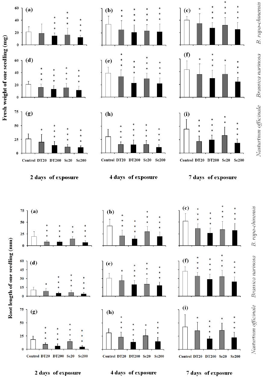 Influences of Cyanobacterial Toxins Microcystins on the Seedling of Plants 38 Figure 2. Fresh weight (mg) of the seedlings (mean value ± SD of n = 40) during incubation. Asterisks indicate significant differenc e between exposures and c ontrol by ANOVA followed by Tukey test (*, p < 0.05; **, p < 0.01; ***, p < 0.001). Figure 3 . Root le ngt h (mm) of t he se edl ing s ( mea n val ue ± SD of n = 40 ) du ring inc ubat ion. As teri sks indi cat e si gnif ic ant dif- ference between exposures and control by ANOVA foll owed by Tukey test (*, p < 0.05; **, p < 0.01; ** *, p < 0.001). OPEN ACCESS JEP 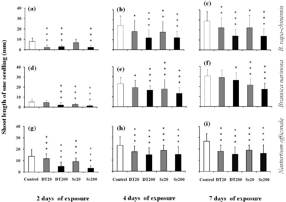 Influences of Cyanobacterial Toxins Microcystins on the Seedling of Plants 39 Figure 4. Shoot length (mm) of the seedlings (mean value ± SD of n = 40) during incubation. Asterisks indicate significant differenc e between exposures and c ontrol by ANOVA followed by Tukey test (*, p < 0.05; **, p < 0.01; ***, p < 0.001). bitors. Therefore, exposure to MC would cause the dis- order of cell development consequently inhibition of root and shoot growth of the exposed seedlings. Also, the water uptake reduction induced by MC as mentioned above would contribute to the decrease of shoot and root prolongation. Besides, MC could cause strong alteration of biotransformation and a ntioxidant e nzyme activ ities in plants [10,12,20] hence some energy is spent on the MC detoxification leading to the reduction of hydrocarbon and nutrient source for root and shoot development. Summing up the adverse effects MC on plants, the seedlings exposed to MC would grow slower than those in the c ontrol as observed i n our experiments . 3.4. Abnormalities of the Seedlings Exposed to Micro cy sti ns Among the MC exposures, abnormalities of leaf shape and color of some seedlings of B. rapa-chinensis were observed. The two young leaves on some seedlings from MC exposure were quite difference in size, and the leaf margin of those seedlings were brown (Figure 5(b)) while young leaves were almost similar in size with green color in the control incubation (Fig ur e 5(a)). Gehringer et a l. [22] found that the leaves of Lepidium sativum exposed to MC were significantly shorter than control samples which supported the unbalanced size of two young leaves observed in our study. It is possible that the brown color on leaf margin of B. rapa-chinensis in this investigation has been involved in the disappear- ance or decrease of chlorophyll content at the margin whi ch was previously reported elsewhere [4]. This phe- nomenon could also be explained as chlorophyll is inhi- bited by MC at the concentrations from 5 - 50 µg L−1 [8] which were in the range with or below the MC concen- trations in our study (20 - 200 µg·L−1). 4. Conclusion Microcystins concentration in water from Dau Tieng Reservoir was extremely high possessing high risk to local residents who daily use the water from the reservoir for their domestic activities. MC from the field water sample and crude extract of cyanobacteria caused ad- verse effects on seedling of the tested plants including signi fica nt d ecr ease s in FW , roo t and shoo t lengt h. T hese effects could be involved in the alteration of regulated (protein phosphatase), energetic (ATPase), biotransfor- mation (glutathione S-transferase) and antioxidant (cata- lase) enzyme activities caused by MC. Besides, abnor- malities of leaf shape and brown color from B. rapa- chinensis seedlings exposed to MC after one week could OPEN ACCESS JEP 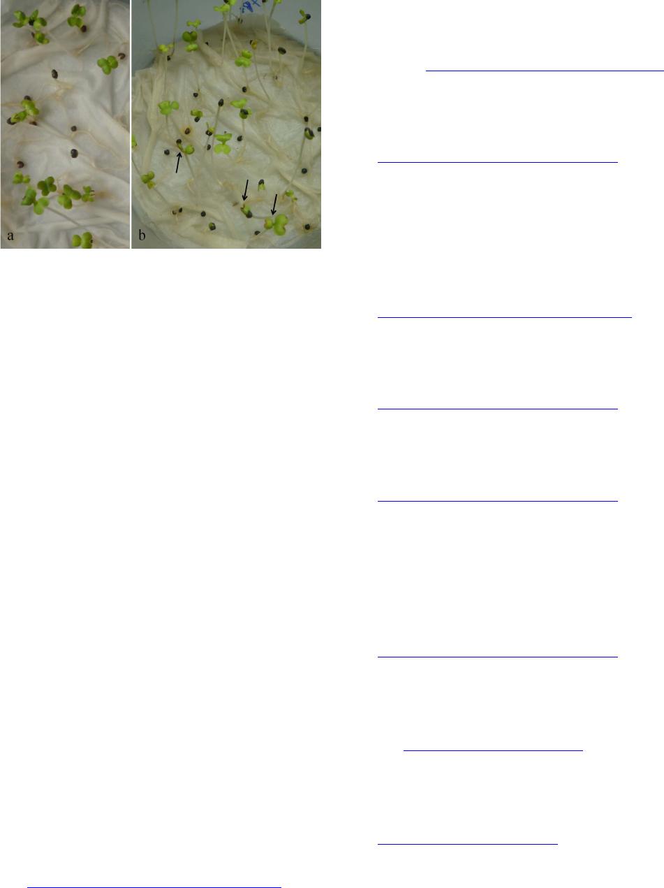 Influences of Cyanobacterial Toxins Microcystins on the Seedling of Plants 40 Figure 5. Normal leaves from control (a) and abnormal leave s from M C ex posure (b) of Brassica rapa -chinensis at 7 days of incubation. Arrows indicate the difference in size and the brown c olor at margin of the yo ung leaves. be attributed to the impact of MC on chlorophyll content. The results of this study confirm the potent toxicity of cyanobacterial toxins from Dau Tieng Reservoir on plants. To our knowledge, this is the first report on the effects of MC on seedlings of B. rapa-chinensis, B. na- rinosa and N. officinale. Ackno wledgements Thi s stud y is fund ed b y the Vietnam National University— Hochiminh City under the granted projects numbers B2012-24-01TD and A2013-48-01. REFERENCES [1] K. Sivonen and G. Jones, “Cyanobacterial Toxins,” In: I. Chorus andJ. Bartram, Eds., Toxic Cyanobacteria in Water—A Guide to Their Public Health Consequences, Monitoring and Management, E & FN Spon, London, 1999, pp . 41-111. [2] S. Pflugmacher, C. Wiegand, K. A. Beattie, G. A. Codd and C. E. W. Steinberg, “Uptake of the Cyanobacterial hepatotoxin Microcystin-LR by Aquatic Macrophytes,” Journal of Applied Botany, Vol. 72, No. 5-6, 1998, pp. 228-232. [3] S. Pflugmacher, C. Wiegand , K. A. Beat tie, E. Krause, C . E. W. Steinberg and G. A. Codd, “Uptake, Effects and Metabolism of Cyanobacterial Toxins in the Emergent Reed Plant Phragmites australis (Cav.) Trin. ex Steud,” Environmental Toxicology and Chemistry, Vol. 20, No. 4, 2001, pp . 846-852. [4] K. Kurki-Helasmo and J. Meriluoto, “Microcystin Uptake Inhibits Growth and Protein Phosphatase Activity in Mustard (Sinapis alba L.) Seedlings,” Toxicon, Vol. 36, No. 12, 1998, pp. 1921-1926. http://dx.doi.org/10.1016/S0041-0101(98)00114-7 [5] S. M. Mitrovic, O. Allis, A. Furey and K. J. James, “Bio- accumulation and Harmful Effects of Microcystin-LR in the Aquatic Plants Lemna minor and Wolffia arrhiza and the Filamentous Alga Chladophora fracta,” Exotoxicolo- gy and Environmental Safety, Vol. 61, No. 3, 2005, pp. 345-352. http://dx.doi.org/10.1016/j.ecoenv.2004.11.003 [6] S. Jarvenpaa, C. Lundberg-Niinisto, L. Spoof, O. Sjovall, E. Tyystjarvi and J. Meriluoto, “Effects of Microcystins on Broccoli and Must ard , and Analysis of Accumulated Toxin by Liquid Chromatography-Mass Spectrometry,” Toxicon, Vol. 49, No. 6, 2007, pp. 865-874. http://dx.doi.org/10.1016/j.toxicon.2006.12.008 [7] G. A. Codd, J. S. Metcalf and K. A. Beattie, “R etentio n of Microcystis aeruginosa and Microcystin b y Salad Lett uce (Lactuca sativa) after Spray Irrigation with Water Con- taini ng Cyano bact eria, ” To xicon, Vol. 37, No. 8, 1999, pp. 1181-1185. [8] J. McElhiney, LA. Lawton and C. Leifert, “Investigations into the Inhibitory Effects of Microcystins on Plant Growt h , and the Toxicity of Plant Tissues Following Ex- posure,” Toxicon , Vol. 39, No. 9, 2001, pp. 1411-1420. http://dx.doi.org/10.1016/S0041-0101(01)00100-3 [9] J. Chen, L. Song, J. Dai and Z. Liu, “ Effect s o f M icro cys - tins on the Growth and the Activity of Superoxide Dis- mutase and Peroxidase of Rape (Brassica napus L. ) and Rice (Oryza sativa L.),” Toxicon, Vol. 43, No. 4, 2004, pp. 393-400. http://dx.doi.org/10.1016/j.toxicon.2004.01.011 [10] S. Pflugmacher, M. Aulhorn and M. Grimm, “Influence of a Cyanobacterial Crude Extract Containing Microcys- tin-LR on the Physiology and Antioxidative Defence Systems o f Differen t Spi nach V ariants, ” New Phyco logist, Vol. 175, No. 3, 2007, pp . 482-489. http://dx.doi.org/10.1016/j.toxicon.2004.01.011 [11] J. Weiss, H. P. Liebert and W. Braune, “Influence on Microcystin-RR on Growth and Photosynthetic Capacity of the Duckweed Lemma minor L.,” Journal of Applied Botany, Vol. 74, No. 3-4, 2000, pp. 10-105. [12] M. H. Ha and S. Pflugmacher, “Phytotoxic Effects of the Cyanobacterial Neurotoxin Anatoxin-a: Morphological, Physiological and Biochemical Responses in Aquatic Macrophyte, Ceratophyllum demersum,” Toxicon, Vol. 70, 2013 , pp. 1-8. http://dx.doi.org/10.1016/j.toxicon.2013.03.021 [13] C. Pietsch, C. Wiegand, M. V. Ame, A. Nicklisch, D. Wunderlin and S. Pflugmacher, “The Effects of Cyano- bacterial Crude Extract on Different Aquatic Organisms: Evidence for Cyanobacterial Toxin Modulating Factors, ” Environmental Toxicology, Vol. 16, No. 6, 2 00 1, pp . 535- 542. http://dx.doi.org/10.1002/tox.10014 [14] C. Wiegand, A. Peuther t, S. Pflug macher and S. Carmeli, “Effects of Microcin SF608 and Microcystin-LR, Two Cyanobacterial Compounds Produced by Microcystis sp., on Aquatic Organisms,” Environmental Toxicology, Vol. 17, No. 4, 2002, pp . 400-406. http://dx.doi.org/10.1002/tox.10065 [15] J. Stuven and S. Pflugmacher, “Antioxidative Stress Re- sponse of Lepidium sativum Due to Exposure to Cyano- bacterial Seco ndar y Metabo li tes,” Toxicon, Vol. 50, No. 1, 2007, pp . 85-93. OPEN ACCESS JEP  Influences of Cyanobacterial Toxins Microcystins on the Seedling of Plants 41 http://dx.doi.org/10.1016/j.toxicon.2007.02.019 [16] L. Yin, J. Huang, W. Huang, D. Li and Y. Liu, “Res- ponses of Antioxidant System in Arabidopsis thaliana Suspension Cells to the Toxicity of Microcystin-RR,” Toxicon, Vol. 46, No. 8, 2005, pp. 8 59-864. http://dx.doi.org/10.1016/j.toxicon.2004.12.025 [17] L. Yin, J. Huang, W. Huang, D. Li, G. Wang and Y. Liu, “Micro cystin-RR-Induced Accumulation of Reactive Oxygen Species and Alteration of Antioxidant Systems in Tobaco BY-2 Cells,” Toxicon, Vol. 46, No. 5, 2005, pp. 507-512. http://dx.doi.org/10.1016/j.toxicon.2005.06.015 [18] T. S. Dao, T. L. Pham, L. C. Do-Hong and B. T. Bui, “Occurrence of Toxic Cyanobacteria and Their Toxins from Freshwater Bodies in Vietnam—A Short Review,” Vietnam Journal of Science and Technology, Vol. 50, No. 1C, 2012, pp. 264-269. [19] A. Mikhailov, A. S. Harmala-Brasken, J. Her man, J. A. O. Meriluoto and J. E. Eriksson, “Identification of ATP- Synthetase as a Novel Intracellular Target for Microcys- tin-LR,” Chemico-Biological Interactions, Vol. 142, No. 3, 2003, pp. 223-237. http://dx.doi.org/10.1016/S0009-2797(02)00075-3 [20] S. Pflugmacher, “Promotion of Oxidative Stress in the Aquatic Macrophyte Ceratophyllum demersum during Biotransformation of the Cyanobacterial Toxin Microcys- tin-LR,” Aquatic Toxicology, Vol. 70, No. 3, 2004, pp. 169-178. http://dx.doi.org/10.1016/j.aquatox.2004.06.010 [21] C. Garbers, A. DeLong, J. Deruere, P. Ber nasconi and D. Soll, “A Mutation in Protein Phosphatase 2A Regulatory Subumit A Affects Auxin Transport in Arabidopsis,” The EMBO Journal, Vol. 15, No. 9, 199 6, pp. 2115-2124. [22] M. M. Gehringer, V. Kewada, N. Coates and T. G. Downing, “The Use of Lepidiium sativum in a Plant Bio- assay System for the Detection of Microcystin-LR,” Tox- icon, Vol. 41, No. 7, 2003, pp. 871-876. http://dx.doi.org/10.1016/S0041-0101(03)00049-7 OPEN ACCESS JEP |