

Paper Menu >>
Journal Menu >>
 Chinese Medicine, 2011, 2, 16-19 doi:10.4236/cm.2011.21003 Published Online March 2011 (http://www.SciRP.org/journal/cm) Copyright © 2011 SciRes . CM Primary Pupillary Margin Cyst of the Iris Pigment Epithelium Rosanna Dammacco1, Giovanni Giancipoli1, Silvana Guerriero1, Domenico P iscite l li2, Nicola Cardascia1 1Department of Ophthalmology and Otorhinolaryngology, Bari University, Bari, Ita ly 2Department of Pathological Anatomy, Bari Unive rsity, Bar i, Italy E-mail: [email protected] Received January 13, 2011; revised February 2, 2011; accepted February 10, 2011 Abstract Purpose: Description of a patient with a solitary cyst of the pupillary margin iris pigment epithelium (IPE). Methods: A 63-year-old man referred a suspected iris-ciliary body melanoma in his left eye. Based on both clinical examination and ultrasound biomicroscopy, melanoma was considered unlikely. Surgery was under- taken to correct recurrent deterioration of vision due to movement of the lesion across the visual axis. Results: The lesion was excised completely. Ultrasound biomicroscopy and histopathological examination ruled out melanoma and allowed a final diagnosis of primary pupillary margin cyst of the IPE, characterized of pig- mented epithelium, with no connective tissue or vessels. No recurrences or fresh lesions appeared during a one-year follow-up. Conclusions: Primary epithelial iris cysts are usually benign. Treatment is required only in symptomatic patients and those with an uncertain diagnosis. Ultrasound biomicroscopy is indispensable to confirm the clinical diagnosis, follow the clinical course and intervene if surgery is required. Keywords: Melanoma, I ris Pigment E pithe lium, P up illary Cyst, S urgical Excision, Ultrasound Biomicroscopy 1. Introduction Primary cysts in t he anterior segment, observed during a routine eye examination, are divided into stromal and pigment epithelial cysts [1 ]. Primary stromal cysts are less common and mostly congenital [2]. Their differen- tial diagnosis from malignant uveal melanoma may sometimes be difficult. Primary cysts are of neuroepi- thelial origin, whereas secondary cysts may result from intraocular implantation of surface epithelium following surgery or a penetrating trauma, or else from metastasis, parasites, or chronic myotic therapy [3]. Neuroepithelial cysts involve the iris pigment epithelium (IPE) and the ciliary bod y [4]. Primary cysts of the IPE are uncommon and sponta- neous. T hey are defined as epithelial-lined struc tur es t hat arise in t he poster ior iris layer [ 5 ] . Shields (1981) distin- guishes four t ypes, according to their po sition in rela tion to the iris [1]: c entral, at the pupillar y mar gin; midzonal, between the pupillary margin and the iris roo t; peripheral, at the iridociliary sulcus; dislodged, in the anterior chamber or the vitreous cavity. The vast majority (76%) is peripheral [1]. This paper describes a case of symptomatic primary central IPE cyst initially mistaken for an iris melanoma. Stress is laid on ultrasound biomicroscopy (UBM) as a noninvasive way of determining the nature of a cyst, establishing its final diagnosis and following its course. 2. Case Report A 63-year-old man presented with a two-month history of disturbed vision in his left eye and a suspected iris melanoma. In distant vision, his best corrected visual acuity (BCVA) was 20/20 in both eyes, but his left-eye disturbance was aggravated by strong daylight, which caused a severe temporary deterioration of vision (≤ 20/50). Slit-lamp examination showed a solitary, homo- geneously pigmented mass hanging at the superior pu- pillary margin of the iris and at 1 o’clock in the anterior chamber (Figure 1). It slowly moved when the patient changed the position of his head. It was smooth, dark-brown and apparently cystic. The pupil was round, readily reacting to light and accomodation. During mi- osis, the lesion often obstructed the pupillary aperture. The slit lamp also revealed a normal left external eye, 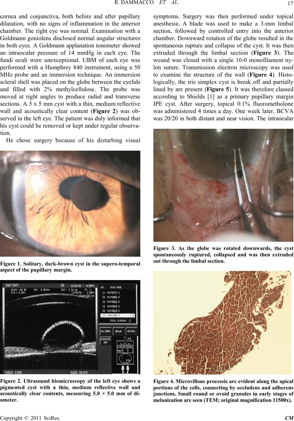 R. DAMMACC O ET AL. Copyright © 2011 SciRes . CM 17 cornea and conjunctiva, both before and after papilla ry dilatation, with no signs of inflammation in the anterior chamber. The right eye was normal. Examination with a Goldmann goniolens disclosed normal angular structures in both eyes. A Gold mann ap planation tonometer sho wed an intraocular pressure of 14 mmHg in each eye. The fundi oculi were unexceptional. UBM of each eye was performed with a Humphrey 840 instrument, using a 50 MHz probe and an immersion technique. An immersion scleral shell was placed on the globe between the eyelids and filled with 2% methylcellulose. The probe was moved at right angles to produce radial and transverse sections. A 5 x 5 mm cyst with a thin, mediu m re flective wall and acoustically clear content (Figure 2) was ob- served in the left eye. The patient was dul y informed that his cyst could be removed or kept under regular observa- tion. He chose surgery because of his disturbing visual Figure 1. Solitary, dark-brow n cyst in the supero-temporal aspect of t he p upillary margin. Figure 2. Ultrasound biomicroscopy of the left eye shows a pigmented cyst with a thin, medium reflective wall and acoustically clear contents, measuring 5.0 × 5.0 mm of di- ameter. symptoms. Surgery was then performed under topical anesthesia. A blade was used to make a 3-mm limbal section, followed by controlled entry into the anterior chamber. Do wnward ro tation of the globe resulted in the spo ntaneous r upture and coll apse of the cyst. I t was the n extruded through the limbal section (Fig ure 3). The wound was closed with a single 10-0 monofilament ny- lon suture. Transmission electron microscopy was used to examine the structure of the wall (Figure 4). Histo- logically, the iris simplex cyst is break off and partially lined by are present (Figure 5). It was therefore classed according to Shields [1] as a primary pupillary margin IPE cyst. After surgery, topical 0.1% fluorometholone was ad ministered 4 times a day. One week later, BCVA was 20/20 in both dis tant a nd near vi sion. T he i ntraoc ular Figure 3. As the globe was rotated downwards, the cyst spontaneously ruptured, collapsed and was then extruded out through t he li mb al se ction. Figure 4 . Microvil lous pr oces sis are evident alo ng t he apical port ions of t he cell s, con necting by occlu dens an d adherens junctions. Small round or ovoid granules in early stages of melanization are seen (TEM; original magnification 11500 x).  R. DAMMACC O ET AL. Copyright © 2011 SciRes . CM 18 Figure 5. Hi stopatholog ical ex amin ation of th e tissue sh ows that it consists entirely of pigmented epithelium without connective tissue or vascular el ements. pressure, the shape of the anterior chamber and the reac- tion of the pupil to light were normal. 3. Discussion Primary IPE cysts are found anywhere along the post- erior surface of the iris from the pupillary margin to the anterior ciliary body. The great majority are stationary and small, and sometimes regress spontaneously. Their presence is occasionally responsible for angle closure glaucoma, plateau iris syndrome and secondary pigment dispersion syndrome. [6-9]. Central cysts account for approximately 3% of all primary IPE cysts [1,5]. They are usually dark brown, round or oval with a thin wall and sonolucent contents [1-5 ] . The high reflectivity of their wall is attributed to the epithelial cells, while their sonolucent core is compatible with a fluid content. The origin of primary central IPE cysts is not clear. Their appearance in early childhood in a few familial cases suggests that they may be hereditary with an autosoma l- dominant pattern [7-10]. Lewis et al. [11] described an association with familial aortic dissection. Sallo and Hatvani [12] reported four cases of primary pupillary IPE cysts in a single family. These patients also suffered fro m transie nt vis ion d istur banc es. O ur pati ent, ho weve r, had no history of associated systemic diseases and no other membe r of hi s fa mi l y ha d a cyst . P r imar y I P E cysts have often been misdiagnosed as iris or ciliary body me- lanoma [13]. Cysts of the iris are more likely to be con- fused with melanoma than those of ciliary body because of their pigmentation and more anterior location. Diag- nosis and monitoring of iris, ciliary body and posterior chamber cysts require the imaging of structures not al- ways visible with a slit lamp and go ni olens, even through a dilated pupil. UBM has thus become indispensable for evaluating anterior segment tumors and cysts and is used to confirm a clinical diagnosis of primary IPE cysts, dif- ferentiate them from cystic and solid tumors (iris-ciliary body melanoma and melanocytoma, adenoma of the IPE and medulloepithelioma), follow their course and pro mptl y inte rvene i f s ur gery is r eq uir e d. I t al so p ro vid e s cross-sectional images illustrating the surface contour, internal reflectivity and borders, and reveals their size, extension and site. Management is determined by a cyst’s site, size, local extent and growth pattern, and the presence of secondary complications. Most primary IPE cysts run a benign clinical course that seldom results in ocular complications. They are usually small and fre- quently regress spontaneously. Those that arise from the IPE layers are stationary, asymptomatic, and detected only incide ntally. Shield s beli eves that most pri mary IPE cysts are ophthalmic curiosities that need prolonged ob- servation and no treatment [1]. A steadily growing cyst, however, may disturb vision by covering the visual axis and provoke an increase in intraocular pressure or even inflammation i f it to uches t he cor neal endotheli u m. Steps must then be taken to prevent or treat complications, such as pupillary obstruction, secondary glaucoma, iri- docyclitis, corneal decompensation and loss of vision. Numerous forms of managements have been employed. Mitomycin injection into the cyst and needle aspiration with endodiathermy were proposed by Kawaguchi et al. [14] and Tsai et al. [15]. Kuchenbecker et al. [16], Öner et al. [17] and Baykara et al. [18] prefer Nd:YAG laser cystotomy as the least invasive procedure. Excision in our case, like in that reported by Verma et al. [19], was dictated by the fact that in strong daylight the cyst ob- scured the visual axis almost completely and caused a severe temporary decrease of visual acuity. There were no complications and no clinical or UBM evidence of recurrences or fresh lesions during a one-year follow-up. In conclusion, in agreement with most authors we be- lieve that stable and asymptomatic primary IPE cysts usual ly requi re no more than regular o bserva tion. P roper treatment depends on the overall clinical picture, which includes both the patient’s general health and the size and location of the cyst. Lastly, the importance of early diagnosis should be stressed. UBM is a useful, readily available and noninvasive tool for this purpose, since it can be employed to both confirm the clinical diagnosis and follow the course of these cysts. We address future studies on anterior chamber OCT to “in vivo” analyze cyst morphology. 4. Referen ces [1] J. A. Sh ields, “Primary Cysts of the Iris,” Transactions of  R. DAMMACC O ET AL. Copyright © 2011 SciRes . CM 19 the American Ophthalmological Society, 1981, Vol. 79, pp. 771-809. [2] C. Auw-Haedrich, G. Schlunck and H. Witsch el, “Pri- mary Iris Stromal Cyst with Unusual Symptoms in an Adult,” Klinische Monatsblätter für Augenheilkunde, 2000, Vol. 216, pp. 420-423. doi:10.1055/s-2000-10589 [3] T. Hildreth, J. Maino and T. Hartong, “Primary and Sec- ondary Iris Cysts,” Journal of the American Optometric Association, 1991, Vol. 62, pp. 588-592. [4] H. Tanihara, J. Akita, M. Honjo and Y. Honda, “Angle Closure Caused by Multiple, Bilateral Iridociliary Cysts,” Acta Ophthalmologica Scandinavica, 1997, Vol. 75, No. 2, pp. 216-217. doi:10.1111/j.1600-0420.1997.tb00128.x [5] N. Lois, C. L. Shields, J. A. Shields and G. Mercado, “Primary Cysts of the Iris Pigment Epithelium. Clinical Features and Natural Course in 234 Patients,” Ophthal- mology, 1998, Vol. 105, No. 10, pp. 1879-1885. doi:10.1016/S0161-6420(98)91034-X [6] P. A. Chandler and H. E. Braconier, “Spontaneous In- tra-Epithelial Cysts of Iris and Ciliary Body with Glau- co ma ,” 1958, Vol. 45(4 Pt II), pp. 64-74. [7] A. Vela, J. C. Rieser an d D. G. Campbell, “The H er edity and Treatment of Angle-Closure Glaucoma Secondary to Iris and Ciliary Body Cysts,” Ophthalmology, 1983, Vol. 91, No. 4, pp. 332-337. [8] A. Azuara-Blanco, G. L. Spaeth, S. V. Araujo, J. J. Augsburger and A. K. Terebuh, “Plateau Iris Syndrome Associated with Multiple Ciliary Body Cysts : Report of 3 Cases,” Arch ives of Ophthalmology, 1996, Vol. 114, No. 6, pp. 666-668. [9] W. L. M. Al war d and K. C. Ossoi nig, “Pigment Disp er- sion Secondary to Cysts of the Iris Pigment Epithelium,” Archives of Ophthalmology, 1995, Vol. 113, No.12, pp. 1574-1575. [10] A. C owa n , “Congenital and Familial Cysts and Flocculi of the Iris,” American Journal of Ophthalmology, 1936. Vol. 19, pp. 287-291. [11] R. A. Le wi s and L. M. Merin, “Iris Flocculi and Familial Aortic Dissection,” Archives of Ophthalmology, 1995, Vol. 113, pp. 1330-1331. [12] F. B. Sallo and I. Hatvani, “Recurring Transitory Blind- ness Cau sed by Primary Marginal Pigment Epithelial Iris C ys t s ,” American Journal of Ophthalmology, 2002, Vol. 133, No. 3, pp. 407-409. doi:10.1016/S0002-9394(01)01332-0 [13] M. Yan off and L. E. Z immerman, “Pseudomelanoma of Anterior Chamber Caused by Implantation of Iris Pig- ment Epithelium,” Archives of ophthalmology, 1965, Vol. 74, No. 3, pp. 302-305. [14] K. Kawaguchi, S. Yamamoto, Y. Nagae, A. Okada, N. Iwa sa ki and Y. Tano, “Treatment of Recurrent Giant Iris Cyst with Intracyst Administration of Mitomycin C,” British Journal of Ophthalmology, 2000, Vol. 84, No. 7, pp. 800-801. doi:10.1136/bjo.84.7.799b [15] J. C. Tsai, E. L Arrindell, D. M. O’Day, “Needle Asp i ra- tion and Endodiathermy Treatment of Epithelial Inclusion Cyst of the Iris,” American Journal of Ophthalmology, 2001, Vol. 131, No. 2, pp . 263-265. doi:10.1016/S0002-9394(00)00707-8 [16] J. Kuchenbecker, M. Motschmann, K. Schmitz and W. Behren s -B aumann, “Laser Iridocystotomy for Bilateral Acu te An gle -Closure Glau coma Seco ndar y to Iris Cysts,” American Journal of Ophthalmology, 2000, Vol. 129, No. 3, pp. 391-393. doi:10.1016/S0002-9394(99)00392-X [17] H. F. Öner, S. Kaynak, N. Koçak and G. Çingil, “Man- agement of Free Floating Iris Cysts in the Anterior Chamber: A Case Report,” European Journal of Oph- thalmology, 2003, Vol. 13, No. 2, pp. 212-214. [18] M. Baykara, S. S ah i n and H. Ertürk, “Free Iris Cyst in the Anterior Chamber,” Ophthalmic Surgery Lasers and Im- aging, 2004, Vol. 35, No. 1, pp. 74-75. [19] L. Verma, P. Venkatesh, S. Sen, N. C. Lakshmaiah and H. K. Tewari, “Surgical Removal of a Free Floating Cyst of the Iris Pigment Epithelium Causing Disturbing Visual Sy mp t o ms ,” Ophthalmic Surgery and Lasers, 1999, Vol. 30, No. 3, pp. 223-225. |