

Paper Menu >>
Journal Menu >>
 Engineering, 2013, 5, 553-560 http://dx.doi.org/10.4236/eng.2013.510B114 Published Online October 2013 (http://www.scirp.org/journal/eng) Copyright © 2013 SciRes. ENG Noninvasive Blood Glucose Monitoring System Based on Distributed Multi-Sensors Information Fusion of Multi-Wavelength NIR* Bo Zeng, Wei Wang#, Na Wang, Funing Li, Fulong Zhai, Lintao Hu School of Information Science and Engineering, Lanzhou Universit y, Lanzhou, China Email: [email protected], #[email protected] Received 2013 ABSTRACT In this research, a near infrared multi-wavelength noninvasive blood glucose monitoring system with distributed laser multi-sensors is applied to monitor human blood glucose concentration. In order to improve the monitoring accuracy, a multi-sensors information fusion model based on Back Propagation Artificial Neural Network is proposed. The Root- Mean-Square Error of Prediction for noninvasive blood glucose measurement is 0.088mmol/L, and the correlation coef- ficient is 0.94. The noninvasive blood glucose monitoring system based on distributed multi-sensors information fusion of multi-wavelength NIR is proved to be of great efficient. And the new proposed idea of measurement based on distri- buted multi-sensors, shows better prediction accuracy. Keywords: Noninvasive Glucose Monitoring; NIR Arrays Signals Fusion; BP-Artificial Neural Network 1. Introduction Diabetes has become a modern disease and more than 150 million people are suffering from it all around the world [1]. In order to prevent complication, the tight blood glucose level control is very essential. Currently, patients are recommended to monitor their blood glucose level via an invasive finger-tip stick method, this way will inevitably bring patients pain and infection [2]. In order to avoid the weakness of invasive method, a num- ber of noninvasive measurements occur r ed, like middle- infrared emission spectroscopy [3,4], Near Infrared (NIR) spectroscopy [5,6]. The main drawback of infrared mea- surement is its accuracy and stability. In our past work, an infrared noninvasive blood glucose measurement sys- tem based on multi-sensors and Mixture of Experts (ME) [7] was designed. ME algorithm greatly improved the precision of noninvasive blood glucose measurement. In the noninvasive blood glucose monitoring system based on distributed multi-sensors information fusion of mul- ti-wavelength NIR, this system was designed on the pur- pose of continuous blood glucose monitoring for patients at home and hospital [2]. Another advantage of this sys- tem was the introduction of distributed multi-sensors id ea, this change has been proved to be better improvement for blood glucose prediction accuracy. In this distributed multi-sensors system, a multi-sensors information fusion model, Back Prop agation Artificial N eural Network ( BP- ANN), was appli e d. 2. Noninvasive Blood Glucose Monitoring Model The experimental data for this research was obtained via NIR based on laser arrays as shown in Figure 1. The NIR light source consists of 3*3 laser diodes arrays op- erating at output powers of 5 mW [8]. 2.1. Hardware Design Wavelengths selection: According to the specialty of glucose molecular structure and absorption specialty, second order times frequency absorption exists between 1100 - 1300 nm, and first order times frequency absorp- tion exists between 1500 - 1800 nm. Other components in blood, such as hemoglobin and water contain groups of hydrogen which can trigger NIR absorption as well. Water absorption peaks are mainly distributed between 1440 - 1460 nm and between 1940 - 1960 nm, those regions should be avoided in wavelength selection. Be- tween 1400 - 1800 nm, water absorption only exists at 1787 nm, fat and protein exist no absorption peak in this region. Therefore, 1400 - 1800 nm region is suitable for measurement wavelength selection. Measurement sites selection: In noninvasive blood * The work is supported b y National Nature Science Fund of China (No 30970876, 81141076). *Corresponding a uthor. 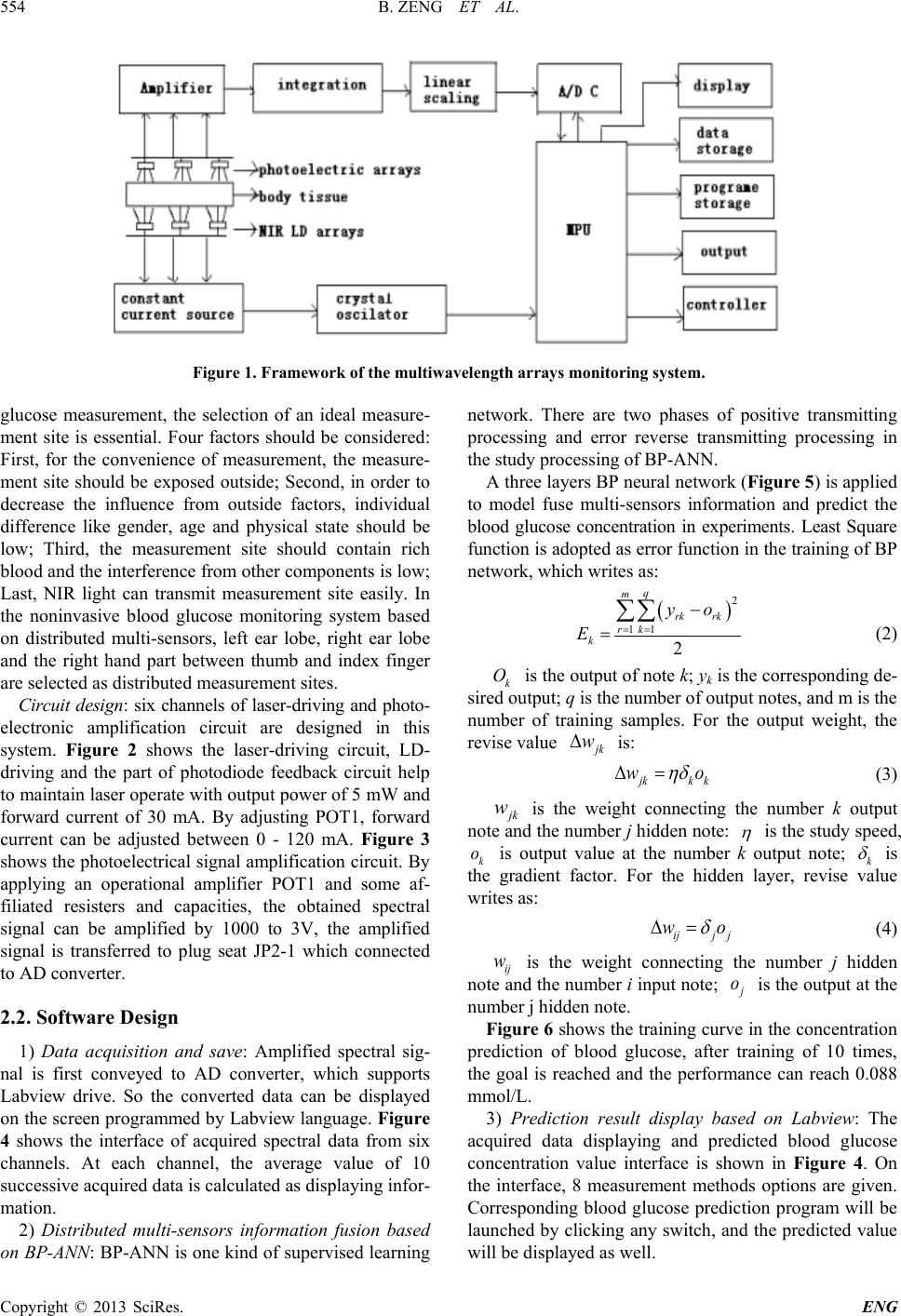 B. ZENG ET AL. Copyright © 2013 SciRes. ENG 554 Figure 1. Framework of the multiwavelength arrays monitoring system. glucose measurement, the selection of an ideal measure- ment site is essential. Four factors should be considered : First, for the convenience of measurement, the measure- ment site should be exposed outside; Second, in order to decrease the influence from outside factors, individual difference like gender, age and physical state should be low; Third, the measurement site should contain rich blood and the interference from other components is low; Last, NIR light can transmit measurement site easily. In the noninvasive blood glucose monitoring system based on distributed multi-sensors, left ear lobe, right ear lobe and the right hand part between thumb and index finger are selected as distributed measurement sites. Circuit design: six channels of laser-driving and photo- electronic amplification circuit are designed in this system. Figure 2 shows the laser-driving circuit, LD- driving and the part of photodiode feedback circuit help to maintain laser operate with output power of 5 mW and forward current of 30 mA. By adjusting POT1, forward current can be adjusted between 0 - 120 mA. Figure 3 shows the photoelectrical signal amplification c ircuit. By applying an operational amplifier POT1 and some af- filiated resisters and capacities, the obtained spectral signal can be amplified by 1000 to 3V, the amplified signal is transferred to plug seat JP2-1 which connected to AD converter. 2.2. Software Design 1) Data acquisition and save: Amplified spectral sig- nal is first conveyed to AD converter, which supports Labview drive. So the converted data can be displayed on the screen programmed by Labview language. Figu r e 4 shows the interface of acquired spectral data from six channels. At each channel, the average value of 10 successive acquired data is calculated as displaying infor- mation. 2) Distributed multi-sensors information fusion based on BP-ANN: BP-ANN is one kind of supervised learning network. There are two phases of positive transmitting processing and error reverse transmitting processing in the study processing of BP-ANN. A three layers BP neural network (Figure 5) is applied to model fuse multi-sensors information and predict the blood glucose concentration in experiments. Least Square function is adopted as error function in the training of BP network, which writes as: ( ) 2 11 2 q m rk rk rk k yo E = = − = ∑∑ (2) k O is the output of note k; yk is the corresponding de- sired output; q is the number of output notes, and m is the number of training samples. For the output weight, the revise value jk w∆ is: jkk k wo ηδ ∆= (3) jk w is the weight connecting the number k output note and the number j hidden note: η is the study speed, k o is output value at the number k output note; k δ is the gradient factor. For the hidden layer, revise value writes as: ijj j wo δ ∆= (4) ij w is the weight connecting the number j hidden note and the number i input note; j o is the output at the number j hi dden note . Figure 6 shows the training curve in the concentration prediction of blood glucose, after training of 10 times, the goal is reached and the performance can reach 0.088 mmol/L. 3) Prediction result display based on Labview: The acquired data displaying and predicted blood glucose concentration value interface is shown in Figure 4. On the interface, 8 measurement methods options are given. Corresponding blood glucose prediction program will be launched by clicking any switch, and the predicted value will be displayed as well. 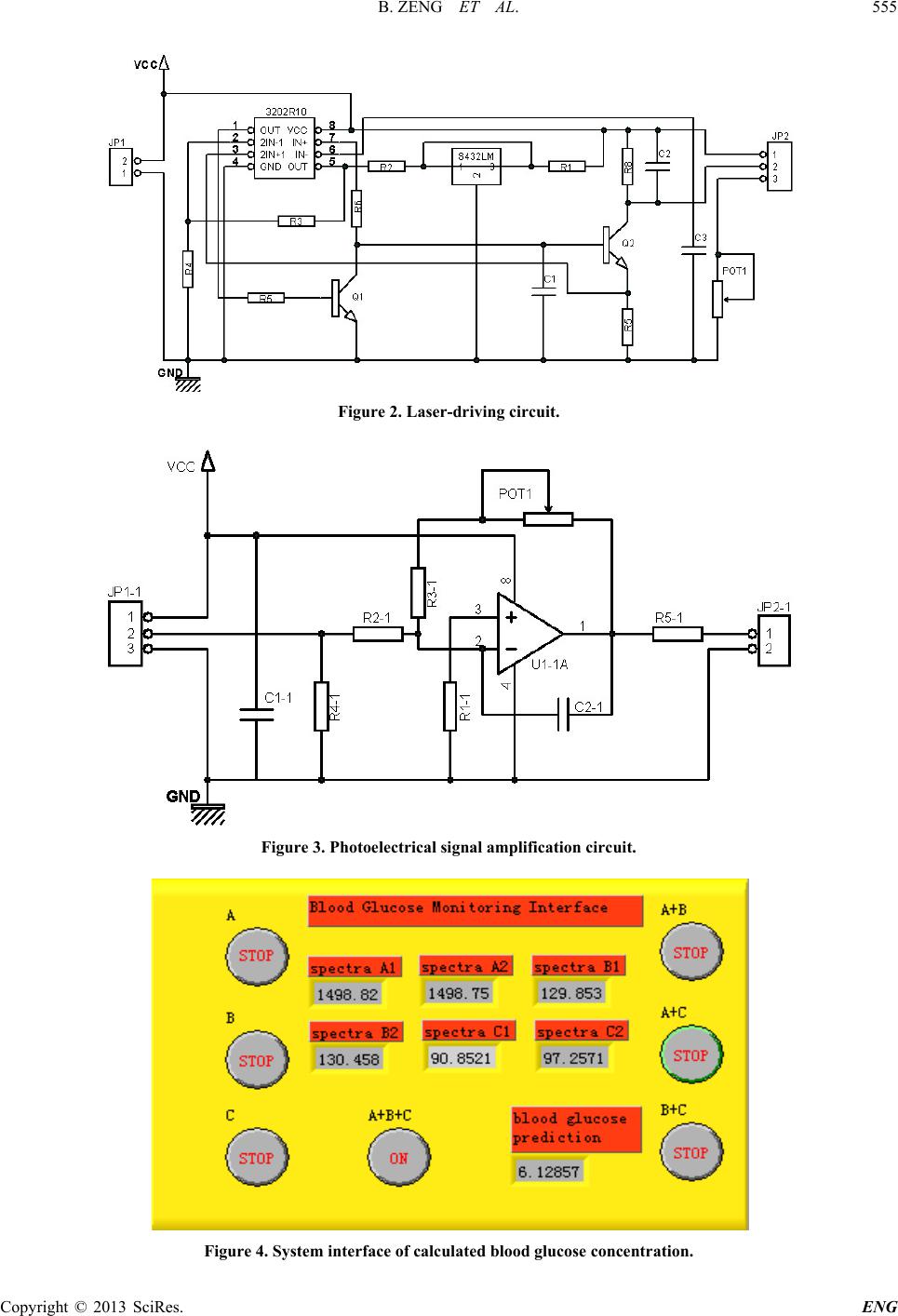 B. ZENG ET AL. Copyright © 2013 SciRes. ENG 555 Figure 2. Laser-driving circuit. Figure 3. Photoelectrical signal amplification circuit. Figure 4. System interface of calculated blood glucose concentration. 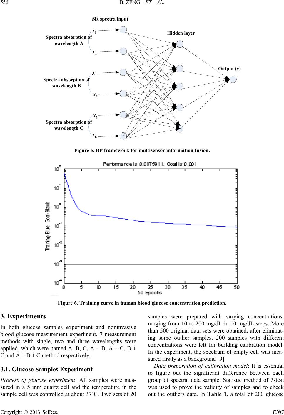 B. ZENG ET AL. Copyright © 2013 SciRes. ENG 556 Figure 5. BP framework for multisensor information fusion. Figure 6. Training curve in human blood glucose concent ration predict i on. 3. Experiments In both glucose samples experiment and noninvasive blood glucose measurement experiment, 7 measurement methods with single, two and three wavelengths were applied, which were named A, B, C, A + B, A + C, B + C and A + B + C method respectively. 3.1. Glucose Samples Experiment Process of glucose experiment: All samples were mea- sured in a 5 mm quartz cell and the temperature in the sample cell was controlled at about 37˚C. Two sets of 2 0 samples were prepared with varying concentrations, ranging from 10 to 200 mg/dL in 10 mg/dL steps. More than 500 original data sets were obtained, after eliminat- ing some outlier samples, 200 samples with different concentrations were left for building calibration model. In the experiment, the spectrum of empty cell was mea- sured firstly as a background [9]. Data preparation of calibration model: It is essential to figure out the significant difference betwe en each group of spectral data sample. Statistic method of T-test was used to prove the validity of samples and to check out the outliers data. In Table 1, a total of 200 glucose 1 x 2 x 3 x 4 x 5 x 6 x Six spectra input Hidden layer Output (y) Spectra absorption of wavelength A Spectra absorption of wavelength B Spectra absorption of wavelength C 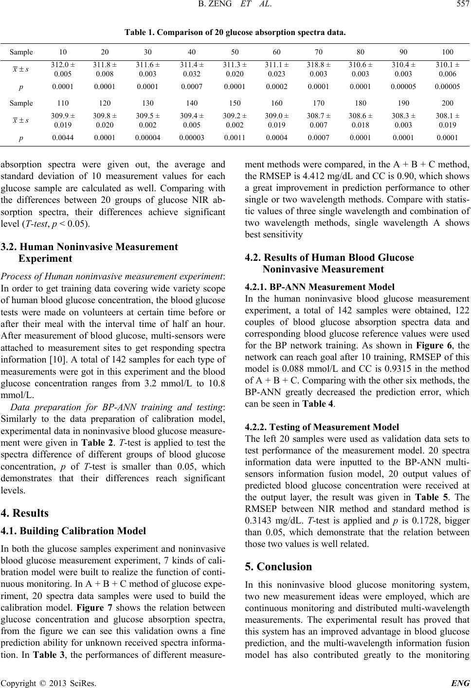 B. ZENG ET AL. Copyright © 2013 SciRes. ENG 557 Table 1. Comparison of 20 glucose absorption spectra data. Sample 10 20 30 40 50 60 70 80 90 100 xs± 312.0 ± 0.005 311.8 ± 0.008 311.6 ± 0.003 311.4 ± 0.032 311.3 ± 0.020 311.1 ± 0.023 318.8 ± 0.003 310.6 ± 0.003 310.4 ± 0.003 310.1 ± 0.006 p 0.0001 0.0001 0.0001 0.0007 0.0001 0.0002 0.0001 0.0001 0.00005 0.00005 Sample 110 120 130 140 150 160 170 180 190 200 xs± 309.9 ± 0.019 309.8 ± 0.020 309.5 ± 0.002 309.4 ± 0.005 309.2 ± 0.002 309.0 ± 0.019 308.7 ± 0.007 308.6 ± 0.018 308.3 ± 0.003 308.1 ± 0.019 p 0.0044 0.0001 0.00004 0.00003 0.0011 0.0004 0.0007 0.0001 0.0001 0.0001 absorption spectra were given out, the average and standard deviation of 10 measurement values for each glucose sample are calculated as well. Comparing with the differences between 20 groups of glucose NIR ab- sorption spectra, their differences achieve significant level (T-test, p < 0.05). 3.2. Human Noninvasive Measurement Experiment Process of Human noninvasive measurement experiment: In order to get training data covering wide variety scope of human blood glucose concentration, the blood glucose tests were made on volunteers at certain time before or after their meal with the interval time of half an hour. After measurement of blood glucose, multi-sensors were attached to measurement sites to get responding spectra information [10]. A total of 142 samples for each type of measurements were got in this experiment and the blood glucose concentration ranges from 3.2 mmol/L to 10.8 mmol/L. Data preparation for BP-ANN training and testing: Similarly to the data preparation of calibration model, experimental data in noninvasive blood glucose measure- ment were given in Table 2. T-test is applied to test the spectra difference of different groups of blood glucose concentration, p of T-test is smaller than 0.05, which demonstrates that their differences reach significant levels. 4. Results 4.1. Building Calibration Model In both the glucose samples experiment and noninvasive blood glucose measurement experiment, 7 kinds of cali- bration model were built to realize the function of conti- nuous monitoring. In A + B + C method of glucose expe- riment, 20 spectra data samples were used to build the calibration model. Figure 7 shows the relation between glucose concentration and glucose absorption spectra, from the figure we can see this validation owns a fine prediction ability for unknown received spectra informa- tion. In Table 3, the performances of different measure- ment methods were compared, in the A + B + C method, the RMSEP is 4.412 mg/dL and CC is 0.90, which shows a great improvement in prediction performance to other single or two wavelength methods. Compare with statis- tic values of three single wavelength and combination of two wavelength methods, single wavelength A shows best sensitivity 4.2. Results of Human Blood Glucose Noninvasive Measurement 4.2.1. BP-ANN Measurement Model In the human noninvasive blood glucose measurement experiment, a total of 142 samples were obtained, 122 couples of blood glucose absorption spectra data and corresponding blood glucose reference values were used for the BP network training. As shown in Figure 6, the network can reach goal after 10 training, RMSEP of this model is 0.088 mmol/L and CC is 0.9315 in the method of A + B + C. Comparing with the other six methods, the BP-ANN greatly decreased the prediction error, which can be seen in Table 4. 4.2.2. Testing of Measurement Model The left 20 samples were used as validation data sets to test performance of the measurement model. 20 spectra information data were inputted to the BP-ANN multi- sensors information fusion model, 20 output values of predicted blood glucose concentration were received at the output layer, the result was given in Table 5. The RMSEP between NIR method and standard method is 0.3143 mg/dL. T-test is applied and p is 0.1728, bigger than 0.05, which demonstrate that the relation between those two values is well related. 5. Conclusion In this noninvasive blood glucose monitoring system, two new measurement ideas were employed, which are continuous monitoring and distributed multi-wavelength measurements. The experimental result has proved that this system has an improved advantage in blood glucose prediction, and the multi-wavelength information fusion model has also contributed greatly to the monitoring 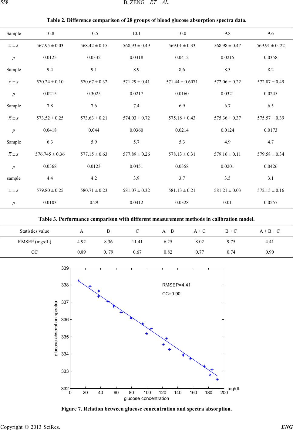 B. ZENG ET AL. Copyright © 2013 SciRes. ENG 558 Table 2. Difference comparison of 28 groups of blood glucose absorption spectra data. Sample 10.8 10.5 10.1 10.0 9.8 9.6 xs± 567.95 ± 0.03 568.42 ± 0.15 568.93 ± 0.49 569.01 ± 0.33 568.98 ± 0.47 569.91 ± 0. 22 p 0.0125 0.0332 0.0318 0.0412 0.0215 0.0358 Sample 9.4 9.1 8.9 8.6 8.3 8.2 xs± 570.24 ± 0.10 570.67 ± 0.32 571.29 ± 0.41 571.44 ± 0.6071 572.06 ± 0.22 572.87 ± 0.49 p 0.0215 0.3025 0.0217 0.0160 0.0321 0.0245 Sample 7.8 7.6 7.4 6.9 6.7 6.5 xs± 573.52 ± 0.25 573.63 ± 0.21 574.03 ± 0.72 575.18 ± 0.43 575.36 ± 0.37 575.57 ± 0.39 p 0.0418 0.044 0.0360 0.0214 0.0124 0.0173 Sample 6.3 5.9 5.7 5.3 4.9 4.7 xs± 576.745 ± 0.36 577.15 ± 0.63 577.89 ± 0.26 578.13 ± 0.31 579.16 ± 0.11 579.58 ± 0.34 p 0.0368 0.0123 0.0451 0.0358 0.0201 0.0426 sa mpl e 4.4 4.2 3.9 3.7 3.5 3.1 xs± 579.80 ± 0.25 580.71 ± 0.23 581.07 ± 0.32 581.13 ± 0.21 581.21 ± 0.03 572.15 ± 0.16 p 0.0103 0.29 0.0412 0.0328 0.01 0.0257 Table 3. Performance comparison with different measurement methods in calibration model. Statistics value A B C A + B A + C B + C A + B + C RMSEP (mg/dL) 4.92 8.36 11.41 6.25 8.02 9.75 4.41 CC 0.89 0. 79 0.67 0.82 0.77 0.74 0.90 Figure 7. Relation between glucose concentration and spectra absorption. 020406080100 120 140160 180200 332 333 334 335 336 337 338 339 glucose concent rat i on glucose abs orpti on spectra RMSEP=4.41 CC=0 .90 mg/dL 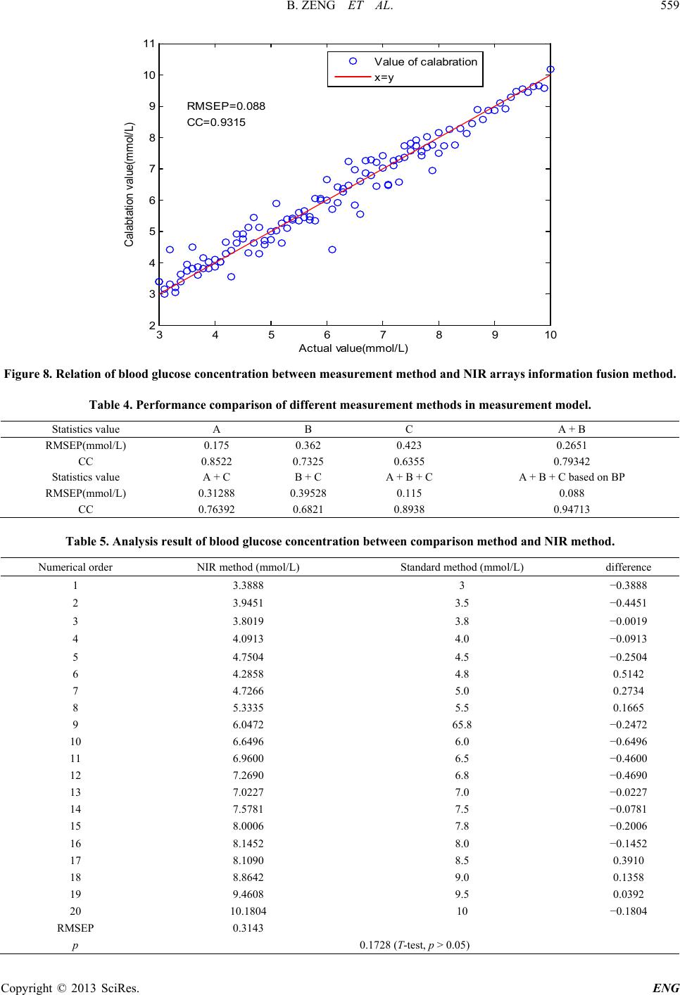 B. ZENG ET AL. Copyright © 2013 SciRes. ENG 559 Figure 8. Rel ation of blood glucose concentration between measurement method and NIR arrays information fusion method. Table 4. Performance comparison of different measurement methods in measurement model. Statistics value A B C A + B RMSEP(mmol/L) 0.175 0.362 0.423 0.2651 CC 0.8522 0.7325 0.6355 0.79342 Statistics value A + C B + C A + B + C A + B + C based on BP RMSEP(mmol/L) 0.31288 0.39528 0.115 0.088 CC 0.76392 0.6821 0.8938 0.94713 Table 5. Analysis result of blood glucose concentration between comparison method and NIR method. Numerical order NIR method (mmol/L) Standard method (mmol/L) difference 1 3.3888 3 −0.3888 2 3.9451 3.5 −0.4451 3 3.8019 3.8 −0.0019 4 4.0913 4.0 −0.0913 5 4.7504 4.5 −0.2504 6 4.2858 4.8 0.5142 7 4.7266 5.0 0.2734 8 5.3335 5.5 0.1665 9 6.0472 65.8 −0.2472 10 6.6496 6.0 −0.6496 11 6.9600 6.5 −0.4600 12 7.2690 6.8 −0.4690 13 7.0227 7.0 −0.0227 14 7.5781 7.5 −0.0781 15 8.0006 7.8 −0.2006 16 8.1452 8.0 −0.1452 17 8.1090 8.5 0.3910 18 8.8642 9.0 0.1358 19 9.4608 9.5 0.0392 20 10.1804 10 −0.1804 RMSEP 0.3143 p 0.1728 (T-test, p > 0.05) 3 4 5678910 2 3 4 5 6 7 8 9 10 11 A ct ual value(mmol /L) Calabtat i on val ue(mm ol /L) RMSEP =0. 088 CC=0.9315 V al ue of c al abrat i on x=y 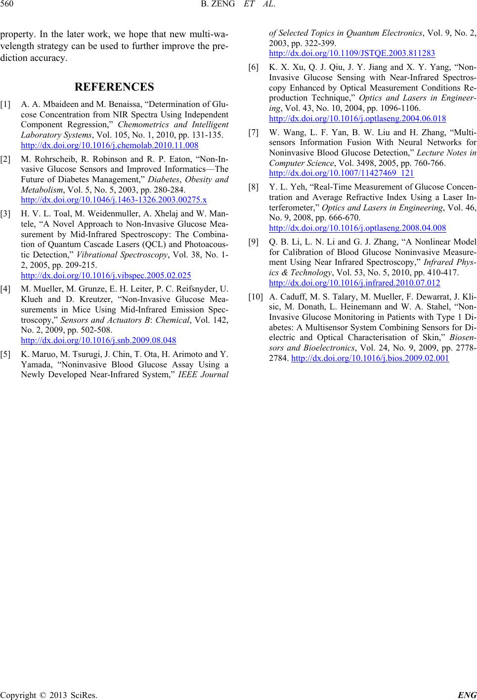 B. ZENG ET AL. Copyright © 2013 SciRes. ENG 560 property. In the later work, we hope that new multi-wa- velength stra tegy can be used to further improve th e pre- diction accuracy. REFERENCES [1] A. A. Mbaideen and M. Benaissa, “Determination of Glu- cose Concentration from NIR Spectra Using Independent Component Regression,” Chemometrics and Intelligent Laboratory Systems, Vol. 105, No. 1, 2010, pp. 131-135. http://dx.doi.org/10.1016/j.chemolab.2010.11.008 [2] M. Rohrscheib, R. Robinson and R. P. Eaton, “Non-In- vasive Glucose Sensors and Improved Informatics—The Future of Diabetes Management,” Diabetes, Obesity and Metabolism, Vol. 5, No. 5, 2003, pp. 280-284. http://dx.doi.org/10.1046/j.1463-1326.2003.00275.x [3] H. V. L. Toal, M. Weidenmuller, A. Xhelaj and W. Man- tele, “A Novel Approach to Non-Invasive Glucose Mea- surement by Mid-Infrared Spectroscopy: The Combina- tion of Quantum Cascade Lasers (QCL) and Photoacous- tic Detection,” Vibrational Spectroscopy, Vol. 38, No. 1- 2, 2005, pp. 209-215. http://dx.doi.org/10.1016/j.vibspec.2005.02.025 [4] M. Mueller, M. Grunze, E. H. Leiter, P. C. Reifsnyder, U. Klueh and D. Kreutzer, “Non-Invasive Glucose Mea- surements in Mice Using Mid-Infrared Emission Spec- troscopy,” Sensors and Actuators B: Chemical, Vol. 142, No. 2, 2009, pp. 502-508. http://dx.doi.org/10.1016/j.snb.2009.08.048 [5] K. Maruo, M. Tsurugi, J. Chin, T. Ota, H. Arimoto and Y. Yamada, “Noninvasive Blood Glucose Assay Using a Newly Developed Near-Infrared System,” IEEE Journal of Selected Topics in Quantum Electronics, Vol. 9, No. 2, 2003, pp. 322-399. http://dx.doi.org/10.1109/JSTQE.2003.811283 [6] K. X. Xu, Q. J. Qiu, J. Y. Jiang and X. Y. Yang, “Non- Invasive Glucose Sensing with Near-Infrared Spectros- copy Enhanced by Optical Measurement Conditions Re- production Technique,” Optics and Lasers in Engineer- ing, Vol. 43, No. 10, 2004, pp. 1096-1106. http://dx.doi.org/10.1016/j.optlaseng.2004.06.018 [7] W. Wang, L. F. Yan, B. W. Liu and H. Zhang, “Multi- sensors Information Fusion With Neural Networks for Noninvasive Blood Glucose Detection,” Lecture Notes in Computer Science, Vol. 3498, 2005, pp. 760-766. http://dx.doi.org/10.1007/11427469_121 [8] Y. L. Yeh, “Real -Time Measurement of Glucose Concen- tration and Average Refractive Index Using a Laser In- terferometer,” Optics and Lasers in Engineering, Vol. 46, No. 9, 2008, pp. 666-670. http://dx.doi.org/10.1016/j.optlaseng.2008.04.008 [9] Q. B. Li, L. N. Li and G. J. Zhang, “A Nonlinear Model for Calibration of Blood Glucose Noninvasive Measure- ment Using Near Infrared Spectroscopy,” Infrared Phys- ics & Technology, Vol. 53, No. 5, 2010, pp. 410-417. http://dx.doi.org/10.1016/j.infrared.2010.07.012 [10] A. Caduff, M. S. Talary, M. Mueller, F. Dewarrat, J. Kli- sic, M. Donath, L. Heinemann and W. A. Stahel, “Non- Invasive Glucose Monitoring in Patients with Type 1 Di- abetes: A Multisensor System Combining Sensors for Di- electric and Optical Characterisation of Skin,” Biosen- sors and Bioelectronics, Vol. 24, No. 9, 2009, pp. 2778- 2784. http://dx.doi.org/10.1016/j.bios.2009.02.001 |