

Paper Menu >>
Journal Menu >>
 Open Journal of Gastroenterology, 2013, 3, 344-348 OJGas http://dx.doi.org/10.4236/ojgas.2013.38060 Published Online December 2013 (http://www.scirp.org/journal/ojgas/) Cirrhotic cardiomyopathy among patients with liver cirrhosis Tilahun Belay1, Todd Gress1, Rameez Sayyed2 1Department of Internal Medicine, Marshall University, Huntington, USA 2Department of Cardiovascular Medicine, Marshall University, Huntington, USA Email: [email protected] Received 8 November 2013; revised 8 December 2013; accepted 19 December 2013 Copyright © 2013 Tilahun Belay et al. This is an open access article distributed under the Creative Commons Attribution License, which permits unrestricted use, distribution, and reproduction in any medium, provided the original work is properly cited. In accor- dance of the Creative Commons Attribution License all Copyrights © 2013 are reserved for SCIRP and the owner of the intellectual property Tilahun Belay et al. All Copyright © 2013 are guarded by law and by SCIRP as a guardian. ABSTRACT Introduction: Cirrhotic cardiomyopathy (CCM) is a clinical syndrome in patients with liver cirrhosis characterized by an abnormal and blunted response in cardiac output and contractility to physiologic, pathologic, or pharmacologic stress but a normal to increased cardiac response at rest [1-4]. Information on the epidemiology and natural history of CCM is limited. Methods: All patients with a diagnosis of cir- rhosis (N = 451) seen at gastroenterology clinic over the four years were evaluated. CCM was defined us- ing echocardiogram (ECHO) and electrocardiogram (ECG) criteria [1]. Patients with structural or ische- mic heart disease or incomplete information were excluded (N = 220). Results: Among the 231 patients with cirrhosis, 118 (51.1%) met criteria for CCM, and no patient had this problem documented in their medical record. Those with CCM were older (62.7 vs 57.8 years; p < 0.001) and more likely to be female (55.8 vs 40.2%; p = 0.02) compared to those without CCM. The likelihood of CCM increased with each quartil e of age (OR 1.6 per quartile; 95% CI 1.2 - 2 .0). Patients with alcoholic and unknown causes of cir- rhosis are more likely to have CCM, (p < 0.001). CCM was more commonly associated with alcohol abuse in men than women (49.1 vs 21.3 %; p = 0.002). Conclusion: CCM, a diagnosis of exclusion, defined by ECHO and ECG criteria is a common problem among cirrhotic patients attending a gastroenterology practice. Advancing age and female gender were as- sociated with a higher prevalence of CCM, but the cause of cirrhosis was not possibly limited by smaller sa mple size within cause-spe cific categories. CCM was not recognized by our clinicians, and routine screen- ing tests were not performed. Provider awareness of CCM is needed since implementation of angiotensin receptor blocker and beta-blocker therapy early in the course of cirrhosis may modify the changes in car- diac function [5 ,6]. Keywords: Cirrhosis; Cardiomyopathy 1. INTRODUCTION CCM is a clinical syndrome in patients with liver cirrho- sis characterized by an abnormal and blunted response to physiologic, pathologic, or pharmacologic stress but nor- mal to increased cardiac output and contractility at rest [7-19]. CCM is a diagnosis of exclusion and other causes of cardiac dysfunction including valvular heart disease, congenital heart disease, ischemic heart disease, Conduc- tion abnormalities, and hypertrophic cardiomyopathy should be excluded. Previous studies on liver transplant candidates have estimated that up to 50% of patients with advanced cirrhosis have features of cardiac dysfunction and 7% - 21% of post-operative deaths were attributed to heart failure [20,21]. The epidemiology and natural history of CCM is not well defined. It is felt that the onset is insidious, latency time is long, and it is often undiagnosed, or a diagnosis is made late in the course of the disease [1-4]. Irrespective of the etiology, symptoms range from mild diastolic dysfunction with progressive exercise limitation, to par- oxysmal atrial fibrillation and ventricular arrhythmias, to fulminant heart failure with biventricular dilatation, and ventricular hypokinesis which becomes evident under increased circulatory demands. It is associated with the hepatorenal syndrome [1,10]. The increase in cardiac output and circulatory in- travascular plasma volume and the hyperdynamic state of cirrhosis induces a volume overload in cardiac muscle OPEN ACCESS 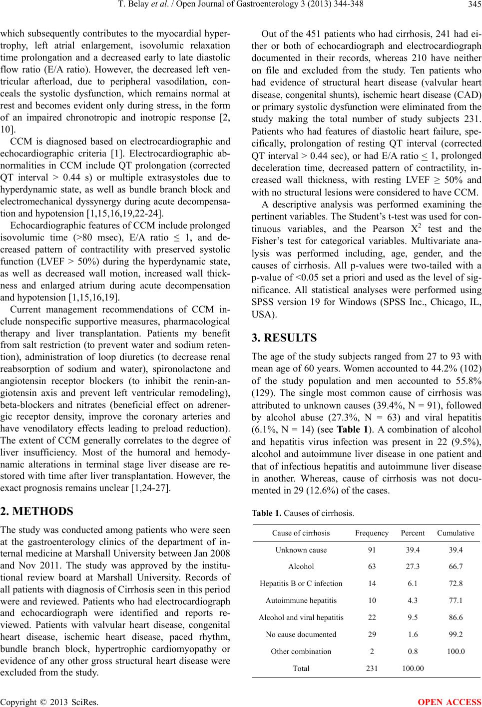 T. Belay et al. / Open Journal of Gastroenterology 3 (2013) 344-348 345 which subsequently contributes to the myocardial hyper- trophy, left atrial enlargement, isovolumic relaxation time prolongation and a decreased early to late diastolic flow ratio (E/A ratio). However, the decreased left ven- tricular afterload, due to peripheral vasodilation, con- ceals the systolic dysfunction, which remains normal at rest and becomes evident only during stress, in the form of an impaired chronotropic and inotropic response [2, 10]. CCM is diagnosed based on electrocardiographic and echocardiographic criteria [1]. Electrocardiographic ab- normalities in CCM include QT prolongation (corrected QT interval > 0.44 s) or multiple extrasystoles due to hyperdynamic state, as well as bundle branch block and electromechanical dyssynergy during acute decompensa- tion and hypotension [1,15,16,19,22-24]. Echocardiographic features of CCM include prolonged isovolumic time (>80 msec), E/A ratio ≤ 1, and de- creased pattern of contractility with preserved systolic function (LVEF > 50%) during the hyperdynamic state, as well as decreased wall motion, increased wall thick- ness and enlarged atrium during acute decompensation and hypotension [1,15,16,19]. Current management recommendations of CCM in- clude nonspecific supportive measures, pharmacological therapy and liver transplantation. Patients my benefit from salt restriction (to prevent water and sodium reten- tion), administration of loop diuretics (to decrease renal reabsorption of sodium and water), spironolactone and angiotensin receptor blockers (to inhibit the renin-an- giotensin axis and prevent left ventricular remodeling), beta-blockers and nitrates (beneficial effect on adrener- gic receptor density, improve the coronary arteries and have venodilatory effects leading to preload reduction). The extent of CCM generally correlates to the degree of liver insufficiency. Most of the humoral and hemody- namic alterations in terminal stage liver disease are re- stored with time after liver transplantation. However, the exact prognosis remains unclear [1,24-27]. 2. METHODS The study was conducted among patients who were seen at the gastroenterology clinics of the department of in- ternal medicine at Marshall University between Jan 2008 and Nov 2011. The study was approved by the institu- tional review board at Marshall University. Records of all patients with diagnosis of Cirrhosis seen in this period were and reviewed. Patients who had electrocardiograph and echocardiograph were identified and reports re- viewed. Patients with valvular heart disease, congenital heart disease, ischemic heart disease, paced rhythm, bundle branch block, hypertrophic cardiomyopathy or evidence of any other gross structural heart disease were excluded from the study. Out of the 451 patients who had cirrhosis, 241 had ei- ther or both of echocardiograph and electrocardiograph documented in their records, whereas 210 have neither on file and excluded from the study. Ten patients who had evidence of structural heart disease (valvular heart disease, congenital shunts), ischemic heart disease (CAD) or primary systolic dysfunction were eliminated from the study making the total number of study subjects 231. Patients who had features of diastolic heart failure, spe- cifically, prolongation of resting QT interval (corrected QT interval > 0.44 sec), or had E/A ratio < 1, prolonged deceleration time, decreased pattern of contractility, in- creased wall thickness, with resting LVEF ≥ 50% and with no structural lesions were considered to have CCM. A descriptive analysis was performed examining the pertinent variables. The Student’s t-test was used for con- tinuous variables, and the Pearson X2 test and the Fisher’s test for categorical variables. Multivariate ana- lysis was performed including, age, gender, and the causes of cirrhosis. All p-values were two-tailed with a p-value of <0.05 set a priori and used as the level of sig- nificance. All statistical analyses were performed using SPSS version 19 for Windows (SPSS Inc., Chicago, IL, USA). 3. RESULTS The age of the study subjects ranged from 27 to 93 with mean age of 60 years. Women accounted to 44.2% (102) of the study population and men accounted to 55.8% (129). The single most common cause of cirrhosis was attributed to unknown causes (39.4%, N = 91), followed by alcohol abuse (27.3%, N = 63) and viral hepatitis (6.1%, N = 14) (see Ta b l e 1 ). A combination of alcohol and hepatitis virus infection was present in 22 (9.5%), alcohol and autoimmune liver disease in one patient and that of infectious hepatitis and autoimmune liver disease in another. Whereas, cause of cirrhosis was not docu- mented in 29 (12.6%) of the cases. Table 1. Causes of cirrhosis. Cause of cirrhosis Frequency Percent Cumulative Unknown cause 91 39.4 39.4 Alcohol 63 27.3 66.7 Hepatitis B or C infection14 6.1 72.8 Autoimmune hepatitis 10 4.3 77.1 Alcohol and viral hepatitis22 9.5 86.6 No cause documented 29 1.6 99.2 Other combination 2 0.8 100.0 Total 231 100.00 Copyright © 2013 SciRes. OPEN ACCESS 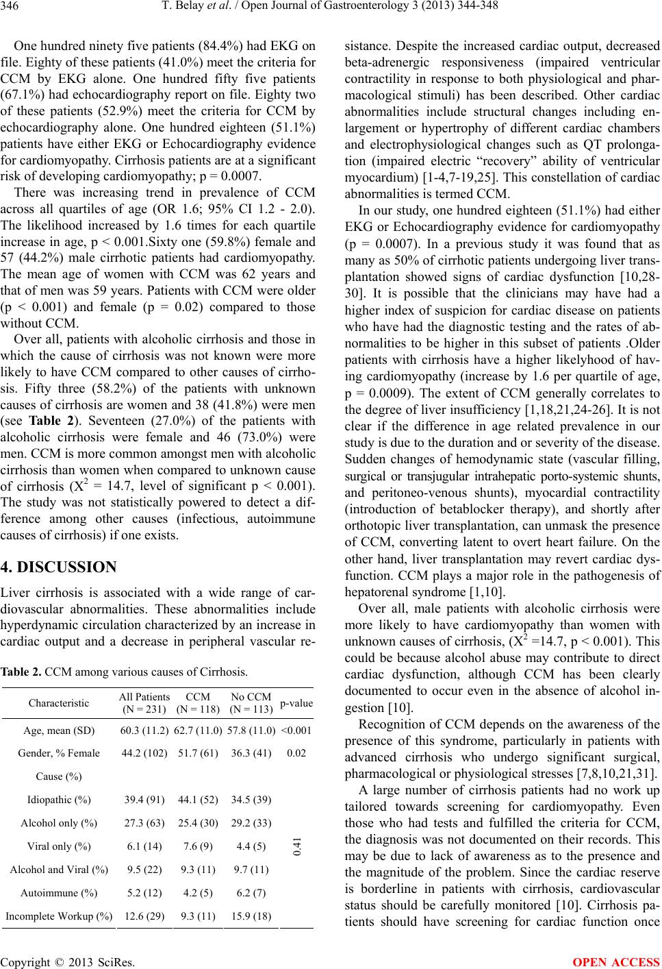 T. Belay et al. / Open Journal of Gastroenterology 3 (2013) 344-348 346 One hundred ninety five patients (84.4%) had EKG on file. Eighty of these patients (41.0%) meet the criteria for CCM by EKG alone. One hundred fifty five patients (67.1%) had echocardiography report on file. Eighty two of these patients (52.9%) meet the criteria for CCM by echocardiography alone. One hundred eighteen (51.1%) patients have either EKG or Echocardiography evidence for cardiomyopathy. Cirrhosis patients are at a significant risk of developing cardiomyopathy; p = 0.0007. There was increasing trend in prevalence of CCM across all quartiles of age (OR 1.6; 95% CI 1.2 - 2.0). The likelihood increased by 1.6 times for each quartile increase in age, p < 0.001.Sixty one (59.8%) female and 57 (44.2%) male cirrhotic patients had cardiomyopathy. The mean age of women with CCM was 62 years and that of men was 59 years. Patients with CCM were older (p < 0.001) and female (p = 0.02) compared to those without CCM. Over all, patients with alcoholic cirrhosis and those in which the cause of cirrhosis was not known were more likely to have CCM compared to other causes of cirrho- sis. Fifty three (58.2%) of the patients with unknown causes of cirrhosis are women and 38 (41.8%) were men (see Table 2). Seventeen (27.0%) of the patients with alcoholic cirrhosis were female and 46 (73.0%) were men. CCM is more common amongst men with alcoholic cirrhosis than women when compared to unknown cause of cirrhosis (X2 = 14.7, level of significant p < 0.001). The study was not statistically powered to detect a dif- ference among other causes (infectious, autoimmune causes of cirrhosis) if one exists. 4. DISCUSSION Liver cirrhosis is associated with a wide range of car- diovascular abnormalities. These abnormalities include hyperdynamic circulation characterized by an increase in cardiac output and a decrease in peripheral vascular re- Table 2. CCM among various causes of Cirrhosis. Characteristic All Patients (N = 231) CCM (N = 118) No CCM (N = 113)p-value Age, mean (SD) 60.3 (11.2)62.7 (11.0) 57.8 (11.0)<0.001 Gender, % Female 44.2 (102)51.7 (61) 36.3 (41)0.02 Cause (%) Idiopathic (%) 39.4 (91)44.1 (52) 34.5 (39) Alcohol only (%) 27.3 (63)25.4 (30) 29.2 (33) Viral only (%) 6.1 (14)7.6 (9) 4.4 (5) Alcohol and Viral (%) 9.5 (22)9.3 (11) 9.7 (11) Autoimmune (%) 5.2 (12)4.2 (5) 6.2 (7) Incomplete Workup (%) 12.6 (29)9.3 (11) 15.9 (18) 0.41 sistance. Despite the increased cardiac output, decreased beta-adrenergic responsiveness (impaired ventricular contractility in response to both physiological and phar- macological stimuli) has been described. Other cardiac abnormalities include structural changes including en- largement or hypertrophy of different cardiac chambers and electrophysiological changes such as QT prolonga- tion (impaired electric “recovery” ability of ventricular myocardium) [1-4,7-19,25]. This constellation of cardiac abnormalities is termed CCM. In our study, one hundred eighteen (51.1%) had either EKG or Echocardiography evidence for cardiomyopathy (p = 0.0007). In a previous study it was found that as many as 50% of cirrhotic patients undergoing liver trans- plantation showed signs of cardiac dysfunction [10,28- 30]. It is possible that the clinicians may have had a higher index of suspicion for cardiac disease on patients who have had the diagnostic testing and the rates of ab- normalities to be higher in this subset of patients .Older patients with cirrhosis have a higher likelyhood of hav- ing cardiomyopathy (increase by 1.6 per quartile of age, p = 0.0009). The extent of CCM generally correlates to the degree of liver insufficiency [1,18,21,24-26]. It is not clear if the difference in age related prevalence in our study is due to the duration and or severity of the disease. Sudden changes of hemodynamic state (vascular filling, surgical or transjugular intrahepatic porto-systemic shunts, and peritoneo-venous shunts), myocardial contractility (introduction of betablocker therapy), and shortly after orthotopic liver transplantation, can unmask the presence of CCM, converting latent to overt heart failure. On the other hand, liver transplantation may revert cardiac dys- function. CCM plays a major role in the pathogenesis of hepatorenal syndrome [1,10]. Over all, male patients with alcoholic cirrhosis were more likely to have cardiomyopathy than women with unknown causes of cirrhosis, (X2 =14.7, p < 0.001). This could be because alcohol abuse may contribute to direct cardiac dysfunction, although CCM has been clearly documented to occur even in the absence of alcohol in- gestion [10]. Recognition of CCM depends on the awareness of the presence of this syndrome, particularly in patients with advanced cirrhosis who undergo significant surgical, pharmacological or physiological stresses [7,8,10,21,31]. A large number of cirrhosis patients had no work up tailored towards screening for cardiomyopathy. Even those who had tests and fulfilled the criteria for CCM, the diagnosis was not documented on their records. This may be due to lack of awareness as to the presence and the magnitude of the problem. Since the cardiac reserve is borderline in patients with cirrhosis, cardiovascular status should be carefully monitored [10]. Cirrhosis pa- tients should have screening for cardiac function once Copyright © 2013 SciRes. OPEN ACCESS  T. Belay et al. / Open Journal of Gastroenterology 3 (2013) 344-348 347 diagnosis is made and at least prior to a scheduled major procedure that can unmask the dysfunction and lead to serious morbidity or death [32]. Regarding the screening method, it appears that echocardiography identifies a larger proportion of patients with cardiomyopathy than EKG. Additional studies should be conducted to identify the number of patients that would require screening di- agnose CCM, the cost effectiveness of undertaking the diagnostic testing as well as how early to start and how frequent to undertake screening evaluation. 5. CONCLUSION CCM is a common problem, but often under diagnosed. A higher prevalence of CCM in our study was noted amongst patients with advancing age, female gender, alcoholic cirrhosis and patients in whom the cause of cirrhosis was not identified. While a higher prevalence of CCM was evident among patients in whom the cause of cirrhosis is unknown and alcoholic cirrhosis, the study was not statistically powered to detect a difference among other causes (infectious, autoimmune causes of cirrhosis) if one exists. A large number of patients have not had screenings for CCM. Awareness should be increased among providers to increase recognition and target ther- apy. 6. ACKNOWLEDGEMENTS We are grateful to Natalie Hartley (RN, BSN) who was the clinical allscripts support and Melissa Marcum, (BSN, CCRC), research coor- dinator for University Cardiovascular Services at Marshall University, Joan C. Edwards School of Medicine for their support at the initial phase of the study. We would like to thank Dr. Larry Dial, chairman of internal medicine department at Marshall University, Joan C. Edwards School of Medicine for his steady support at all times of the study. REFERENCES [1] Zardi, E.M., et al. (2010) Cirrhotic cardiomyopathy. Journal of the American College of Cardiology, 56, 539- 549. http://dx.doi.org/10.1016/j.jacc.2009.12.075 [2] Al Hamoudi, W. and Lee, S.S. (2006) Cirrhotic cardio- myopathy. Annals of Hepatology, 5, 132-139. [3] Baik, S.K., Fouad, T.R. and Lee, S.S. (2007) Cirrhotic cardiomyopathy. Orphanet Journal of Rare Diseases, 2, 15. http://dx.doi.org/10.1186/1750-1172-2-15 [4] Christos, A., Fourlas, C.A. and Alexopoulou, A.A. (2004) Cirrhotic cardiomyopathy. Hellenic Journal of Cardiol- ogy, 45, 114-120. [5] Liu, H., Song, D. and Lee, S.S. (2002) Cirrhotic cardio- myopathy. Gastroentérologie Clinique et Biologique, 26, 842-847. [6] Henriksen, J.H., Bendtsen, F., Hansen, E.F. and Møller, S. (2004) Acute non-selective beta-adrenergic blockade re- duces prolonged frequency-adjusted Q-T interval (QTc) in patients with cirrhosis. Journal of Hepatology, 40, 239-246. http://dx.doi.org/10.1016/j.jhep.2003.10.026 [7] Grose, R.D., Noan, J., Dillon, J.F., Errington, M., et al. (1995) Exercise induced let ventricular dysfunction in alcoholic and non alcoholic cirrhosis. Journal of Hepa- tology, 22, 326-332. http://dx.doi.org/10.1016/0168-8278(95)80286-X [8] Wong, F., et al. (2001) The cardiac response to exercise in cirrhosis. Gut, 49, 268-275. http://dx.doi.org/10.1136/gut.49.2.268 [9] Murray, J.G., Dawson, A.M. and Sherlock, S. (1958) Cir- culatory changes in chronic liver disease. American Jour- nal of Medicine, 24, 358-367. http://dx.doi.org/10.1016/0002-9343(58)90322-X [10] Ma, Z. and Lee, S.S. (1996) Cirrhotic cardiomyopathy: Getting to the heart of the matter. Hepatology, 24, 451- 459. http://dx.doi.org/10.1002/hep.510240226 [11] Liu, H. and Lee, S.S. (1999) Cardiopulmonary dysfunc- tion in cirrhosis. Journal of Gastroenterology and Hepa- tology, 14, 600-608. http://dx.doi.org/10.1046/j.1440-1746.1999.01920.x [12] Grose, R.D., et al. (1995) Exercise-induced left ventricu- lar dysfunction in alcoholic and non-alcoholic cirrhosis. Journal of Hepatology, 22, 326-332. http://dx.doi.org/10.1016/0168-8278(95)80286-X [13] Finucci, G., et al. (1996) Left ventricular diastolic func- tion in liver cirrhosis. Scandinavian Journal of Gastroen- terology, 31, 279-284. http://dx.doi.org/10.3109/00365529609004879 [14] Pozzi, M., et al. (1997) Evidence of functional and struc- tural cardiac abnormalities in cirrhotic patients with and without ascites. Hepatology, 26, 1131-1137. [15] Wong, F., Liu, P., Lilly, L., Bomzon, A. and Blendis, L. (1999) Role of cardiac structural and functional abnor- malities in the pathogenesis of hyperdynamic circulation and renal sodium retention in cirrhosis. Clinical Science (London), 97, 259-267. http://dx.doi.org/10.1042/CS19990026 [16] Wong, F., et al. (2001) The cardiac response to exercise in cirrhosis. Gut, 49, 268-275. http://dx.doi.org/10.1136/gut.49.2.268 [17] Abelmann, W.H., Kowalski, H.J. and McNeely, W.F. (1955) The hemodynamic response to exercise in patients with laennec’s cirrhosis. Journal of Clinical Investiga- tion, 34, 690-695. http://dx.doi.org/10.1172/JCI103120 [18] Kim, M.Y. and Baik, S.K. (2007) Cirrhotic cardio- myopathy. Korean Journal of Hepatology, 13, 22-26. [19] Liu, H.Q., Gaskari, S.A. and Lee, S.S. (2006) Cardiac and vascular changes in cirrhosis: Pathogenic mechanisms. World Journal of Gastroenterology, 12, 837-842. [20] Donovan, C.L., et al. (1996) Two dimensional and dobu- tamine stress echocardiography in the prospective as- sessment of patients with end stage liver disease prior to orthotopic liver transplantation. Transplantation, 61, 1180-1188. http://dx.doi.org/10.1097/00007890-199604270-00011 [21] Myers, R.P. and Lee, S.S. (2000) Cirrhotic cardiomyopa- thy and liver transplantation. Liver Transplantation, 6, Copyright © 2013 SciRes. OPEN ACCESS  T. Belay et al. / Open Journal of Gastroenterology 3 (2013) 344-348 Copyright © 2013 SciRes. 348 OPEN ACCESS S44-S52. http://dx.doi.org/10.1002/lt.500060510 [22] Kempler, P., et al. (1993) Prolongation of the QTc-in- terval reflects the severity of autonomic neuropathy in primary biliary cirrhosis and in other non-alcoholic liver diseases. Zeitschrift für Gastroenterologie, 31, 96-98. [23] Bernardi, M., et al. (1998) Q-T interval prolongation in cirrhosis: Prevalence, relationship with severity and eti- ology of the disease and possible pathogenetic factors. Hepatology, 27, 28-34. http://dx.doi.org/10.1002/hep.510270106 [24] Finucci, G., et al. (1998) Q-T interval prolongation in liver cirrhosis. Reversibility after orthotopic liver trans- plantation. Japanese Heart Journal, 39, 321-329. http://dx.doi.org/10.1536/ihj.39.321 [25] Blendis, L. and Wong, F. (2000) Is there a cirrhotic car- diomyopathy? American Journal of Gastroenterology, 95, 3026-3028. http://dx.doi.org/10.1111/j.1572-0241.2000.03249.x [26] Navasa, M., et al. (1993) Hemodynamic and humoral changes after liver transplantation in patients with cirrho- sis. Hepatology, 17, 355-360. http://dx.doi.org/10.1002/hep.1840170302 [27] Henriksen, J.H., Bendtsen, F., Hansen, E.F. and Møller, S. (2004) Acute non-selective beta-adrenergic blockade re- duces prolonged frequency-adjusted Q-T interval (QTc) in patients with cirrhosis. Journal of Hepatology, 40, 239- 246. http://dx.doi.org/10.1016/j.jhep.2003.10.026 [28] Gould, L., Shariff, M., Zahir, M. and Di Lieto, M. (1969) Cardiac haemodynamics in alcoholic patients with chro- nic liver disease and presystolic gallop. Journal of Clini- cal Investigation, 48, 860-868. http://dx.doi.org/10.1172/JCI106044 [29] Hendrickse, M.T. and Triger, D.R. (1994) Vagal dys- function and impaired urinary sodium and water excre- tion in cirrhosis. American Journal of Gastroenterology, 89, 750-757. [30] Hansen, S., Møller, S., Bendtsen, F., Jensen, G. and Hen- riksen, J.H. (2007) Diurnal variation and dispersion in QT interval in cirrhosis: Relation to haemodynamic changes. Journal of Hepatology, 47, 373-380. http://dx.doi.org/10.1016/j.jhep.2007.03.013 [31] Møller, S., et al. (2012) New insights into cirrhotic car- diomyopathy. International Journal of Cardiology, 167, 1101-1108. [32] Møller, S. and Henriksen, J.H. (2009) Cardiovascular complications of cirrhosis. Postgraduate Medical Jour- nal, 85, 44-54. |