

Paper Menu >>
Journal Menu >>
 International Journal of Clinical Medicine, 2013, 4, 543-547 Published Online December 2013 (http://www.scirp.org/journal/ijcm) http://dx.doi.org/10.4236/ijcm.2013.412094 Open Access IJCM 543 Short Term Prognostic Utility of Tc-99m DMSA Renal Imaging in Sepsis Induced Acute Renal Failure; Provisional Data Amr Amin1, Hatem Nasr1, Gehan Younis1, Hatem Gamal2 1Nuclear Medicine Unit, Faculty of Medicine, Cairo University , Cairo, Egypt; 2Intensive Care Unit, Faculty of Medicine, Cairo Uni- versity, Cairo, Egypt. Email: [email protected] Received October 8th, 2013; revised November 3rd, 2013; accepted December 2nd, 2013 Copyright © 2013 Amr Amin et al. This is an open access article distributed under the Creative Commons Attribution License, which permits unrestricted use, distribution, and reproduction in any medium, provided the original work is properly cited. ABSTRACT Background: Sound prognostic data in sepsis induced acute renal failure (SARF) are lacking especially on the short term outcome [STO] in the intensive care unit [ICU]. We addressed the use of Tc-99m DMSA [2,3-dimercaptosuc- cinicacid] renal cortical imaging as a prognostic tool in SARF. Methods: Forty patients with acute renal failure due to sepsis [age range 15 - 74 years; median 44.5] were subjected for full history taking complete physical examination, rou- tine ICU monitoring, routine laboratory investigations, APACHE II [Acute Physiolog y and Chronic Health Evaluation] and SOFA [Sequential Organ Failure Assessment] together with Tc-99m DMSA cortical renal scintigraphy. Patients’ death in the ICU or discharge was considered as the end point of the study representing the so-called short term out- come [STO]. Results: 25% mortality rate [10/40] was found along the admission period in the ICU. All non-survivors were abnormal with DMSA imaging [NPV & PPV 100% & 66.7% respectively]. Abnormal DMSA cases showed sig- nificant positive associations with serum creatinine at admission [r = 0.5 ; P 0.02]; admission duration [r = 0.4; P 0.002]; APACHE II score [r = 0.5; P 0.004] and STO [r = 0.4; P 0.03]. Statistically significant difference was elicited between subjects with normal and abnormal DMSA regarding the same parameters. Conclusion: This preliminary data could raise Tc-99m DMSA renal imaging as a prognostic tool in SARF that could allow influential interference to prohibit dramatic outcomes as mortality. Keywords: Acute Renal Failure; Tc-99m DMSA; Sep sis Induced Acute R e n a l Failure 1. Introduction Acute renal failure [ARF] is a sudden sustained decline in glomerular filtration rate [GFR] usually associated with uremia and a fall in urine output with a high mortality rate [1]. Factors affecting its incidence and outcomes are unclear therefore making optimal decision for patients’ care is hindered [2]. Tc-99m labeled DMSA (2,3-dimercaptosuccinicacid) is avidly taken up by cells of the proximal tubule, with about 35% of the injected activity being localized in the renal cortex by one hour [3]. The renal uptake of Tc-99m DMSA parallels the renal function as determined b y GFR and accordingly its uptake is significantly decreased in nephropathy [4,5] which is accepted as a poor prognostic sign [6]. Bern- heim et al. in 1976 proposed renal scintigraphic scanning as a tool to assess the prognosis of ARF using hippuran and concluded that its uptake was a parameter which authorized the prognosis of a favorable course whereas its absence permitted one to envisage an unfavorable course [7]. However, in a case report published in 1986; a 35-year-old patient developed ARF on top of acute tu- bular necrosis requiring hemodialysis and they stated that failure to visualize acutely injured kidneys with Tc-99m DMSA did not preclude recoverable function [4], while the reverse was elicited in another two studies using hip- puran [8,9]. Besides, much research is still needed be- cause our understanding of what happens to the kidney in ARF is still poor [10]. Hence, we aimed to evaluate the predictive role of Tc-99m DMSA cortical scanning in predicting the short term outcome [STO] of the ad- mission period in the intensive care in sepsis-induced ARF patie n ts [SARF] . 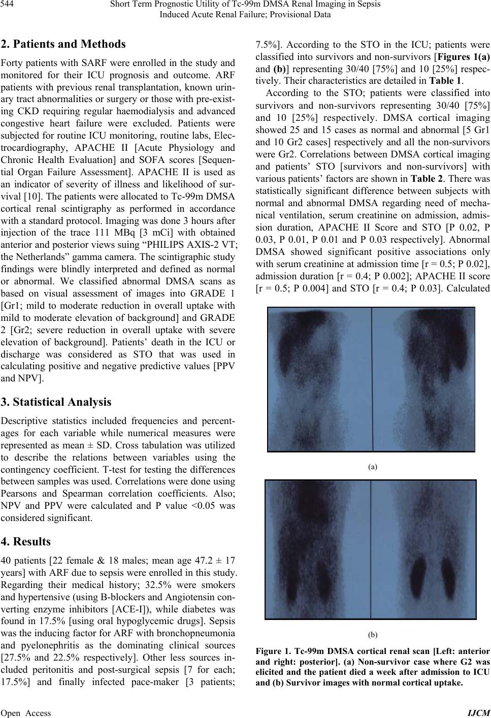 Short Term Prognostic Utility of Tc-99m DMSA Renal Imaging in Sepsis Induced Acute Renal Failure; Provisional Data 544 2. Patients and Methods Forty patients with SARF were enrolled in the study and monitored for their ICU prognosis and outcome. ARF patients with previous renal transplantation, known urin- ary tract abnormalities or surgery or those with pre-exist- ing CKD requiring regular haemodialysis and advanced congestive heart failure were excluded. Patients were subjected for routine ICU monitoring, routine lab s, Elec- trocardiography, APACHE II [Acute Physiology and Chronic Health Evaluation] and SOFA scores [Sequen- tial Organ Failure Assessment]. APACHE II is used as an indicator of severity of illness and likelihood of sur- vival [10]. The patients were allocated to Tc-99m DMSA cortical renal scintigraphy as performed in accordance with a standard protocol. Imaging was done 3 hours after injection of the trace 111 MBq [3 mCi] with obtained anterior and poster ior views su ing “PHILIPS AX IS-2 VT; the Netherlands” gamma camera. The scintigraph ic study findings were blindly interpreted and defined as normal or abnormal. We classified abnormal DMSA scans as based on visual assessment of images into GRADE 1 [Gr1; mild to moderate reduction in overall uptake with mild to moderate elevation of background] and GRADE 2 [Gr2; severe reduction in overall uptake with severe elevation of background]. Patients’ death in the ICU or discharge was considered as STO that was used in calculating positive and negative predictive values [PPV and NPV]. 3. Statistical Analysis Descriptive statistics included frequencies and percent- ages for each variable while numerical measures were represented as mean ± SD. Cross tabulation was utilized to describe the relations between variables using the contingency coefficient. T-test for testing the differences between samples was used. Correlations were done using Pearsons and Spearman correlation coefficients. Also; NPV and PPV were calculated and P value <0.05 was considered significant. 4. Results 40 patients [22 female & 18 males; mean age 47.2 ± 17 years] with ARF due to sepsis were enrolled in th is study. Regarding their medical history; 32.5% were smokers and hypertensive (using B-blockers and Angiotensin con- verting enzyme inhibitors [ACE-I]), while diabetes was found in 17.5% [using oral hypoglycemic drugs]. Sepsis was the inducing factor for ARF with bronchopn eumonia and pyelonephritis as the dominating clinical sources [27.5% and 22.5% respectively]. Other less sources in- cluded peritonitis and post-surgical sepsis [7 for each; 17.5%] and finally infected pace-maker [3 patients; 7.5%]. According to the STO in the ICU; patients were classified into survivors and non-surv ivors [Figures 1(a) and (b)] representing 30/40 [75%] and 10 [25%] respec- tively. Their characteristics are detailed in Table 1. According to the STO; patients were classified into survivors and non-survivors representing 30/40 [75%] and 10 [25%] respectively. DMSA cortical imaging showed 25 and 15 cases as normal and abnormal [5 Gr1 and 10 Gr2 cases] respectively and all the non-survivors were Gr2. Correlations between DMSA cortical imaging and patients’ STO [survivors and non-survivors] with various patients’ factors are shown in Table 2. There w a s statistically significant difference between subjects with normal and abnormal DMSA regarding need of mecha- nical ventilation, serum creatinine on admission, admis- sion duration, APACHE II Score and STO [P 0.02, P 0.03, P 0.01, P 0.01 and P 0.03 respectively]. Abnormal DMSA showed significant positive associations only with serum creatinine at admission time [r = 0.5; P 0.02], admission duration [r = 0.4; P 0.002]; APACHE II score [r = 0.5; P 0.004] and STO [r = 0.4; P 0.03]. Calculated (a) (b) Figure 1. Tc-99m DMSA cortical renal scan [Left: anterior and right: posterior]. (a) Non-survivor case where G2 was elicited and the patient died a week after admission to ICU nd (b) Survivor images with normal cortical uptake. a Open Access IJCM 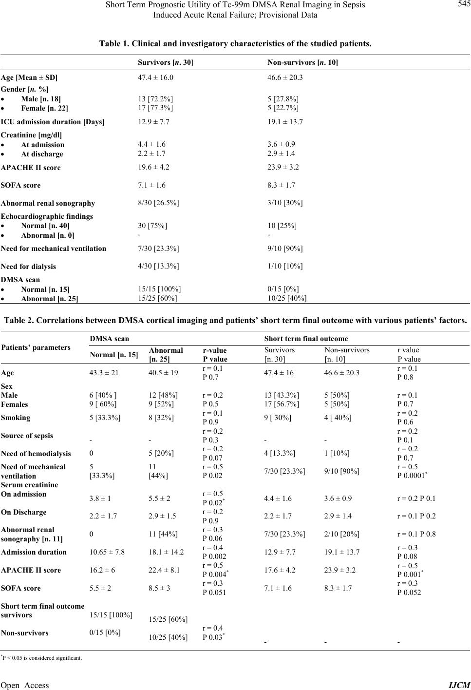 Short Term Prognostic Utility of Tc-99m DMSA Renal Imaging in Sepsis Induced Acute Renal Failure; Provisional Data Open Access IJCM 545 Table 1. Clinical and investigatory characteristics of the studied patients. Survivors [n. 30] Non-survivors [n. 10] Age [Mean ± SD] 47.4 ± 16.0 46.6 ± 20.3 Gender [n. %] Male [n. 18] Female [n. 22] 13 [72.2%] 17 [77.3%] 5 [27.8%] 5 [22.7%] ICU admission duration [Days] 12.9 ± 7.7 19.1 ± 13.7 Creatinine [mg/dl] At admission At discharge 4.4 ± 1.6 2.2 ± 1.7 3.6 ± 0.9 2.9 ± 1.4 APACHE II score 19.6 ± 4.2 23.9 ± 3.2 SOFA score 7.1 ± 1.6 8.3 ± 1.7 Abnormal renal sonography 8/30 [26.5%] 3/10 [30%] Echocardiographic findings Normal [n. 40] Abnormal [n. 0] 30 [75%] - 10 [25%] - Need for mechanical ventilation 7/30 [23.3%] 9/10 [90%] Need for dialysis 4/30 [13.3%] 1/10 [10%] DMSA scan Normal [n. 15] Abnormal [n. 25] 15/15 [100%] 15/25 [60%] 0/15 [0%] 10/25 [40%] Table 2. Correlations betw ee n DMSA c o r t ical imaging and patients’ short term final outcome with various patients’ factors. DMSA scan Short term final outcome Patients’ parameters Normal [n. 15] Abnormal [n. 25] r-value P value Survivors [n. 30] Non-survivors [n. 10] r value P value Age 43.3 ± 21 40.5 ± 19 r = 0.1 P 0.7 47.4 ± 16 46.6 ± 20.3 r = 0.1 P 0.8 Sex Male Females 6 [40% ] 9 [ 60%] 12 [48%] 9 [52%] r = 0.2 P 0.5 13 [43.3%] 17 [56.7%] 5 [50%] 5 [50%] r = 0.1 P 0.7 Smoking 5 [33.3%] 8 [32%] r = 0.1 P 0.9 9 [ 30%] 4 [ 40%] r = 0.2 P 0.6 Source of sepsis - - r = 0.2 P 0.3 - - r = 0.2 P 0.1 Need of hemodialysis 0 5 [20%] r = 0.2 P 0.07 4 [13.3%] 1 [10%] r = 0.2 P 0.7 Need of mechanical ventilation 5 [33.3%] 11 [44%] r = 0.5 P 0.02 7/30 [23.3%] 9/10 [90%] r = 0.5 P 0.0001* Serum creatinine On admission On Discharge 3.8 ± 1 2.2 ± 1.7 5.5 ± 2 2.9 ± 1.5 r = 0.5 P 0.02* r = 0.2 P 0.9 4.4 ± 1.6 2.2 ± 1.7 3.6 ± 0.9 2.9 ± 1.4 r = 0.2 P 0.1 r = 0.1 P 0.2 Abnormal renal sonography [n. 11] 0 11 [44%] r = 0.3 P 0.06 7/30 [23.3%] 2/10 [ 2 0%] r = 0.1 P 0.8 Admission duration 10.65 ± 7.8 18.1 ± 14.2 r = 0.4 P 0.002 12.9 ± 7.7 19.1 ± 13.7 r = 0.3 P 0.08 APACHE II score 16.2 ± 6 22.4 ± 8.1 r = 0.5 P 0.004* 17.6 ± 4.2 23.9 ± 3.2 r = 0.5 P 0.001* SOFA score 5.5 ± 2 8. 5 ± 3 r = 0.3 P 0.051 7.1 ± 1.6 8.3 ± 1.7 r = 0. 3 P 0.052 Short term final outcome survivors Non-survivors 15/15 [100%] 0/15 [0%] 15/25 [60%] 10/25 [40%] r = 0.4 P 0.03* - - - *P < 0.05 is considered si gnificant.  Short Term Prognostic Utility of Tc-99m DMSA Renal Imaging in Sepsis Induced Acute Renal Failure; Provisional Data 546 PPV and NPV for DMSA imaging were 66.7% and 100% respectively. 5. Discussion This study was carried out on 40 SARF patients and showed a mortality rate of 25% along the ICU admission period. Abnormal Tc-99m DMSA revealed a significant positive asso ciation with STO [r = 0.4; P 0.03]. Although different modalities for this clinically relevant task has been proposed, to our knowledge no previous studies have reported using Tc-99m DMSA cortical imaging as a prognostic tool; hence, this prospective study was ad- dressed. Our patients were recruited from ICU unit, as in Egypt ARF is usually treated in ICU and attributed mostly to sepsis. The latter was reported as the commonest cau- sative factor of ARF (up to 50%) [2,10,11]. In fact, ARF is nowadays mostly observed as part of the multi-organ dysfunction syndrome in severe sepsis and septic shock [12]. 10/40 [25%] of our patients died in the ICU ad- mission period and this agrees with Prescott et al. study as many of his deaths [20% - 30%] were very early where ARF was sepsis-induced. They concluded that the presence of sepsis increased the risk of death; both as an etiological factor and poor indicator of prognosis in a startling manner [2]. Also, we have chosen the German Prevalence Study [GPS] [13] for comparison with our elicited data as they studied a cohort of ARF patients with and without sepsis and compared both groups regarding patients’ various factors and reported significant differences between serum creatinine at admission, APACHE II and SOFA scores. They reported a higher mortality rate [64.4% vs. 25%]. This disagreement could be explained by our small patients’ number and the relatively younger age of our cases that carries in general an expected better outcome. Also, pulmonary infection was the most frequent source for sepsis as in our study [59% vs. 40%]. In our study significant statistical associations were found between DMSA and STO, serum creatinine at admission, APACHE II and need for mechanical ventilation in concordance with the same significant parameters of GPS. We tried to find an explanation for our findings; as DMSA is avidly taken up by cells of the proximal tubule and with sepsis damage of the proximal tubules might occur and [3], so the higher the damage the lower is DMSA uptake and the worst is outcome. Hence in Gr2 pattern extensive damage of the proximal tubules was found in non-survivors. Finally, since kidney injury in SARF plays an im- portant role in prognosis; this preliminary data could raise Tc-99m DMSA cortical imaging as a tool to predict STO in such patients with 100% and 66.7% NPV and PPV respectively. As said by Niccolo Machiavelli [The Prince, 1513], “It ought to be remembered that there is nothing more difficult to take in hand, more perilous to conduct or more uncertain in its success than to take the lead in the introduction of a new order of things” hence, we tried to introduce DMSA renal imaging as a pro- mising prognostic radiopharmaceutical with subsequent interference to ban serious sequences as mortality. How- ever, we recommend further larger studies to support these provisional data. 6. Conclusion Tc-99m DMSA cortical imaging could be raised as a diagnostic tool to predict short term outcome in such sepsis induced acute renal failure patients with 100% NPV and 66.7% PPV respectively that could allow in- fluential interference to prohibit dramatic outcomes as mortality. 7. Acknowledgements The authors thank Dr. Zeinab Nawito for contributions in the finalization of this work regarding grammatical aspects. REFERENCES [1] A. R. Nissenson, “Acute Renal Failure: Definition and Pathogenesis,” Kidney International Supplements, Vol. 66, 1998, pp. S7-S10. [2] G. J. Prescott, W. Metcalfe, J. Baharani, I. H. Khan, K. Simpson, W. C. Smith, et al., “A Prospective National Study of Acute Renal Failure Treated with RRT: Inci- dence, Aetiology and Outcomes,” Nephrology Dialysis Transplantation, Vol. 22, No. 9, 2007, pp. 2513-2519. http://dx.doi.org/10.1093/ndt/gfm264 [3] P. F. Sharp, H. G. Gemmell and A. D. Murray, “Practical Nuclear Medicine Third Edition Chapter,” the Urinary Tract Philip S. Cosgriff, Springer, 2006, pp. 210-224. [4] A. Taylor Jr., F. Akiya and M. C. Gregory, “Failure to Visualize Acutely Injured Kidneys with Technetium-99m DMSA Does Not Preclude Recoverable Function,” Jour- nal of Nuclear Medicine, Vol. 27, No. 3, 1986, pp. 377- 379. [5] W. Shelfhout, M. Simons, W. Oosterlink and W. A. De Sy, “Evaluation of Tc-99m-DMSA Renal Uptake as an Index of Individual Kidney Function after Acute Ureteral Obstruction and Desobstruction: An Experimental Study in Rats,” European Urology, Vol. 9, 1983, pp. 221-226. [6] I. A. Becker, R. Kutcher and N. Solomon, “The Radiol- ogy of Renal Failure,” In: E. A. Friedman, Ed., Strategy in Renal Failure, John Wiley and Sons, New York, 1978. [7] J. Bernheim, M. Collard, M. Westphall, A. Guey and J. Traeger, “Usefulness of Renal Scinitigraphic Scanning in the Prognosis of Acute Renal Failure (Author’s Transl),” Journal de Radiologie, d’Électrologie, et de Médecine Open Access IJCM  Short Term Prognostic Utility of Tc-99m DMSA Renal Imaging in Sepsis Induced Acute Renal Failure; Provisional Data 547 Nucléaire, Vol. 57, No. 1, 1976, pp. 63-68. [8] R. A. Sherman and M. D. Blaufox, “Obstructive Uropa- thy in Patients with Non-Visualization on Renal Scan,” Nephron, Vol. 25, No. 2, 1980, pp. 82-86. http://dx.doi.org/10.1159/000181758 [9] R. Romero, A. Caralps, A. Brulles, J. Andreu, J. Griño and J. Matin-Comin, “The Significance of the Absence of 131I-Hippuran Uptake by a Kidney Graft,” Nephron, Vol. 39, No. 4, 1985, pp. 306-308. http://dx.doi.org/10.1159/000183395 [10] F. G. Brivet, D. J. Kleinknecht, P. Loirat and P. J. Lan- dais, “Acute Renal Failure in Intensive Care Units— Causes, Outcome, and Prognostic Factors of Hospital Mortality: A Prospective Multicenter Study,” Critical Care Medicine, Vol. 24, No. 2, 1996, pp. 192-198. http://dx.doi.org/10.1097/00003246-199602000-00003 [11] S. Uchino, J. A. Kellum, R. Bellomo, G. S. Doig, H. Mo- rimatsu, S. Morgera, et al., “Beginning and Ending Sup- portive Therapy for the Kidney (BEST Kidney) Investi- gators. Acute Renal Failure in Critically ill Patients: A Multinational, Multicenter Study,” JAMA, Vol. 294, 2005, pp. 813-818. [12] R. W. Schrier and W. Wang, “Acute Renal Failure and Sepsis,” New England Journal of Medicine, Vol. 351, 2004, pp. 159-169. http://dx.doi.org/10.1056/NEJMra032401 [13] M. Oppert, C. Engel, F. M. Brunkhorst, H. Bogatsch, K. Reinhart, U. Frei, et al., “German Competence Network Sepsis (Sepnet). Acute Renal Failure in Patients with Se- vere Sepsis and Septic Shock—A Significant Independent Risk Factor for Mortality: Results from the German Pre- valence Study,” Nephrology Dialysis Transplantation, Vol. 23, No. 3, 2008, pp. 904-909. http://dx.doi.org/10.1093/ndt/gfm610 Open Access IJCM |