

Paper Menu >>
Journal Menu >>
 International Journal of Clinical Medicine, 2013, 4, 1-4 Published Online December 2013 (http://www.scirp.org/journal/ijcm) http://dx.doi.org/10.4236/ijcm.2013.412A1001 Open Access IJCM 1 IgG4-Related Disease Presenting as a Soft Tissue Tumor Affecting Skeletal Muscle: A Case Report David Sáez Martínez1, Felipe López Oliva1, María Jesús Fernández Aceñero2, Elena Fontoira Moyer3, Juan Luis Arranz Cozar4 1Ortrhopedic Surgery Department, Fundación Jiménez Díaz, Madrid, Spain; 2Pathology Department, Fundación Jiménez Díaz, Mad- rid, Spain; 3Radiology Department, Fundación Jiménez Díaz, Madrid, Spain; 4Oncology Department, Fundación Jiménez Díaz, Mad- rid, Spain. Email: [email protected] Received October 30th, 2013; revised November 28th, 2013; accepted December 15th, 2013 Copyright © 2013 David Sáez Martínez et al. This is an open access article distributed under the Creative Commons Attribution License, which permits unrestricted use, distribution, and reproduction in any medium, provided the original work is properly cited. In accordance of the Creative Commons Attribution License all Copyrights © 2013 are reserved for SCIRP and the owner of the intellectual property David Sáez Martínez et al. All Copyright © 2013 are guarded by law and by SCIRP as a guardian. ABSTRACT Background: IgG4-related disease is a systemic lymphoproliferative syndrome that shows IgG4-producing plasma cell expansion in affected organs with fibrotic or sclerotic changes. The lacrimal glands, salivary glands and pancreas are typically affected. We report a case of IgG4-related disease presenting a soft tissue tumor affecting skeletal muscle. Case Report: A 32-year-old man presented a soft tissue mass in his left arm. Magnetic resonance imaging revealed a spindle like, peripheral mass, in the lateral head of the triceps of his left arm. Tru-Cut Biopsy provided the diagnosis of IgG4-related disease affecting skeletal muscle. Glucocorticoid treatment was effective. Conclusion: To our knowledge, this is the first reported case of IgG4-related disease affecting skeletal muscle and presenting a soft tissue mass. Keywords: IgG4-Related Disease; Soft Tissue Mass; Skeletal Muscle 1. Introduction Immunoglobulin G4-related disease (IgG4-RD) is an increasingly recognized lymphoproliferative syndrome of unknown etiology. Patients show tumor-like swelling of involved organs, lymphoplasmacytic and IgG4 producing plasma cells infiltrating and affected organs with fibrotic or sclerotic changes. Elevated serum concentrations of IgG4 are found in 60 % of pat ie nt s wi t h Ig G4 -R D. The lacrimal glands, salivary glands and pancreas are the major affected organs. Another feature of IgG4-RD is good glucocorticoid responsiveness. We report the first case to our knowledge of IgG4-RD presenting a soft tissue tu mor affecting skeletal muscle in the arm. 2. Case Report A 32-year-old man was referred to our institution pre- senting a soft tissue mass in his left arm. He had a medical history of asthma, left hypertrophic pachymeningitis and an orbital pseudotumor waiting for surgery. Physical examination found a hard, nontender and mobile mass, with well defined borders, in the lateral aspect of his left arm, measuring 3 × 3 cm in size. It showed mild growing during the last two years. Tinel Test was negative. He also presented a right exophthal- mos. Complete blood count and serum chemistry, including IgG levels (787 mg/dl), were all within their normal lim- its (Table 1). The MRI described a spindle like, peripheral mass, in the lateral head of the triceps, with well demarcated bor- ders, T1 and T2 hyperintense and hypercaptant with con- trast. It had a central hypointense region in all seq uences that could correspond to fibrosis. It measured 2 cm × 17 mm × 2.5 cm (Figure 1). An ultrasound guided Tru-Cut Biopsy was performed, referring 4 cylinders of material to Soft Tissue Pathology Department. The biopsy showed a fibro inflammatory condition, with abundant plasma cells, dissecting and 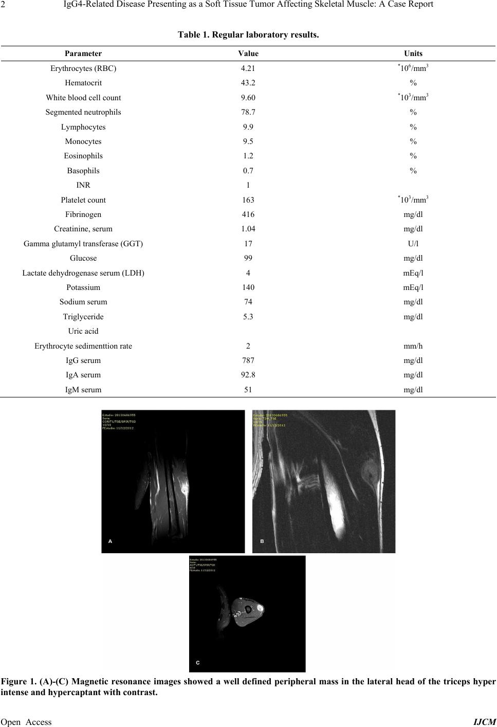 IgG4-Related Disease Presenting as a Soft Tissue Tumor Affecting Skeletal Muscle: A Case Report 2 Table 1. Regular laboratory results. Parameter Value Units Erythrocytes (RBC) 4.21 *106/mm3 Hematocrit 43.2 % White blood cell count 9.60 *103/mm3 Segmented neutrophils 78.7 % Lymphocytes 9.9 % Monocytes 9.5 % Eosinophils 1.2 % Basophils 0.7 % INR 1 Platelet count 163 *103/mm3 Fibrinogen 416 mg/dl Creatinine, serum 1.04 mg/d l Gamma glutamyl transferase (GGT) 17 U/l Glucose 99 mg/d l Lactate dehydrogenase serum (LDH) 4 mEq/ l Potassium 140 mEq/l Sodium serum 74 mg/d l Triglyceride 5.3 mg/dl Uric acid Erythrocy te sedimenttion rate 2 mm/h IgG serum 787 mg/dl IgA serum 92.8 mg/d l IgM serum 51 mg /dl Figure 1. (A)-(C) Magnetic resonance images showed a well defined peripheral mass in the lateral head of the triceps hyper ntense and hypercaptant with contrast. i Open Access IJCM 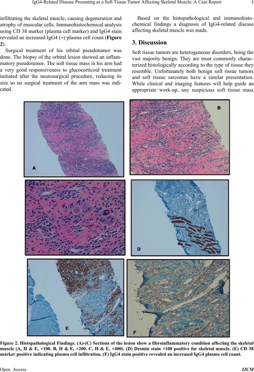 IgG4-Related Disease Presenting as a Soft Tissue Tumor Affecting Skeletal Muscle: A Case Report 3 infiltrating the skeletal muscle, causing degeneration and atrophy of muscular cells. Immunohistochemical analysis using CD 38 marker (plasma cell marker) and IgG4 stain revealed an increased IgG4 (+) plasma cell count (Figure 2). Surgical treatment of his orbital pseudotumor was done. The biopsy of the orbital lesion showed an inflam- matory pseudotumor. The so ft tissue mass in his arm had a very good responsiveness to glucocorticoid treatment initiated after the neurosurgical procedure, reducing its size so no surgical treatment of the arm mass was indi- cated. Based on the histopathological and immunohisto- chemical findings a diagnosis of IgG4-related disease affecting skeletal muscle was made. 3. Discussion Soft tissue tumors are heterogeneous disorders, being the vast majority benign. They are most commonly charac- terized histologically according to the type of tissue they resemble. Unfortunately both benign soft tissue tumors and soft tissue sarcomas have a similar presentation. While clinical and imaging features will help guide an appropriate work-up, any suspicious soft tissue mass Figure 2. Histopathological Findings. (A)-(C) Sections of the lesion show a fibroinflammatory condition affecting the skeletal muscle (A, H & E, ×100. B, H & E, ×200. C, H & E, ×400). (D) Desmin stain ×100 positive for skeletal muscle. (E) CD 38 marker positive indicating plasma cell infiltration. (F) IgG4 stain positive revealed an increased IgG4 plasma cell count. Open Access IJCM  IgG4-Related Disease Presenting as a Soft Tissue Tumor Affecting Skeletal Muscle: A Case Report 4 must be biopsied [1]. IgG4-RD is a novel lymphoproliferative syndrome of unknown etiology including a collection of disorders that share clinical and pathological characteristics. Several of the manifestations can occur in the same patient and comprise: Type 1 autoinmune pancreatitis and IgG4-re- lated sclerosing cholangitis, inflammatory orbital pseu- dotumor, retroperitoneal fibrosis, thyroiditis, and other sclerosing or IgG4-related disorders as dacryoadenitis, sialadenitis, aortitis and periaortitis, interstitial pneumo- nitis, tubulointerstitial nephritis, hypertrophic pachyme- ningitis [2]. The principal symptoms and signs of IgG4-RD are a slow growing mass or diffuse enlargement of an organ. To our knowledge this is the first reported case of local- ized IgG4-related disease affecting skeletal muscle and presenting as a soft tissue tumor. IgG4-RD can also pre- sent with clinical findings related to affected organs with fibrotic or sclerotic changes [2]. Pathologically the disease is characterized by IgG4 positive plasma cells and lymphocytes tissue infiltration which may be accompanied by sclerosis. Elevated serum levels of IgG4 may be present, being an important aid in diagnosis, although they are not diagnostic [3]. The diagnosis of IgG4-RD is made with biopsy find- ings demonstrating t he characterist ic hist opathology . Core- Needle Biopsy or Tru-Cut Biopsy is most often indicated for superficial or accessible deep extremity soft tissue tumors that are of sufficient size to n eedle placement (>3 cm) [1]. In our case this was the technique of biopsy in- dicated. A good therapeutic response to glucocorticoids is also characteristic of IgG4-RD, by symptomatic and organ function improvement or reductions in the size of masses, as it happened in our case [4,5]. Glucocorticoid therapy has been suggested as the ini- tial treatment of IgG4-RD. Azathioprine, mycophenolate mofetil and rituximab have been used to treat resistant patients [5,6]. The natural history and prognosis of IgG4-related dis- ease are not well described. Despite that good respon- siveness to glucocorticoid treatment, relapses are com- mon and organ dysfunctio n may arise from inflammato ry and fibrotic changes. The possibility of increased risk of malignancy is not clear [5,6]. 4. Conclusion Despite its extreme rarity, IgG4-related disease may be presen ted as a sof t tissue tu mor affecting skeletal muscle. Biopsy may be the only diagnostic procedure. A good therapeutic response to glucocorticoids should be ex- pected. REFERENCES [1] T. A. Damron, “Oncology and Basic Science,” Lippincott Williams & Wilkins, Philadelphia, 2008. [2] J. H. Stone, Y. Zen and V. Deshpande, “Mechanisms of disease: IgG4-related disease,” The New England Journal of Medici ne, Vol. 366, No. 6, 2012, pp. 539-551. http://dx.doi.org/10.1056/NEJMra1104650 [3] H. Umehara, K. Okazaki, Y. Masaki, et al., “Comprehen- sive Diagnostic Criteria for IgG4-Related Disease (IgG4- RD), 2011,” Modern Rheumatology, Vol. 22, No. 1, 2012, pp. 21-30. http://dx.doi.org/10.1007/s10165-011-0571-z [4] M. Yasufumi, K. Nozomu and U. Hisanori, “IgG4-Rela- ted Disease: A Novel Lymphoproliferative Disorder Dis- covered and Established in Japan in the 21st Century,” Journal of Clinical and Experimental Hematopathology, Vol. 51, No. 1, 2011, pp. 13-20. http://dx.doi.org/10.3960/jslrt.51.13 [5] H. M. Moutsopoulos, G. E. Fragoulis and J. H. Stone, “Overview of IgG4-Related Disease,” 2013. http://www.uptodate.com/contents/overview-of-igg4-relat ed-disease [6] M. N. Carruthers, J. H. Stone and A. Khosroshahi, “The Latest on IgG4-RD: A Rapidly Emerging Disease,” Cur- rent Opinion in Rheumatology, Vol. 24, No. 1, 2012, pp. 60-69. http://dx.doi.org/10.1097/BOR.0b013e32834ddb4a Open Access IJCM |