

Paper Menu >>
Journal Menu >>
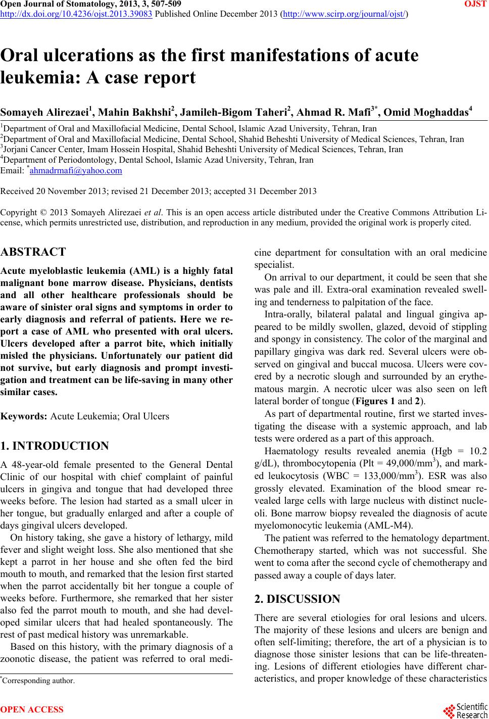 Open Journal of Stomatology, 2013, 3, 507-509 OJST http://dx.doi.org/10.4236/ojst.2013.39083 Published Online December 2013 (http://www.scirp.org/journal/ojst/) Oral ulcerations as the first manifestations of acute leukemia: A case report Somayeh Alirezaei1, Mahin Bakhshi2, Jamileh -Bigom Taheri2, Ahmad R. Mafi3*, Omid Moghaddas4 1Department of Oral and Maxillofacial Medicine, Dental School, Islamic Azad University, Tehran, Iran 2Department of Oral and Maxillofacial Medicine, Dental School, Shahid Beheshti University of Medical Sciences, Tehran, Iran 3Jorjani Cancer Center, Imam Hossein Hospital, Shahid Beheshti University of Medical Sciences, Tehran, Iran 4Department of Periodontology, Dental School, Islamic Azad University, Tehran, Iran Email: *[email protected] Received 20 November 2013; revised 21 December 2013; accepted 31 December 2013 Copyright © 2013 Somayeh Alirezaei et al. This is an open access article distributed under the Creative Commons Attribution Li- cense, which permits unrestricted use, distribution, and reproduction in any medium, provided the original work is properly cited. ABSTRACT Acute myeloblastic leukemia (AML) is a highly fatal malignant bone marrow disease. Physicians, dentists and all other healthcare professionals should be aware of sinister oral signs and symptoms in order to early diagnosis and referral of patients. Here we re- port a case of AML who presented with oral ulcers. Ulcers developed after a parrot bite, which initially misled the physicians. Unfortunately our patient did not survive, but early diagnosis and prompt investi- gation and treatment can be life-saving in many other similar cases. Keywords: Acute Leukemia; Oral Ulcers 1. INTRODUCTION A 48-year-old female presented to the General Dental Clinic of our hospital with chief complaint of painful ulcers in gingiva and tongue that had developed three weeks before. The lesion had started as a small ulcer in her tongue, but gradually enlarged and after a couple of days gingival ulcers developed. On history taking, she gave a history of lethargy, mild fever and slight weight loss. She also mentioned that she kept a parrot in her house and she often fed the bird mouth to mouth, and remarked that the lesion first started when the parrot accidentally bit her tongue a couple of weeks before. Furthermore, she remarked that her sister also fed the parrot mouth to mouth, and she had devel- oped similar ulcers that had healed spontaneously. The rest of past medical history was unremarkable. Based on this history, with the primary diagnosis of a zoonotic disease, the patient was referred to oral medi- cine department for consultation with an oral medicine specialist. On arrival to our department, it could be seen that she was pale and ill. Extra-oral examination revealed swell- ing and tenderness to palpitation of the face. Intra-orally, bilateral palatal and lingual gingiva ap- peared to be mildly swollen, glazed, devoid of stippling and spongy in consistency. The color of the marginal and papillary gingiva was dark red. Several ulcers were ob- served on gingival and buccal mucosa. Ulcers were cov- ered by a necrotic slough and surrounded by an erythe- matous margin. A necrotic ulcer was also seen on left lateral border of tongue (Figures 1 and 2). As part of departmental routine, first we started inves- tigating the disease with a systemic approach, and lab tests were ordered as a part of this approach. Haematology results revealed anemia (Hgb = 10.2 g/dL), thrombocytopenia (Plt = 49,000/mm3), and mark- ed leukocytosis (WBC = 133,000/mm3). ESR was also grossly elevated. Examination of the blood smear re- vealed large cells with large nucleus with distinct nucle- oli. Bone marrow biopsy revealed the diagnosis of acute myelomonocytic leukemia (AML-M4). The patient was referred to the hematology department. Chemotherapy started, which was not successful. She went to coma after the second cycle of chemotherapy and passed away a couple of days later. 2. DISCUSSION There are several etiologies for oral lesions and ulcers. The majority of these lesions and ulcers are benign and often self-limiting; therefore, the art of a physician is to diagnose those sinister lesions that can be life-threaten- ing. Lesions of different etiologies have different char- acteristics, and proper knowledge of these characteristics *Corresponding author. OPEN ACCESS 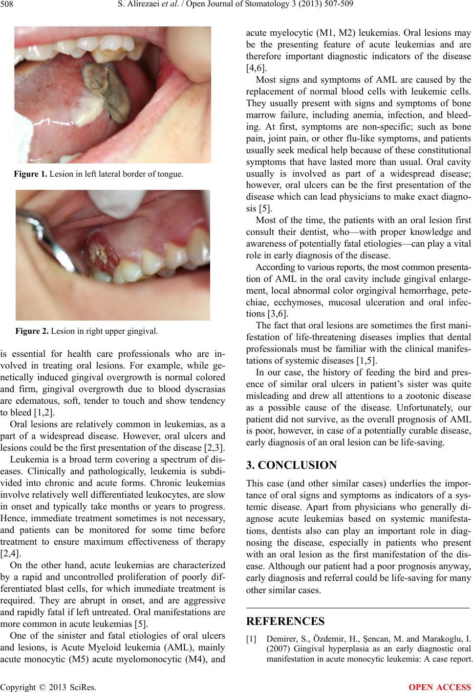 S. Alirezaei et al. / Open Journal of Stomatology 3 (2013) 507-509 508 Figure 1. Lesion in left lateral border of tongue. Figure 2. Lesion in right upper gingival. is essential for health care professionals who are in- volved in treating oral lesions. For example, while ge- netically induced gingival overgrowth is normal colored and firm, gingival overgrowth due to blood dyscrasias are edematous, soft, tender to touch and show tendency to bleed [1,2]. Oral lesions are relatively common in leukemias, as a part of a widespread disease. However, oral ulcers and lesions could be the first presentation of the disease [2,3]. Leukemia is a broad term covering a spectrum of dis- eases. Clinically and pathologically, leukemia is subdi- vided into chronic and acute forms. Chronic leukemias involve relatively well differentiated leukocytes, are slow in onset and typically take months or years to progress. Hence, immediate treatment sometimes is not necessary, and patients can be monitored for some time before treatment to ensure maximum effectiveness of therapy [2,4]. On the other hand, acute leukemias are characterized by a rapid and uncontrolled proliferation of poorly dif- ferentiated blast cells, for which immediate treatment is required. They are abrupt in onset, and are aggressive and rapidly fatal if left untreated. Oral manifestations are more common in acute leukemias [5]. One of the sinister and fatal etiologies of oral ulcers and lesions, is Acute Myeloid leukemia (AML), mainly acute monocytic (M5) acute myelomonocytic (M4), and acute myelocytic (M1, M2) leukemias. Oral lesions may be the presenting feature of acute leukemias and are therefore important diagnostic indicators of the disease [4,6]. Most signs and symptoms of AML are caused by the replacement of normal blood cells with leukemic cells. They usually present with signs and symptoms of bone marrow failure, including anemia, infection, and bleed- ing. At first, symptoms are non-specific; such as bone pain, joint pain, or other flu-like symptoms, and patients usually seek medical help because of these constitutional symptoms that have lasted more than usual. Oral cavity usually is involved as part of a widespread disease; however, oral ulcers can be the first presentation of the disease which can lead physicians to make exact diagno- sis [5]. Most of the time, the patients with an oral lesion first consult their dentist, who—with proper knowledge and awareness of potentially fatal etiologies—can play a vital role in early diagnosis of the disease. According to various reports, the most common presenta- tion of AML in the oral cavity include gingival enlarge- ment, local abnormal color orgingival hemorrhage, pete- chiae, ecchymoses, mucosal ulceration and oral infec- tions [3,6]. The fact that oral lesions are sometimes the first mani- festation of life-threatening diseases implies that dental professionals must be familiar with the clinical manifes- tations of systemic diseases [1,5]. In our case, the history of feeding the bird and pres- ence of similar oral ulcers in patient’s sister was quite misleading and drew all attentions to a zootonic disease as a possible cause of the disease. Unfortunately, our patient did not survive, as the overall prognosis of AML is poor, however, in case of a potentially curable disease, early diagnosis of an oral lesion can be life-saving. 3. CONCLUSION This case (and other similar cases) underlies the impor- tance of oral signs and symptoms as indicators of a sys- temic disease. Apart from physicians who generally di- agnose acute leukemias based on systemic manifesta- tions, dentists also can play an important role in diag- nosing the disease, especially in patients who present with an oral lesion as the first manifestation of the dis- ease. Although our patient had a poor prognosis anyway, early diagnosis and referral could be life-saving for many other similar cases. REFERENCES [1] Demirer, S., Özdemir, H., Şencan, M. and Marakoglu, I. (2007) Gingival hyperplasia as an early diagnostic oral manifestation in acute monocytic leukemia: A case report. Copyright © 2013 SciRes. OPEN ACCESS  S. Alirezaei et al. / Open Journal of Stomatology 3 (2013) 507-509 Copyright © 2013 SciRes. OPEN ACCESS 509 European Journal of Dentistry, 12, 111-114. [2] Parisi, E., Draznin, J., Stoopler, E., Schuster, S.J., Porter, D. and Sollecito, T.P. (2002) Acute myelogenous leuke- mia: advances and limitations of treatment. Oral Surgery, Oral Medicine, Oral Pathology, Oral Radiology and En- dodontology, 93, 257-263. http://dx.doi.org/10.1067/moe.2002.121988 [3] Kinane, D.F. and Marshall, G.J. (2001) Periodontal man- ifestations of systemic disease. Australian Dental Jour- nal, 46, 2-12. http://dx.doi.org/10.1111/j.1834-7819.2001.tb00267.x [4] Wu, J., Fantasia, J.E. and Kaplan, R. (2002) Oral mani- festations of acute myelomonocytic leukemia: A case re- port and review of the classification of leukemias. Jour- nal of Periodontology, 73, 664-668. http://dx.doi.org/10.1902/jop.2002.73.6.664 [5] Chavan, M., Subramaniam, A., Jhaveri, H., Khedkar, S., Durkar, S. and Agrwal, A. (2010) Acute myeloid leuke- mia: A case report with palatal and lingual gingival al- terations. Brazilian Journal Of Oral Sciences, 9, 67-69. [6] Dean, A.K., Ferguson, J.W. and Marvan, E.S. (2003) Acute leukaemia presenting as oral ulceration to a dental emergency service. Australian Dental Journal, 48, 195- 197. http://dx.doi.org/10.1111/j.1834-7819.2003.tb00032.x |