

Paper Menu >>
Journal Menu >>
 Open Journal of Stomatology, 2013, 3, 465-470 OJST http://dx.doi.org/10.4236/ojst.2013.39077 Published Online December 2013 (http://www.scirp.org/journal/ojst/) Tumours of salivary glands in the stomatology and maxillo-facial surgery unit at the university hospital centre —Yalgado Ouédraogo Mathieu Millogo1*, Tarcissus Konsem1, Haréfétéguéna Bissa2, Rasm a né Béogo 3, Dieudonné Ouédraogo1 1Service of Stomatology and Maxillo-Facial Surgery of the University Hospital Yalgado Ouédraogo, Ouagadougou, Burkina Faso 2Service of Stomatology and Maxillo-Facial Surgery of the University Hospital of Tokoin, Lomé, Togo 3Service of Stomatology and Maxillo-Facial Surgery of the University Hospital Souro SANOU, Bobo-Dioulasso, Burkina Faso Email: *[email protected] Received 20 September 2013; revised 1 November 2013; accepted 19 November 2013 Copyright © 2013 Mathieu Millogo et al. This is an open access article distributed under the Creative Commons Attribution License, which permits unrestricted use, distribution, and reproduction in any medium, provided the original work is properly cited. ABSTRACT The aim of this study was to contribute to strengthen- ing the efficiency in the treatment of the salivary gland tumours in the Stomatology and Maxillo-facial surgery service at the University Hospital Centre/Yal- gado OUEDRAOGO of Ouagadougou. It has been a retrospective and descriptive study covering seven (7) years [January 2006-December 2012] on epidemiol- ogy and the treatment of 54 cases of the salivary glands tumours histologically confirmed. Benign tu- mours (non-mal ignant tu mour s were pr eval ent (61.11 % of cases). The annual incidences were 4.71 cases for non malignant tumours and 3 cases for malignant tumours. The average age was 34 - 45 years for non malignant tumours and 44 - 33 years for malignant ones. In both groups, the tumour was discovered lately with an average 32 - 48 months before consult- ing in case of non malignant tumours and 18 - 29 mo n- ths for malignant tumours. The location of non ma- lignant tumours was very often the parotid (42.42%), under mandible gland (27.28%). Malignant tumours very often concerned the parotid (76.18%) and the under mandible gland (14.29%). The histology has revealed that pleomorphic adenoma was the leading type of non-malignant tumours (93.94% of cases) whereas carcinoma was predominant in malignant tumours (80.95% of cases). Long term track keeping was difficult and it has been complicated by the loss of contact with some patients. The low living stand- ard, the time wasted before consulting, limited human resources and therapeutic means constitute handicaps to the treatment in our African context. Keywords: Tumours; Salivary Glands; Epide miolo gy; Histology; Treatment 1. INTRODUCTION The tumours developed at the expense of the main sali- vary glands (parotid, under-mandible, sub-lingual) and accessory salivary glands are relatively frequent in every- day medical practice. Indeed, according to Ansari [1], in the Republic of Iran and Boko [2] in Togo, they represent respectively 3% to 6% of all tumours of the head and neck and 17.19% of Oto-Rhino-laryngology sphere. Salivary gland tumours have a diverse histology, but the non-malignant forms are prevalent with pleomorphic adenoma as the leading type [3-5]. However, tumours are not rare and they remain the haunting memory for any doctor. Only anatomo-phatologic study enables to diag- nose these tumours efficiently and therefore to suggest the appropriate therapeu tic attitude [6]. The earlier diagnose of salivary gland tumours is the guaranty for an optimal treatment. In our under-devel- oped African context, many difficulties are met impeding seriously diagnostic and therapeutic procedure. As a matter of fact, most patients come to consult at a very advanced stage of the disease, with very developed tu- mours called historic ones. Incidentally, their precari- ousness limits seriously diagnostic investigations and therapeutic responds. The aim of this study is to do the inventory of salivary gland tumours in our context in or- der to strengthen the efficiency of their treatment. 2. PATIENTS AND METHOD It was a retrospective study concerning medical files of patients’ suffering from tumours of salivary glands in the *Corresponding a uthor. OPEN ACCESS 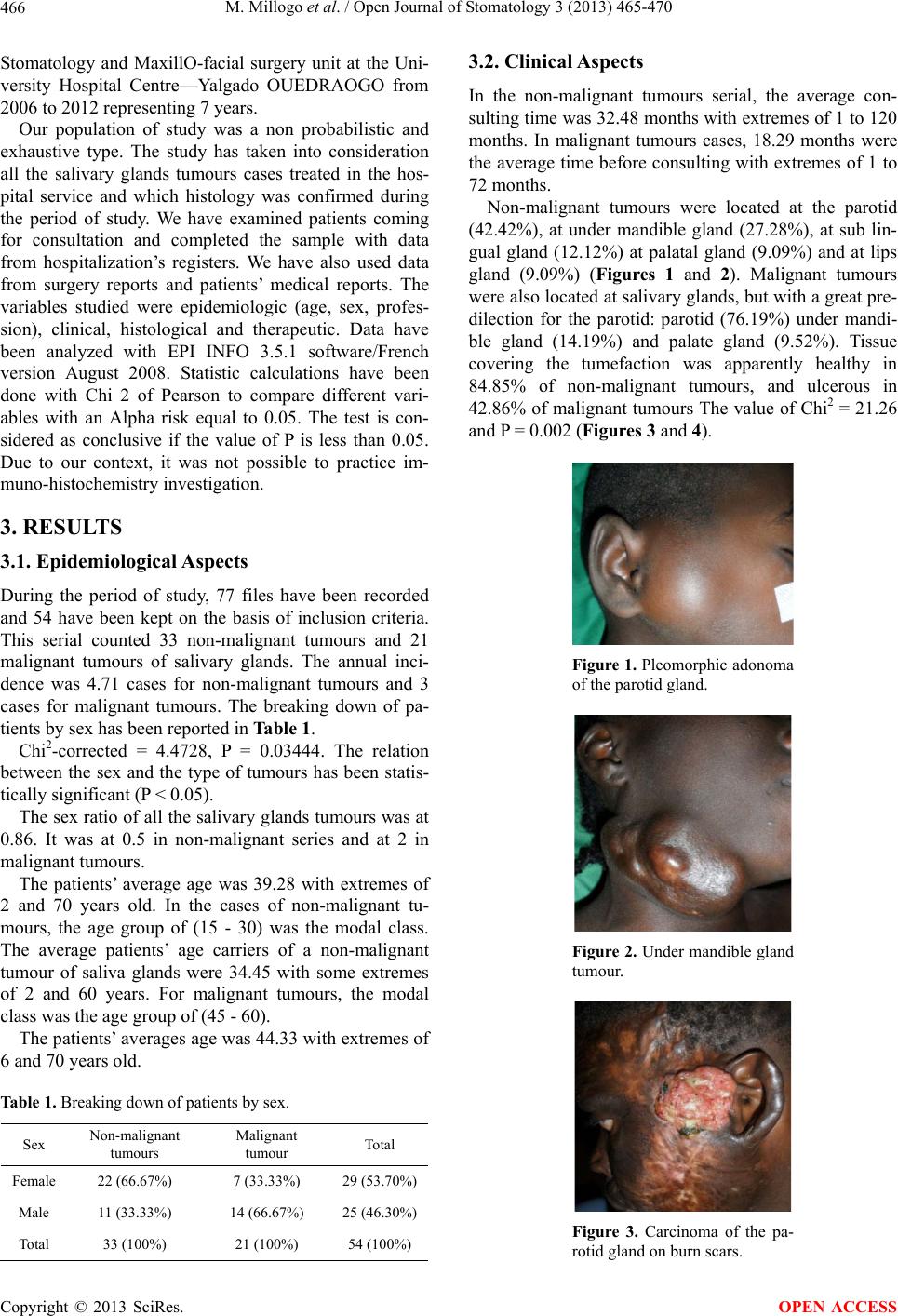 M. Millogo et al. / Open Journal of Stomatology 3 (2013) 465-470 466 Stomatology and MaxillO-facial surgery unit at the Uni- versity Hospital Centre—Yalgado OUEDRAOGO from 2006 to 2012 representing 7 years. Our population of study was a non probabilistic and exhaustive type. The study has taken into consideration all the salivary glands tumours cases treated in the hos- pital service and which histology was confirmed during the period of study. We have examined patients coming for consultation and completed the sample with data from hospitalization’s registers. We have also used data from surgery reports and patients’ medical reports. The variables studied were epidemiologic (age, sex, profes- sion), clinical, histological and therapeutic. Data have been analyzed with EPI INFO 3.5.1 software/French version August 2008. Statistic calculations have been done with Chi 2 of Pearson to compare different vari- ables with an Alpha risk equal to 0.05. The test is con- sidered as conclusive if the value of P is less than 0.05. Due to our context, it was not possible to practice im- muno-histochemistry investigation. 3. RESULTS 3.1. Epidemiological Aspects During the period of study, 77 files have been recorded and 54 have been kept on the basis of inclusion criteria. This serial counted 33 non-malignant tumours and 21 malignant tumours of salivary glands. The annual inci- dence was 4.71 cases for non-malignant tumours and 3 cases for malignant tumours. The breaking down of pa- tients by sex has been reported in Table 1. Chi2-corrected = 4.4728, P = 0.03444. The relation between the sex and the type of tumours has been statis- tically significant (P < 0.05). The sex ratio of all the salivary glands tumours was at 0.86. It was at 0.5 in non-malignant series and at 2 in malignant tumours. The patients’ average age was 39.28 with extremes of 2 and 70 years old. In the cases of non-malignant tu- mours, the age group of (15 - 30) was the modal class. The average patients’ age carriers of a non-malignant tumour of saliva glands were 34.45 with some extremes of 2 and 60 years. For malignant tumours, the modal class was the age group of (45 - 60). The patients’ averages age was 44.33 with extremes of 6 and 70 years old. Table 1. Breaking down of patients by sex. Sex Non-malignant tumours Malignant tumour Total Female 22 (66.67%) 7 (33.33%) 29 (53.70%) Male 11 (33.33%) 14 (66.67%) 25 (46.30%) Total 33 (100%) 21 (100%) 54 (100%) 3.2. Clinical Aspects In the non-malignant tumours serial, the average con- sulting time was 32.48 months with extremes of 1 to 120 months. In malignant tumours cases, 18.29 months were the average time before consulting with extremes of 1 to 72 months. Non-malignant tumours were located at the parotid (42.42%), at under mandible gland (27.28%), at sub lin- gual gland (12.12%) at palatal gland (9.09%) and at lips gland (9.09%) (Figures 1 and 2). Malignant tumours were also located at salivary glands, but with a great pre- dilection for the parotid: parotid (76.19%) under mandi- ble gland (14.19%) and palate gland (9.52%). Tissue covering the tumefaction was apparently healthy in 84.85% of non-malignant tumours, and ulcerous in 42.86% of malignant tumours The value of Chi2 = 21.26 and P = 0.002 (Figures 3 and 4). Figure 1. Pleomorphic adonoma of the parotid gland. Figure 2. Under mandible gland tumour. Figure 3. Carcinoma of the pa- rotid gland on burn scars. Copyright © 2013 SciRes. OPEN ACCESS 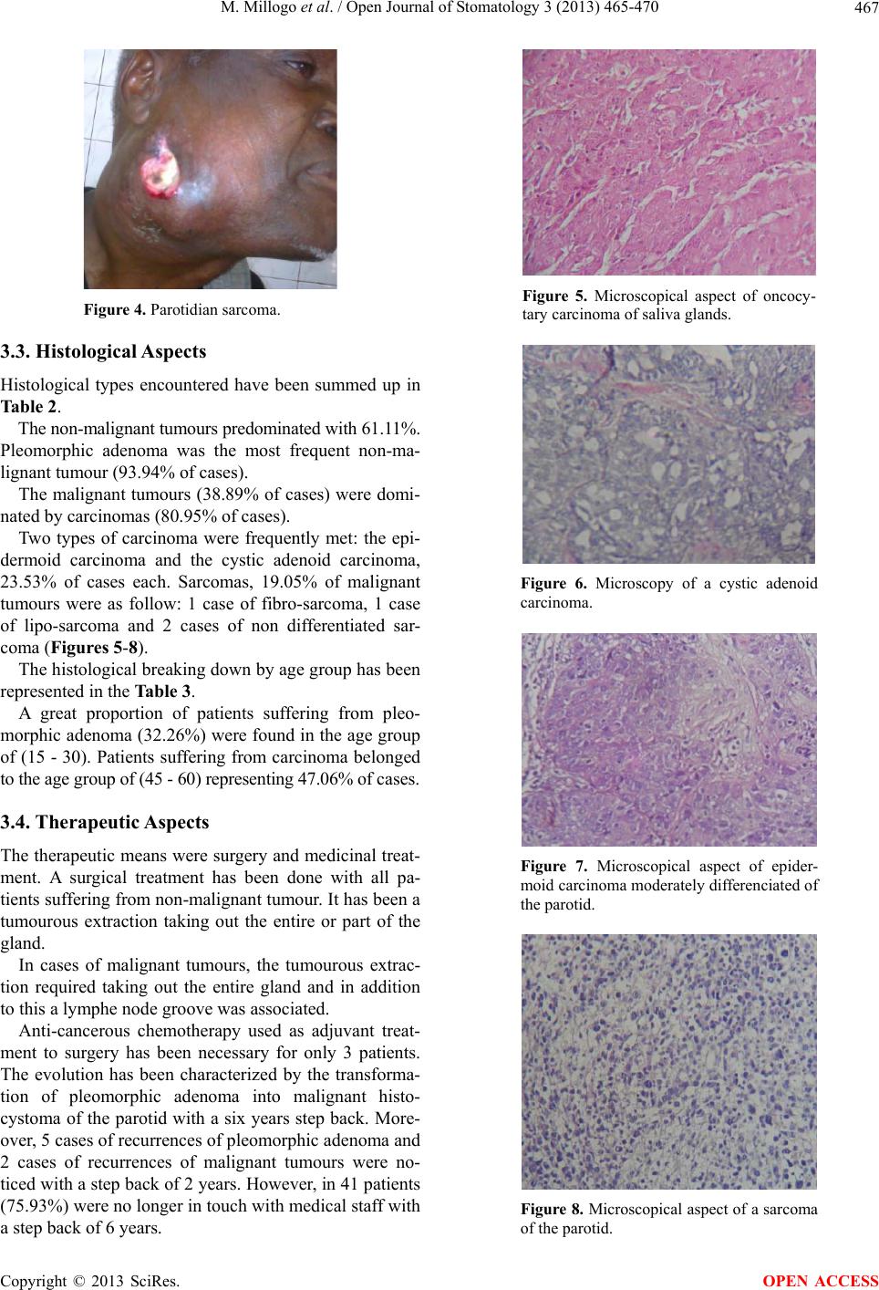 M. Millogo et al. / Open Journal of Stomatology 3 (2013) 465-470 467 Figure 4. Parotidian sarcoma. 3.3. Histological Aspects Histological types encountered have been summed up in Table 2. The non-malignant tumours predominated with 61.11%. Pleomorphic adenoma was the most frequent non-ma- lignant tumour (93.94% of cases). The malignant tumours (38.89% of cases) were domi- nated by carcinomas (80.95% of cases). Two types of carcinoma were frequently met: the epi- dermoid carcinoma and the cystic adenoid carcinoma, 23.53% of cases each. Sarcomas, 19.05% of malignant tumours were as follow: 1 case of fibro-sarcoma, 1 case of lipo-sarcoma and 2 cases of non differentiated sar- coma (Figures 5-8). The histological breaking down by age group has been represented in the Table 3. A great proportion of patients suffering from pleo- morphic adenoma (3 2.26%) were found in the age group of (15 - 30). Patients suffering from carcinoma belonged to the age group of (45 - 60) re presenting 47. 06% of cases. 3.4. Therapeutic A spects The therapeutic means were surgery and medicinal treat- ment. A surgical treatment has been done with all pa- tients suffering from non-malignant tumour. It has been a tumourous extraction taking out the entire or part of the gland. In cases of malignant tumours, the tumourous extrac- tion required taking out the entire gland and in addition to this a lymphe node groove was associated. Anti-cancerous chemotherapy used as adjuvant treat- ment to surgery has been necessary for only 3 patients. The evolution has been characterized by the transforma- tion of pleomorphic adenoma into malignant histo- cystoma of the parotid with a six years step back. More- over, 5 cases of recurrences of pleomorphic adenoma and 2 cases of recurrences of malignant tumours were no- ticed with a step back of 2 years. Howe ver, in 41 patients (75.93%) were no longer in touc h with medical staff with a step back of 6 years. Figure 5. Microscopical aspect of oncocy- tary carcinoma of saliva glands. Figure 6. Microscopy of a cystic adenoid carcinoma. Figure 7. Microscopical aspect of epider- moid carcinoma moderately differenciated of the parotid. Figure 8. Microscopical aspect of a sarcoma of the parotid. Copyright © 2013 SciRes. OPEN ACCESS 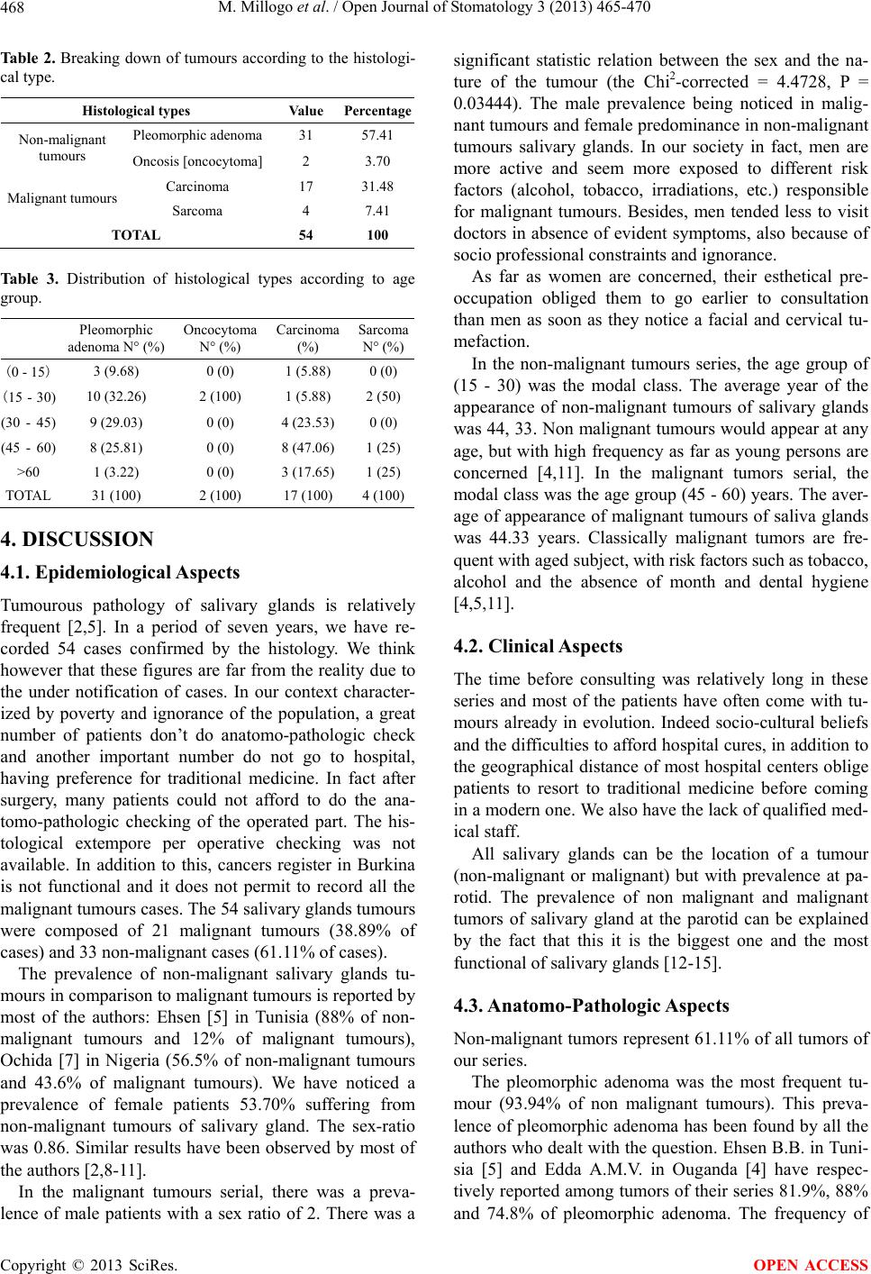 M. Millogo et al. / Open Journal of Stomatology 3 (2013) 465-470 468 Tab le 2 . Breaking down of tumours according to the histologi- cal type. Histological types Value Percentage Pleomorphic adenoma 31 57.41 Non-malignant tumours Oncosis [o nc oc ytom a] 2 3.70 Carcinoma 17 31.48 Malignant tumours Sarcoma 4 7.41 TOTAL 54 100 Table 3. Distribution of histological types according to age group. Pleomorphic adenoma N° (%) Oncocytoma N° (%) Carcinoma (%) Sarcoma N° (%) (0 - 15) 3 (9.68) 0 (0) 1 (5.88) 0 (0) (15 - 30) 10 (32.26) 2 (100) 1 (5.88) 2 (50) (30 - 45)9 (29.03) 0 (0) 4 (23.53) 0 (0) (45 - 60)8 (25.81) 0 (0) 8 (47.06) 1 (25) >60 1 (3.22) 0 (0) 3 (17.65) 1 (25) TOTAL 31 (100) 2 (100) 17 (100) 4 (100) 4. DISCUSSION 4.1. Epidemiological Aspects Tumourous pathology of salivary glands is relatively frequent [2,5]. In a period of seven years, we have re- corded 54 cases confirmed by the histology. We think however that these figures are far from the reality due to the under notification of cases. In our context character- ized by poverty and ignorance of the population, a great number of patients don’t do anatomo-pathologic check and another important number do not go to hospital, having preference for traditional medicine. In fact after surgery, many patients could not afford to do the ana- tomo-pathologic checking of the operated part. The his- tological extempore per operative checking was not available. In addition to this, cancers register in Burkina is not functional and it does not permit to record all the malignant tumours cases. The 54 salivary glands tumours were composed of 21 malignant tumours (38.89% of cases) and 33 non-malignant cases (61.11% of cases). The prevalence of non-malignant salivary glands tu- mours in comparison to malignant tumours is reported by most of the authors: Ehsen [5] in Tunisia (88% of non- malignant tumours and 12% of malignant tumours), Ochida [7] in Nigeria (56.5% of non-malignant tumours and 43.6% of malignant tumours). We have noticed a prevalence of female patients 53.70% suffering from non-malignant tumours of salivary gland. The sex-ratio was 0.86. Similar results have been observed by most of the authors [2,8-11]. In the malignant tumours serial, there was a preva- lence of male patients with a sex ratio of 2. There was a significant statistic relation between the sex and the na- ture of the tumour (the Chi2-corrected = 4.4728, P = 0.03444). The male prevalence being noticed in malig- nant tumours and female predominance in non-malignant tumours salivary glands. In our society in fact, men are more active and seem more exposed to different risk factors (alcohol, tobacco, irradiations, etc.) responsible for malignant tumours. Besides, men tended less to visit doctors in absence of evident symptoms, also because of socio professional constraints and ignorance. As far as women are concerned, their esthetical pre- occupation obliged them to go earlier to consultation than men as soon as they notice a facial and cervical tu- mefaction. In the non-malignant tumours series, the age group of (15 - 30) was the modal class. The average year of the appearance of non-malignant tumours of salivary glands was 44, 33. Non malignant tumours would appear at any age, but with high frequency as far as young persons are concerned [4,11]. In the malignant tumors serial, the modal class was the age group (45 - 60) years. The aver- age of appearance of malignant tumours of saliva glands was 44.33 years. Classically malignant tumors are fre- quent with aged subject, wit h risk f actor s su ch a s tobacco, alcohol and the absence of month and dental hygiene [4,5,11]. 4.2. Clinical Aspects The time before consulting was relatively long in these series and most of the patients have often come with tu- mours already in evolution. Indeed socio-cultural beliefs and the difficulties to afford hospital cures, in addition to the geographical distance of most hospital centers oblige patients to resort to traditional medicine before coming in a modern one. We also have the lack of qualified med- ical staff. All salivary glands can be the location of a tumour (non-malignant or malignant) but with prevalence at pa- rotid. The prevalence of non malignant and malignant tumors of salivary gland at the parotid can be explained by the fact that this it is the biggest one and the most functional of salivary glands [12-15]. 4.3. Anatomo-Pathologic Aspects Non-malignant tumors represent 61.11% of all tumors of our series. The pleomorphic adenoma was the most frequent tu- mour (93.94% of non malignant tumours). This preva- lence of pleomorphic adenoma has been found by all the authors who dealt with the qu estion. Ehsen B.B. in Tuni- sia [5] and Edda A.M.V. in Ouganda [4] have respec- tively reported among tumors of th eir series 81.9%, 88% and 74.8% of pleomorphic adenoma. The frequency of Copyright © 2013 SciRes. OPEN ACCESS  M. Millogo et al. / Open Journal of Stomatology 3 (2013) 465-470 469 the pleomorphic adenoma among non malignant tumors of salivary glands could be explained by the cellular di- versity of salivary glandular tissue. The pleomorphic adenoma is indeed a mixed tumor. Its microscopy shows epithelial and myoepithelial cells in an abundant stroma with some chondroma or myxoid areas [2,5,16,17]. Ma- lignant tumours represented 38.89% of all the tumours (80.95% of carcinomas and 19.05% of sarcomas). The epithelial nature predominating in salivary glands cells could explain the predominance of carcinoma [18,19]. We have noticed in our series 9 different types of car- cinomas dominated by the epidermoid carcinoma (23%, 53%) and adenoid cystic carcinoma (23.53%). This histological diversity among malignant tumours of salivary glands could be explained by the diversified characteristic of salivary glands cells (epithelial interme- diaries, acinous, oncosis, malpighian). There seems to be a general agreement on the prevalence of carcinomas among malignant tumours of salivary glands and on the histological diversity of carcinomas. However, no histo- logical form among carcinomas of salivary glands has been considered as leading type of the file [5,7,18 - 20]. Sarcomas were less frequent (19.05% of malignant tumours), but with also varied histolog ic types. This con- cerns rare nesenchymatous tumours appearing at all age and representing nearly 1% of all cancers [8,12,15,17, 20]. 4.4. Therapeutic A spects The treatment of non malignant tumours of salivary glands is exclusively surgical [6]. The treatment of malignant tumours of salivary glands is multidisciplinary. It combines oncology, surgery, ra- diotherapy and psychology. It takes into consideration the histopathology of the tumour, its state of evolution, its location, the availability of medical platform and the patient. Chemotherapy and radiotherapy are often used to complete surgery in the therapeutic protocol. The radio- therapy was not availab le in our context. In addition, th e immuno-histochemistry could not be done, which has certainly limited the choice of the drugs in the chemo- therapy. The particular case is the one of parotid gland. As a matter of fact, therapeutic indication depends on the location, the volu me of the tumour, the age of the patient, but also the results of the histological examination ex- tempore and the infiltration of adjacent structures by the tumour [6,10,18]. An evoluated malignant tumour will require a surgical extraction of the gland (partial or en- tire), extended to adjacent structures (facial nerves, mus- cular plan). The histological type and grade of malignant tumour as well as its clinical stage will necessitate a lymph node groove. Radiotherapy in association with surgery improves the prognostic of treated tumours and decreases the rate of local and regional recurrence [18]. The evolution of salivary glands tumours in our con- text has been characterized by recurrences (5 cases of pleomorphic adenomas and 25 cases of malignant tu- mours) and a malignant transformation of plesiomorphic adonema. We think however that there is an under noti- fication of recurrent cases in our study due to the diffi- culty to follow patients in long term. The great number of patients who did not keep in touch with medical staff can be a justification. Recurrences are often linked to an incomplete resection of the initial tumour, especially in case of exo facial parotidectomy. Generally, the pleomor- phic adenoma has a high tendency to recurrence [3,8]. 5. CONCLUSION The pathology of salivary glands tumours is relatively frequent but less known by our population. An accurate diagnosis rests upon the histological examination. The very concern with salivary gland tumours in our context is their treatment. As a matter of fact, most patients con- sult lately or wander about because of ignorance or socio-cultural beliefs. The difficulties at the therapeutic level are particularly due to an insufficiency of qualified human resources, but also because of the medical plat- form under acceptable standard. All these situations do not permit an adequate treatment. In addition to this, we have the ignorance of the population and the precarious- ness. REFERENCES [1] Ansari, M.H. (2007) Salivary gland tumours in an Iranian population: A retrospective study of 130 cases. Journal of Oral and Maxillofacial Surgery, 65, 2187-2194. http://dx.doi.org/10.1016/j.joms.2006.11.025 [2] Boko, E., Napo-Koura, G., Kpemissi, E. and Boko-Bessi, L. (2004) Tumours of secondary salivary glands: Epide- miologic and histological aspects. Journal of Oto-Rhino- Laryngology, 125, 233-237. [3] Akkari, K., Chnititir, S. and Sethom, A. (2007) Parotid tumours: About 43 cases. Tunisian Journal of Oto-Rhino- Laryngology, 18, 29-33. [4] Edda, A.M.V. (2004) Salivary gland tumours in Uganda: Clinical pathological of 268 cases. African Health Sci- ences, 4, 15-23. [5] Ehsen, B.B., Malek, F. and Olfa, K. (2010) Salivary gland tumours; histological and epidemiologic study of a 180 cases serial. Tunisia Medical Journal, 88, 240-244. [6] Bensadoun, R., Dassonville, O. and Rousmans, S. (2008) 2008 update of standars, options: Recommendations for management of patients with salivary gland malignant tumors. Bulletin du Cancer, 95, 735-749. [7] Ochicha, O., Malami, S., Aminu, M. and Akinfenwa, A. (2009) A histopathologic study of salivary gland tumors in Kano, Northen Nigeria. Indian Journal of Pathology Copyright © 2013 SciRes. OPEN ACCESS  M. Millogo et al. / Open Journal of Stomatology 3 (2013) 465-470 Copyright © 2013 SciRes. 470 OPEN ACCESS and Microbiology, 52, 473-476. http://dx.doi.org/10.4103/0377-4929.56121 [8] Flavia, A.O., Eliza, C.B.D. and Claudia, T.T. (2009) Sali- vary gland tumour: A revew of 599 cases in a Brazilian population. Head and Neck Pathology, 3, 271-275. [9] Jansisyanont, P. and Blanchaert, R.H. (2002) Salivary glands tumours of the oral cavity: About 80 cases. Inter- national Journal of Oral and Maxillofacial Surgery, 31, 257-261. http://dx.doi.org/10.1054/ijom.2002.0223 [10] Ouoba, K., Dao, M., Sakande, B., Kabre, M., Cisse, R. and Ouedraogo, I. (1998) Salivary glands tumours: About 48 cases opereted. Burkina Medical Journal, 2, 55-59. [11] Solange, S.L., Andréa, F.S., Rivadado, F., Batista, A. and Roseana, A.F. (2005) Epidemiologic profile of salivary gland neoplasms: Analysis of 245 cases. Revista Bra- sileira de Otorrinolaringologia, 71, 335-340. [12] Sadetzki, S. (2007) Cellular phone use and risk of benign and malignant parotid gland tumors: A nationwide case- control study. American Journal of Epidemiology, 167, 457-467. http://dx.doi.org/10.1093/aje/kwm325 [13] Fonseca, F.P., de Vasconcelos, C.M. and de Almeida, O.P. (2012) Clinicopathologic analysis of 493 cases of salivary gland tumors in a Southern Brazilian population. Oral Surgery, Oral Medicine, Oral Pathology and Oral Radiology, 114, 230-239. http://dx.doi.org/10.1016/j.oooo.2012.04.008 [14] Mahmoud, S., Mohamed, J.A., Negar, A., Bijan, K., Basir, H. and Amir, A. (2011) Maxillo-facial salivary glands tumours: A retrospective study about 130 cases in south iran population. Pathology Research International, 87, 934-938. [15] Moatemri, R., Belajouza, H. and Farroukh, U. (2008) Epidemiologic profile of salivary glands tumours at a Tu- nisian academic hospital: About 156 cases. Stomatology and Maxillo-Facial Surgery Journal, A, 109, 148-152. [16] Oukabli, M., Boudhas, A. and Setti, K. (2012) Salivary glands tumours: An histological study about 105 cases. African Cancer Journal, A, 4, 30-36. [17] Toure, S., Sonko, L., Diallo, B., Diop, A., Diouf, R. and Diop, E.M. (2005) Epidemiologic profile of oral cavity cancers in Senegal. Revue de Stomatologie et de Chirur- gie Maxillo-Faciale, 106, 17-26. [18] Halimi, P., Gardner, M. and Petit, F. (2005) Salivary glands tumours. Cancer/Radiothérapie, 9, 251-260. http://dx.doi.org/10.1016/j.canrad.2005.06.003 [19] Kokemuller, H., Bruggemann, N., Brachvogel, P. and Eckardt, A. (2004) Epithelial Malignant tumours of Sali- vary glands. A twenty years clinical study. Mund Kiefer Gesichtschir, 8, 191-201. [20] Védrine, P.O., Toussaint, B., Lapeyre, M., Coffinet, L., Sommelet, D. and Jankowski, R. (2004) Children salivary glands cancers. Annales d Oto-Laryngologie et de Chi- rurgie Cervico-Faciale (Masson, Paris), 121, 257-265. |