

Paper Menu >>
Journal Menu >>
 Engineering, 2013, 5, 545-548 http://dx.doi.org/10.4236/eng.2013.510B112 Published Online October 2013 (http://www.scirp.org/journal/eng) Copyright © 2013 SciRes. ENG The Research on Acupuncture Anesthesia Based on fMRI Jijun Tong1*, Jin Liu1, Yingying Lv2, Huade Chen2 1College of Information, Zhejiang Sci-Tech University, Hangzhou, Zhejiang 2The Third Clinical Medicine College, Zhejiang University of Traditional Chinese Medicine, Hangzhou, Zhejiang Email: *[email protected] Received 2013 ABSTRACT Pain is one of the most important sensations in daily life, and the traditional Chinese medicine such as acupuncture has shown great potential in pain relief. The principle research of acupuncture anesthesia has been the focus of modern science. In this paper the noninvasive brain imaging technique fMRI (functional magnetic reso nance imaging) was used. Eight volunteers were enrolled and received pain stimulation, and the brain response under TEAS (transcutaneous elec- trical acupoint stimulation) and manual needle stimulation were observed. The research finds pain has specific sensation region and the acupuncture anesthesia is realized through modulating the corresponding brain function region, but the manual needle has wider brai n r e sponse region tha n TE A S. Keywords: Acupuncture; Anesthesia; fMRI 1. Introduction Pain is one of the most important sensations in daily life. Pain perception is much more complicated than any other human perceptions. Much research results in many fields have shown that pain is not only physical experience but also closely related to many other factors such as physi- ology, social background, cultural differences and mental state [1]. The official definition of pain by the Interna- tional Association for the Study of Pain (IASP) consists of three elements: 1) it is associated with “injury” and “threat of injury”; 2) it is an “unpleasant” and “emotional” experience; and 3) it is “subjective” [2]. Since the Inter- national Association for the Study of Pain was founded in 1974, the research on pain developed steadily. But up to present the research and control on pain is still quite deficient. In contemporary medicine, acupuncture is con- sidered an important adjuvant treatment and has attracted more and more attention. Especially in anesthesia, acu- puncture was used more widely. Acupuncture was first successfully used in surgical analgesia in the 1950s [3]. In recent decades multidisciplinary experts explore the principle of acupuncture analgesia from the multi-level (from molecular and cellular level into gene level) and multiple perspectives (nerve, body fluids, meridian) [4]. The research of mechanism of acupuncture analgesia has become more and more important. The Traditional Chinese Medicine is difficult to explain many acupunc- ture phenomenons, so the use of modern medical tech- nical in acupuncture treatment has become the research focus in recent years, especially the various noninvasive brain imaging technologies and their application in illu- strating the cerebral mechanism of acupuncture anesthe- sia. It is a huge progress in revealing the scientific es- sence of acupuncture therapy effect [5-8]. These tech- niques mainly include Electroencephalograph (EEG), Magnetoencephalograph (MEG), Positron Emission To- mography (PET), Functional Magnetic Resonance Im- aging (fMRI) and s o on. The paper applies Functional Magnetic Resonance Imaging to study brain activity changes in the different stimulation modes in LI.4 (Hegu acupoint). From a new aspect to discusses the analgesic effect in different acu- puncture stimulation modes, to provide the visible and objective criterion for clinical application of acupuncture analgesia. 2. Methods 2.1. Subjects Eight healthy volunteers were enrolled in the experiment (four male and four female), they are right handed, 22 - 28 years old. All volunteers were students in the univer- sity, none of them had neurological or psychological dis- orders and they were not taking medicine. All volunteers were informed and received acupuncture treatment, signed an informed consent of this experiment. The vo- lunteers had no fMRI contraindications, such as metal prosthet i c l i mbs, joint, heart pacemakers, etc. *Corresponding a uthor. 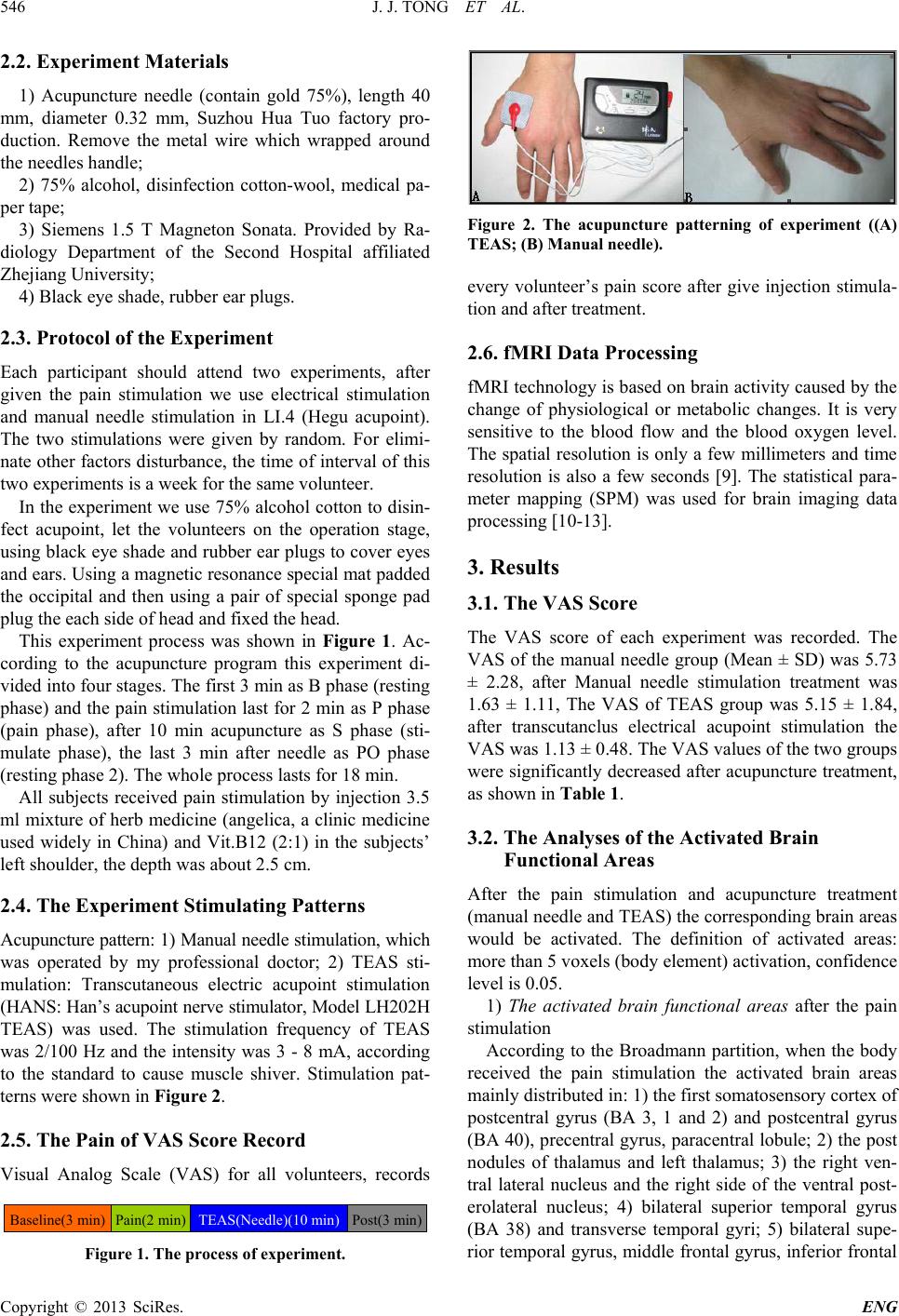 J. J. TONG ET AL. Copyright © 2013 SciRes. ENG 546 2.2. Experiment Materials 1) Acupuncture needle (contain gold 75%), length 40 mm, diameter 0.32 mm, Suzhou Hua Tuo factory pro- duction. Remove the metal wire which wrapped around the needl e s handle; 2) 75% alcohol, disinfection cotton-wool, medical pa- per tape; 3) Siemens 1.5 T Magneton Sonata. Provided by Ra- diology Department of the Second Hospital affiliated Zhejiang University; 4) Black eye shade, rubber ear plugs. 2.3. Protocol of the Experiment Each participant should attend two experiments, after given the pain stimulation we use electrical stimulation and manual needle stimulation in LI.4 (Hegu acupoint). The two stimulations were given by random. For elimi- nate other factors disturbance, the time of interv al of this two experiments is a week for the same volunteer. In the experiment we use 75% alcohol cotton to disin- fect acupoint, let the volunteers on the operation stage, using black eye shade and rubber ear plugs to cover eyes and ears. Using a magnetic resonance special mat padded the occipital and then using a pair of special sponge pad plug the each side of head and fixed the head. This experiment process was shown in Figure 1. Ac- cording to the acupuncture program this experiment di- vided into four stages. The first 3 min as B phase (resting phase) and the pain stimulation last for 2 min as P phase (pain phase), after 10 min acupuncture as S phase (sti- mulate phase), the last 3 min after needle as PO phase (resting phase 2). The whole process lasts for 18 min. All subjects received pain stimulation by injection 3.5 ml mixture of herb medicine (angelica, a clinic medicine used widely in China) and Vit.B12 (2:1) in the subjects’ left shoulder , the depth w a s about 2.5 cm. 2.4. The Experiment Stimulating Patterns Acupuncture pattern: 1) Manual needle sti mulation, which was operated by my professional doctor; 2) TEAS sti- mulation: Transcutaneous electric acupoint stimulation (HANS: Han’s acupoint nerve stimulator, Model LH202H TEAS) was used. The stimulation frequency of TEAS was 2/100 Hz and the intensity was 3 - 8 mA, according to the standard to cause muscle shiver. Stimulation pat- terns were shown in Figure 2. 2.5. The Pain of VAS Score Record Visual Analog Scale (VAS) for all volunteers, records Baseline(3 min) Pain(2 min) TEAS(Needle)(1 0 min) Post(3 min) Figure 1. The process of experiment. Figure 2. The acupuncture patterning of experiment ((A) TEAS; (B) Manual needle). every volunteer’s pain score after give injection stimula- tion and after treatment. 2.6. fMRI Data Processin g fMRI techno logy is based on brain activity caused by the change of physiological or metabolic changes. It is very sensitive to the blood flow and the blood oxygen level. The spatial resolution is only a few millimeters and time resolution is also a few seconds [9]. The statistical para- meter mapping (SPM) was used for brain imaging data processing [10-13]. 3. Results 3.1. The VAS Score The VAS score of each experiment was recorded. The VAS of the manual needle group (Mean ± SD) was 5.73 ± 2.28, after Manual needle stimulation treatment was 1.63 ± 1.11, The VAS of TEAS group was 5.15 ± 1.84, after transcutanclus electrical acupoint stimulation the VAS was 1.1 3 ± 0.48. The VAS values of the two groups were significantly decreased after acupuncture treatment, as shown in Table 1. 3.2. The Analyses of the Activated Brain Functional Areas After the pain stimulation and acupuncture treatment (manual needle and TEAS) the corresponding brain areas would be activated. The definition of activated areas: mor e than 5 voxels (body element) activation, confidence level is 0.05. 1) The activated brain functional areas after the pain stimulation According to the Broadmann partition, when the body received the pain stimulation the activated brain areas mainly distributed in: 1) the first somatosensory cortex of postcentral gyrus (BA 3, 1 and 2) and postcentral gyrus (BA 40), precentral gyrus, paracentral lobule; 2) the post nodules of thalamus and left thalamus; 3) the right ven- tral lateral nucleus and the right side of the ventral post- erolateral nucleus; 4) bilateral superior temporal gyrus (BA 38) and transverse temporal gyri; 5) bilateral supe- rior temporal gyrus, middle frontal gyrus, inferior frontal 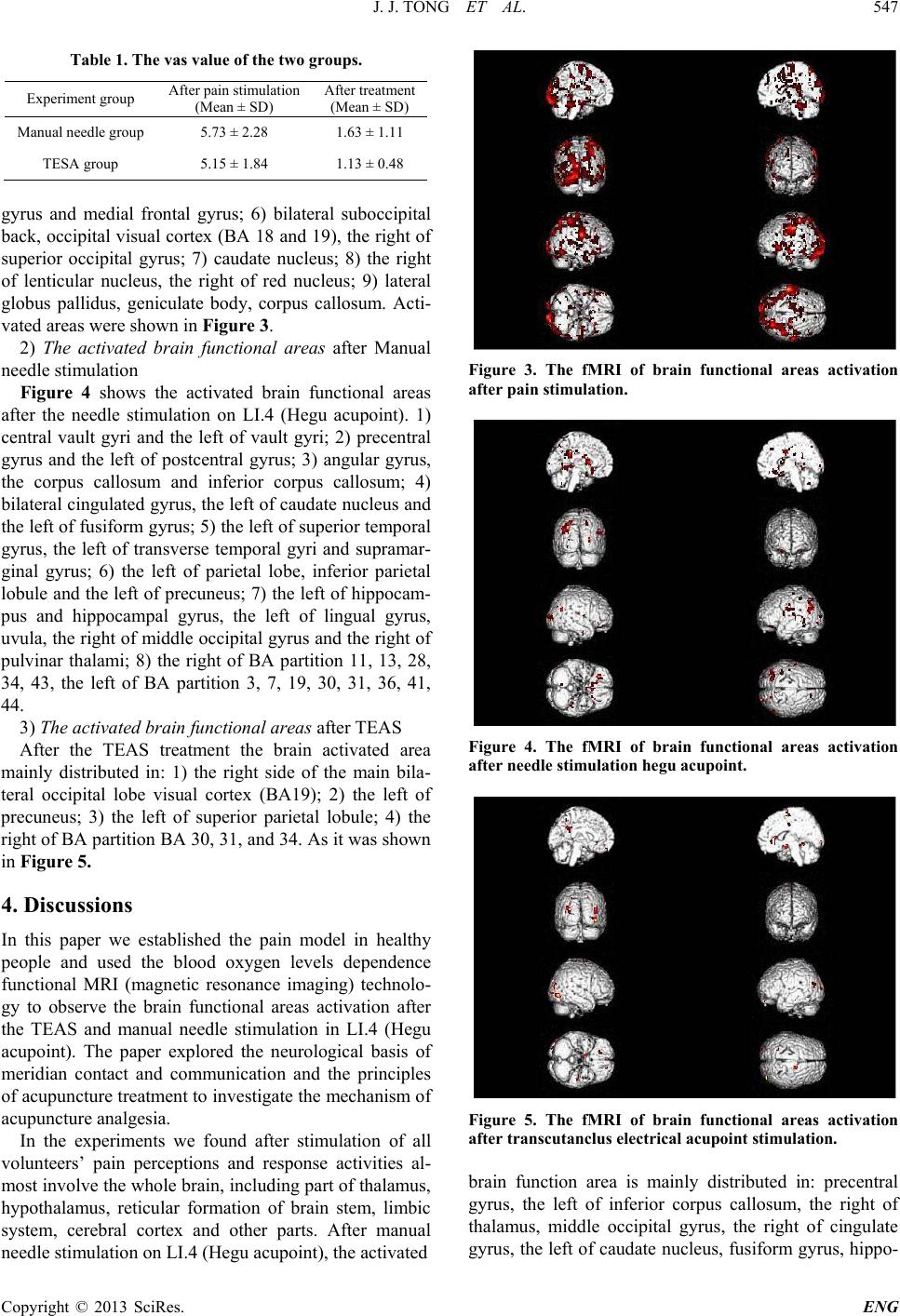 J. J. TONG ET AL. Copyright © 2013 SciRes. ENG 547 Table 1. The vas value of the two groups. Experiment group After pain stimulation (Mean ± SD) After treatment (Mean ± SD) Manual needle group 5.73 ± 2.28 1.63 ± 1.11 TESA group 5.15 ± 1.84 1.13 ± 0.48 gyrus and medial frontal gyrus; 6) bilateral suboccipital back, occipital visual cor tex (BA 18 and 19), the right of superior occipital gyrus; 7) caudate nucleus; 8) the right of lenticular nucleus, the right of red nucleus; 9) lateral globus pallidus, geniculate body, corpus callosum. Acti- vated areas were shown in Figure 3. 2) The activated brain functional areas after Manual needle stimulation Figure 4 shows the activated brain functional areas after the needle stimulation on LI.4 (Hegu acupoint). 1) central vault gyri and the left of vault gyri; 2) precentral gyrus and the left of postcentral gyrus; 3) angular gyrus, the corpus callosum and inferior corpus callosum; 4) bilateral cingulated gyrus, the left of caudate nucleus and the left of fusiform gyrus; 5) the left of superior temporal gyrus, the left of transverse temporal gyri and supramar- ginal gyrus; 6) the left of parietal lobe, inferior parietal lobule and the left of precuneus; 7) the left of hippocam- pus and hippocampal gyrus, the left of lingual gyrus, uvula, the right of middle occipital gyrus and the right of pulvinar thalami; 8) the right of BA partition 11, 13, 28, 34, 43, the left of BA partition 3, 7, 19, 30, 31, 36, 41, 44. 3) The acti v ated brain functio nal areas after TEAS After the TEAS treatment the brain activated area mainly distributed in: 1) the right side of the main bila- teral occipital lobe visual cortex (BA19); 2) the left of precuneus; 3) the left of superior parietal lobule; 4) the right of BA partition BA 30, 31, and 34. As it was shown in Figure 5. 4. Discussions In this paper we established the pain model in healthy people and used the blood oxygen levels dependence functional MRI (magnetic resonance imaging) technolo- gy to observe the brain functional areas activation after the TEAS and manual needle stimulation in LI.4 (Hegu acupoint). The paper explored the neurological basis of meridian contact and communication and the principles of acupuncture treatment to investigate the mechanism of acupuncture analgesia. In the experiments we found after stimulation of all volunteers’ pain perceptions and response activities al- most involve the whole brain, including part of thalamus, hypothalamus, reticular formation of brain stem, limbic system, cerebral cortex and other parts. After manual needle stimul a tion on LI.4 (Hegu acup oint), t he activated Figure 3. The fMRI of brain functional areas activation after pain stimulation. Figure 4. The fMRI of brain functional areas activation after needle stimulation hegu acupoint. Figure 5. The fMRI of brain functional areas activation after transcutanclus electrical acupoint stimulation. brain function area is mainly distributed in: precentral gyrus, the left of inferior corpus callosum, the right of thalamus, middle occipital gyrus, the right of cingulate gyrus, the left of caudate nucleus, fusiform gyrus, hippo- 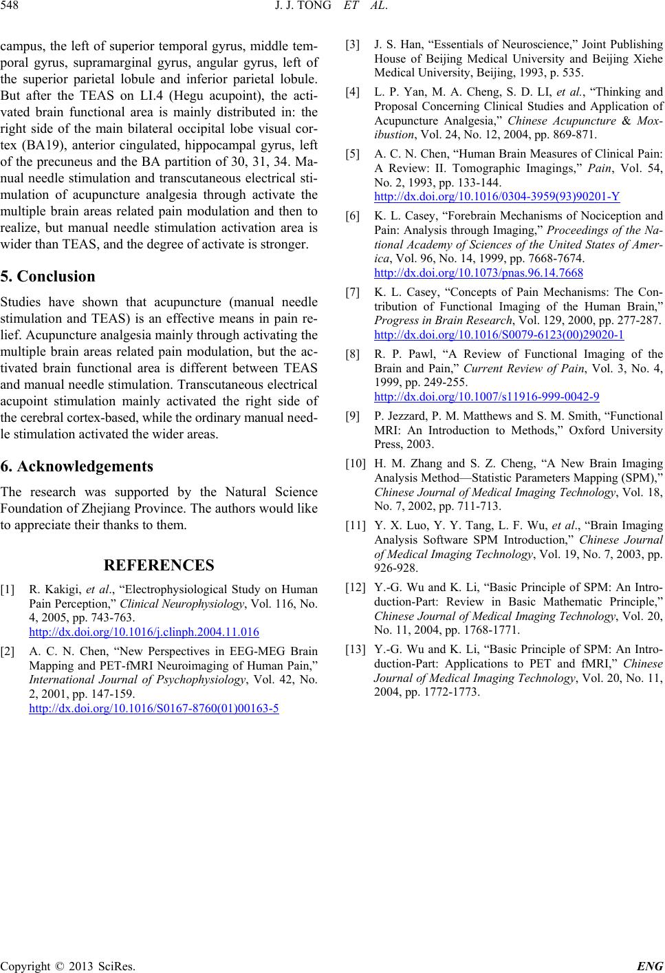 J. J. TONG ET AL. Copyright © 2013 SciRes. ENG 548 campus, the left of superior temporal gyrus, middle tem- poral gyrus, supramarginal gyrus, angular gyrus, left of the superior parietal lobule and inferior parietal lobule. But after the TEAS on LI.4 (Hegu acupoint), the acti- vated brain functional area is mainly distributed in: the right side of the main bilateral occipital lobe visual cor- tex (BA19), anterior cingulated, hippocampal gyrus, left of the precuneus and the BA partition of 30, 31, 34. Ma- nual needle stimulation and transcutaneous electrical sti- mulation of acupuncture analgesia through activate the multiple brain areas related pain modulation and then to realize, but manual needle stimulation activation area is wider than TEAS, and the degree of activate is stronger. 5. Conclusion Studies have shown that acupuncture (manual needle stimulation and TEAS) is an effective means in pain re- lief. Acupuncture analgesia mainly through activating the multiple brain areas related pain modulation, but the ac- tivated brain functional area is different between TEAS and manual needle stimulation. Transcutaneou s electrical acupoint stimulation mainly activated the right side of the cerebral cortex-based, while the ordinary manual need- le stimulation activated the wider areas. 6. Acknowledgements The research was supported by the Natural Science Foundation of Zhejiang Province. The authors would like to appreciate their thanks to them. REFERENCES [1] R. Kakigi, et al., “Electrophysiological Study on Human Pain Perception,” Cli nical Neurophysiology, Vol. 116, No. 4, 2005, pp. 743-763. http://dx.doi.org/10.1016/j.clinph.2004.11.016 [2] A. C. N. Chen, “New Perspectives in EEG-MEG Brain Mapping and PET-fMRI Neuroimaging of Human Pain,” International Journal of Psychophysiology, Vol. 42, No. 2, 2001, pp. 147-159. http://dx.doi.org/10.1016/S0167-8760(01)00163-5 [3] J. S. Han, “Essentials of Neuroscience,” Joint Publishing House of Beijing Medical University and Beijing Xiehe Medical University, Beijing, 1993, p. 535. [4] L. P. Yan, M. A. Cheng, S. D. LI, et al., “Thinking and Proposal Concerning Clinical Studies and Application of Acupuncture Analgesia,” Chinese Acupuncture & Mox- ibustion, Vol. 24, No. 12, 2004, pp. 869-871. [5] A. C. N. Chen, “Human Brain Measures of Clinical Pain: A Review: II. Tomographic Imagings,” Pain, Vol. 54, No. 2, 1993, pp. 133-144. http://dx.doi.org/10.1016/0304-3959(93)90201-Y [6] K. L. Casey, “Forebrain Mechanisms of Nociception and Pain: Analysis through Imaging,” Proceedings of the Na- tional Academy of Sciences of the United States of Amer- ica, Vol. 96, No. 14, 1999, pp. 7668-7674. http://dx.doi.org/10.1073/pnas.96.14.7668 [7] K. L. Casey, “Concepts of Pain Mechanisms: The Con- tribution of Functional Imaging of the Human Brain,” Progress in Brain R e search, Vol. 129, 2000, pp. 277-287. http://dx.doi.org/10.1016/S0079-6123(00)29020-1 [8] R. P. Pawl, “A Review of Functional Imaging of the Brain and Pain,” Current Review of Pain, Vol. 3, No. 4, 1999, pp. 249-255. http://dx.doi.org/10.1007/s11916-999-0042-9 [9] P. Jezzard, P. M. Matthews and S. M. Smith, “Functional MRI: An Introduction to Methods,” Oxford University Press, 2003. [10] H. M. Zhang and S. Z. Cheng, “A New Brain Imaging Analysis Method—Statistic Parameters Mapping (SPM),” Chinese Journal of Medical Imaging Technology , Vol. 18, No. 7, 2002, pp. 711-713. [11] Y. X. Luo, Y. Y. Tang, L. F. Wu, et al., “Brain Imaging Analysis Software SPM Introduction,” Chinese Journal of Medical Imaging Technology, Vol. 19, No. 7, 2003, pp. 926-928. [12] Y.-G. Wu and K. Li, “Basic Principle of SPM: An Intro- duction-Part: Review in Basic Mathematic Principle,” Chinese Journal of Medical Imaging Technol ogy, Vol. 20, No. 11, 2004, pp. 1768-1771. [13] Y.-G. Wu and K. Li, “Basic Principle of SPM: An Intro- duction-Part: Applications to PET and fMRI,” Chinese Journal of Medical Imaging Technology, Vol. 20, No. 11, 2004, pp. 1772-1773. |