

Paper Menu >>
Journal Menu >>
 Advances in Infectious Diseases, 2013, 3, 295-299 Published Online December 2013 (http://www.scirp.org/journal/aid) http://dx.doi.org/10.4236/aid.2013.34045 Open Access AID 295 Tenosynovitis and Sporotrichoid Disease Due to M. marinum on a Patien t u n d e r A n t i - TNFα Therapy*# Diana Póvoas†, J. Machado, F. Maltez Hospital de Curry Cabral, Centro Hospitalar de Lisboa Central, EPE, Lisboa, Portugal. Email: †[email protected] Received September 26th, 2013; revised October 26th, 2013; accepted November 4th, 2013 Copyright © 2013 Diana Póvoas et al. This is an open access article distributed under the Creative Commons Attribution License, which permits unrestricted use, distribution, and reproduction in any medium, provided the original work is properly cited. ABSTRACT There have been several reports of tuberculosis (TB) and, less frequently, of nontuberculous mycobacterial (NTM) in- fections in association with tumor necrosis factor α inhibitor (anti-TNFα) therapy. Mycobacterium marinum is a NTM with a distinct epidemiology and is infrequently responsible for disease in humans. Most commonly, it causes localized skin infections, but in 20% to 40% of cases, it involves deeper structures. Disseminated disease is exceptional and has been reported to occur only in immunocompromised patients. The authors report a clinical case of tenosynovitis and sporotrichoid disease due to M. marinum in a 45-year-old male patient under anti-TNFα therapies for spondyloarthro- pathy. Along antimicrobial therapy, the patient underwent surgical debridement and after two years he is still on treat- ment but substantially improved. A few cases of M. marinum infection occurring in patients treated with anti-TNFα drugs have been reported. The diagnosis of infection due to M. marinum requires a high index of suspicion from a properly obtained exposure history and is important so that efficient diagnostic approach and treatment are ensured. Keywords: Mycobacterium marinum; Anti-TNFα Drugs 1. Introduction Human infection due to Mycobacterium marinum was reported as a tuberculoid infection and historically recog- nized in people using public swimming pool. Most infec- tions occur after contact with contaminated water from “swimming pool” or “fish tank”, most frequently after minor abrasion, laceration, puncture or bite wounds. Hu- man infections with M. marinum are localized primarily to the skin; most commonly, M. marinum presents as a solitary papulonodular lesion on a finger or hand, and in some cases, it takes on a “sporotrichoid” form with one or more nodules in the proximal skin along paths of pre- sumed lymphatic spread. Deep infections such as teno- synovitis (the most frequent), osteomyelitis, arthritis and bursitis occur in 20% to 40% of cases. A few cases of M. marinum in patients treated with anti-TNFα therapy have been reported. 2. Clinical Case On December 2008, a 45-year-old white man, with a pre- vious history of spondyloarthropathy, with long lasting complaints of articular inflammatory pain on the elbows, shoulders, hip and knees treated with non-steroidal anti- inflammatory drugs (NSAIDs), developed two papulo- nodular erythematous lesions on the second and the third finger of the right hand (Figure 1). He was responsible for the maintenance of his domestic fish tank. Although he had no recollection of trauma, he exhib- ited a puncture-like wound on his right thumb. He was medicated with flucloxacillin. On the following six months, the papulonodular lesions remained the same but since he reported worsening of articular complaints on the right hand, local injection with corticosteroid was done, without improvement. Later, he had worsening of his spondyloarthropathy refractory to conventional ther- apy, so he was started on etanercept after two months of isoniazid due to positivity of both purified protein de- rivative (PPD) tuberculin skin testing and gamma inter- *Statements: All persons gave their informed consent and authorized p ublication of this case report. All authors declare that the submitted work has not been published before (neither in English nor in any other language) and that the work is not under consideration for publication elsewhere. #Conflict of interest: On behalf of all authors, the corresponding autho r states that there is no conflict of interest. †Corresponding author. 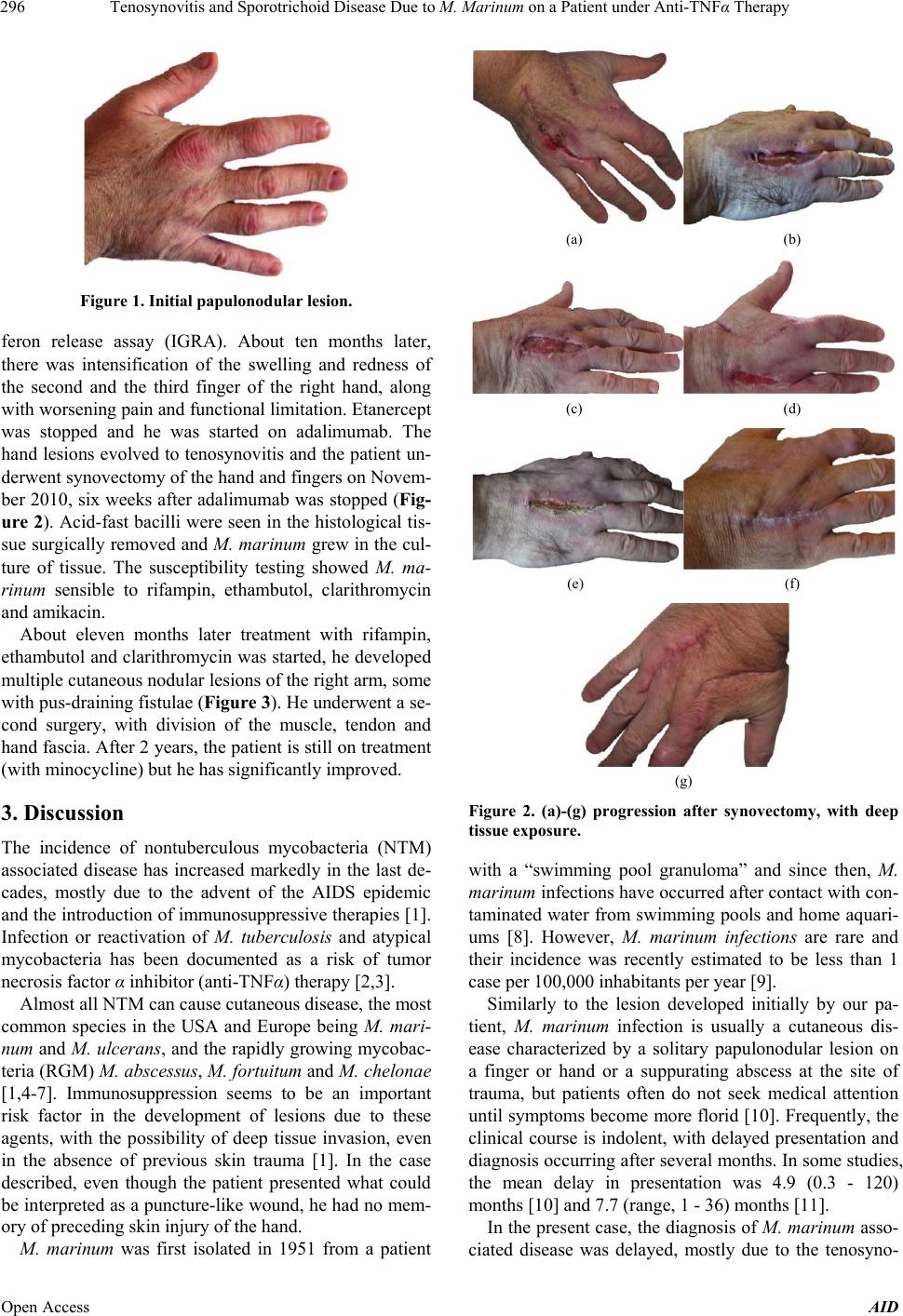 Tenosynovitis and Sporotrichoid Disease Due to M. Marinum on a Patient under Anti-TNFα Therapy 296 Figure 1. Initial papulonodular lesion. feron release assay (IGRA). About ten months later, there was intensification of the swelling and redness of the second and the third finger of the right hand, along with worsening pain and functional limitation. Etanercept was stopped and he was started on adalimumab. The hand lesions evolved to tenosynovitis and the patient un- derwent synovectomy of the hand and fingers on Novem- ber 2010, six weeks after adalimumab was stopped (Fig- ure 2). Acid-fast bacilli were seen in the histological tis- sue surgically removed and M. marinum grew in the cul- ture of tissue. The susceptibility testing showed M. ma- rinum sensible to rifampin, ethambutol, clarithromycin and amikacin. About eleven months later treatment with rifampin, ethambutol and clarithromycin was started, he developed multiple cutaneous nodular lesions of the right arm, some with pus-draining fistulae (Figure 3). He underwent a se- cond surgery, with division of the muscle, tendon and hand fascia. After 2 years, the patient is still on treatment (with minocycline) but he has significantly improved. 3. Discussion The incidence of nontuberculous mycobacteria (NTM) associated disease has increased markedly in the last de- cades, mostly due to the advent of the AIDS epidemic and the introduction of immunosuppressive therapies [1]. Infection or reactivation of M. tuberculosis and atypical mycobacteria has been documented as a risk of tumor necrosis factor α inhibitor (anti-TNFα) therapy [2,3]. Almost all NTM can cause cutaneous disease, the most common species in the USA and Europe being M. mari- num and M. ulcerans, and the rapidly growing mycobac- teria (RGM) M. abscessus, M. fortuitum and M. chelonae [1,4-7]. Immunosuppression seems to be an important risk factor in the development of lesions due to these agents, with the possibility of deep tissue invasion, even in the absence of previous skin trauma [1]. In the case described, even though the patient presented what could be interpreted as a puncture-like wound, he had no mem- ory of preceding skin injury of the hand. M. marinum was first isolated in 1951 from a patient (a) (b) (c) (d) (e) (f) (g) Figure 2. (a)-(g) progression after synovectomy, with deep tissue exposure. with a “swimming pool granuloma” and since then, M. marinum infections have occurred after contact with con- taminated water from swimming pools and home aquari- ums [8]. However, M. marinum infections are rare and their incidence was recently estimated to be less than 1 case per 100,000 inhabitants per year [9]. Similarly to the lesion developed initially by our pa- tient, M. marinum infection is usually a cutaneous dis- ease characterized by a solitary papulonodular lesion on a finger or hand or a suppurating abscess at the site of trauma, but patients often do not seek medical attention until symptoms become more florid [10]. Frequently, the clinical course is indolent, with delayed presentation and diagnosis occurring after several months. In some studies, the mean delay in presentation was 4.9 (0.3 - 120) months [10] and 7.7 (range, 1 - 36) months [11]. In the present case, the diagnosis of M. marinum asso- ciated disease was delayed, mostly due to the tenosyno- Open Access AID 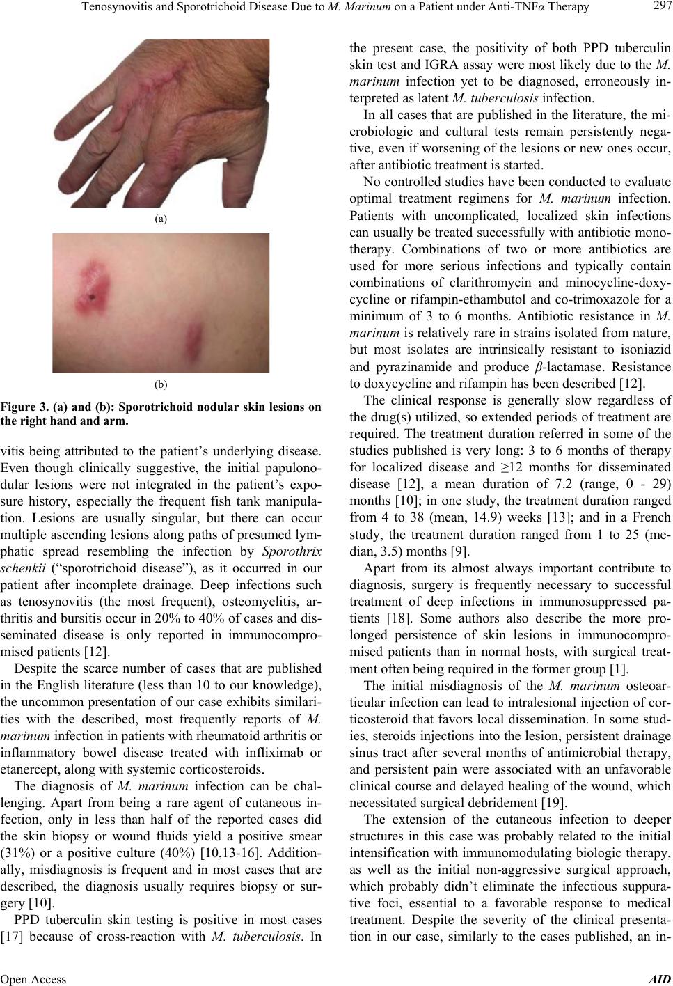 Tenosynovitis and Sporotrichoid Disease Due to M. Marinum on a Patient under Anti-TNFα Therapy 297 (a) (b) Figure 3. (a) and (b): Sporotr ichoid nodular skin lesions on the right hand and arm. vitis being attributed to the patient’s underlying disease. Even though clinically suggestive, the initial papulono- dular lesions were not integrated in the patient’s expo- sure history, especially the frequent fish tank manipula- tion. Lesions are usually singular, but there can occur multiple ascending lesions along paths of presumed lym- phatic spread resembling the infection by Sporothrix schenkii (“sporotrichoid disease”), as it occurred in our patient after incomplete drainage. Deep infections such as tenosynovitis (the most frequent), osteomyelitis, ar- thritis and bursitis occur in 20% to 40% of cases and dis- seminated disease is only reported in immunocompro- mised patients [12]. Despite the scarce number of cases that are published in the English literature (less than 10 to our knowledge), the uncommon presentation of our case exhibits similari- ties with the described, most frequently reports of M. marinum infection in patients with rheumatoid arthritis or inflammatory bowel disease treated with infliximab or etanercept, along with systemic corticosteroids. The diagnosis of M. marinum infection can be chal- lenging. Apart from being a rare agent of cutaneous in- fection, only in less than half of the reported cases did the skin biopsy or wound fluids yield a positive smear (31%) or a positive culture (40%) [10,13-16]. Addition- ally, misdiagnosis is frequent and in most cases that are described, the diagnosis usually requires biopsy or sur- gery [10]. PPD tuberculin skin testing is positive in most cases [17] because of cross-reaction with M. tuberculosis. In the present case, the positivity of both PPD tuberculin skin test and IGRA assay were most likely due to the M. marinum infection yet to be diagnosed, erroneously in- terpreted as latent M. tuberculosis infection. In all cases that are published in the literature, the mi- crobiologic and cultural tests remain persistently nega- tive, even if worsening of the lesions or new ones occur, after antibiotic treatment is started. No controlled studies have been conducted to evaluate optimal treatment regimens for M. marinum infection. Patients with uncomplicated, localized skin infections can usually be treated successfully with antibiotic mono- therapy. Combinations of two or more antibiotics are used for more serious infections and typically contain combinations of clarithromycin and minocycline-doxy- cycline or rifampin-ethambutol and co-trimoxazole for a minimum of 3 to 6 months. Antibiotic resistance in M. marinum is relatively rare in strains isolated from nature, but most isolates are intrinsically resistant to isoniazid and pyrazinamide and produce β-lactamase. Resistance to doxycycline and rifampin has been described [12]. The clinical response is generally slow regardless of the drug(s) utilized, so extended periods of treatment are required. The treatment duration referred in some of the studies published is very long: 3 to 6 months of therapy for localized disease and ≥12 months for disseminated disease [12], a mean duration of 7.2 (range, 0 - 29) months [10]; in one study, the treatment duration ranged from 4 to 38 (mean, 14.9) weeks [13]; and in a French study, the treatment duration ranged from 1 to 25 (me- dian, 3.5) months [9]. Apart from its almost always important contribute to diagnosis, surgery is frequently necessary to successful treatment of deep infections in immunosuppressed pa- tients [18]. Some authors also describe the more pro- longed persistence of skin lesions in immunocompro- mised patients than in normal hosts, with surgical treat- ment often being required in the former group [1]. The initial misdiagnosis of the M. marinum osteoar- ticular infection can lead to intralesional injection of cor- ticosteroid that favors local dissemination. In some stud- ies, steroids injections into the lesion, persistent drainage sinus tract after several months of antimicrobial therapy, and persistent pain were associated with an unfavorable clinical course and delayed healing of the wound, which necessitated surgical debridement [19]. The extension of the cutaneous infection to deeper structures in this case was probably related to the initial intensification with immunomodulating biologic therapy, as well as the initial non-aggressive surgical approach, which probably didn’t eliminate the infectious suppura- tive foci, essential to a favorable response to medical treatment. Despite the severity of the clinical presenta- tion in our case, similarly to the cases published, an in- Open Access AID 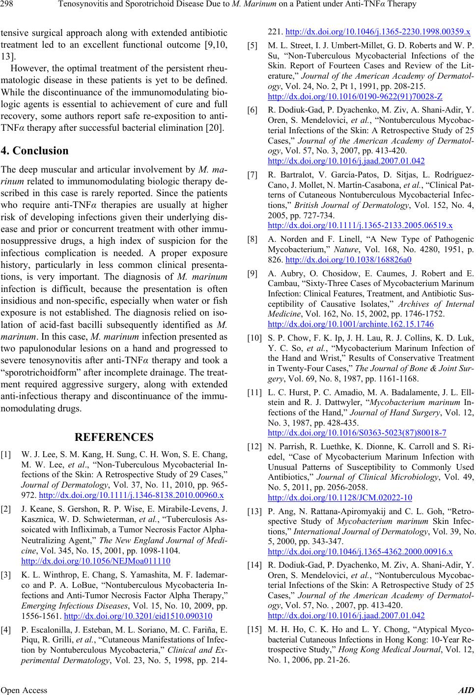 Tenosynovitis and Sporotrichoid Disease Due to M. Marinum on a Patient under Anti-TNFα Therapy 298 tensive surgical approach along with extended antibiotic treatment led to an excellent functional outcome [9,10, 13]. However, the optimal treatment of the persistent rheu- matologic disease in these patients is yet to be defined. While the discontinuance of the immunomodulating bio- logic agents is essential to achievement of cure and full recovery, some authors report safe re-exposition to anti- TNFα therapy after successful bacterial elimination [20]. 4. Conclusion The deep muscular and articular involvement by M. ma- rinum related to immunomodulating biologic therapy de- scribed in this case is rarely reported. Since the patients who require anti-TNFα therapies are usually at higher risk of developing infections given their underlying dis- ease and prior or concurrent treatment with other immu- nosuppressive drugs, a high index of suspicion for the infectious complication is needed. A proper exposure history, particularly in less common clinical presenta- tions, is very important. The diagnosis of M. marinum infection is difficult, because the presentation is often insidious and non-specific, especially when water or fish exposure is not established. The diagnosis relied on iso- lation of acid-fast bacilli subsequently identified as M. marinum. In this case, M. marinum infection presented as two papulonodular lesions on a hand and progressed to severe tenosynovitis after anti-TNFα therapy and took a “sporotrichoidform” after incomplete drainage. The treat- ment required aggressive surgery, along with extended anti-infectious therapy and discontinuance of the immu- nomodulating drugs. REFERENCES [1] W. J. Lee, S. M. Kang, H. Sung, C. H. Won, S. E. Chang, M. W. Lee, et al., “Non-Tuberculous Mycobacterial In- fections of the Skin: A Retrospective Study of 29 Cases,” Journal of Dermatology, Vol. 37, No. 11, 2010, pp. 965- 972. http://dx.doi.org/10.1111/j.1346-8138.2010.00960.x [2] J. Keane, S. Gershon, R. P. Wise, E. Mirabile-Levens, J. Kasznica, W. D. Schwieterman, et al., “Tuberculosis As- soicated with Infliximab, a Tumor Necrosis Factor Alpha- Neutralizing Agent,” The New England Journal of Medi- cine, Vol. 345, No. 15, 2001, pp. 1098-1104. http://dx.doi.org/10.1056/NEJMoa011110 [3] K. L. Winthrop, E. Chang, S. Yamashita, M. F. Iademar- co and P. A. LoBue, “Nontuberculous Mycobacteria In- fections and Anti-Tumor Necrosis Factor Alpha Therapy,” Emerging Infectious Diseases, Vol. 15, No. 10, 2009, pp. 1556-1561. http://dx.doi.org/10.3201/eid1510.090310 [4] P. Escalonilla, J. Esteban, M. L. Soriano, M. C. Fariña, E. Piqu, R. Grilli, et al., “Cutaneous Manifestations of Infec- tion by Nontuberculous Mycobacteria,” Clinical and Ex- perimental Dermatology, Vol. 23, No. 5, 1998, pp. 214- 221. http://dx.doi.org/10.1046/j.1365-2230.1998.00359.x [5] M. L. Street, I. J. Umbert-Millet, G. D. Roberts and W. P. Su, “Non-Tuberculous Mycobacterial Infections of the Skin. Report of Fourteen Cases and Review of the Lit- erature,” Journal of the American Academy of Dermatol- ogy, Vol. 24, No. 2, Pt 1, 1991, pp. 208-215. http://dx.doi.org/10.1016/0190-9622(91)70028-Z [6] R. Dodiuk-Gad, P. Dyachenko, M. Ziv, A. Shani-Adir, Y. Oren, S. Mendelovici, et al., “Nontuberculous Mycobac- terial Infections of the Skin: A Retrospective Study of 25 Cases,” Journal of the American Academy of Dermatol- ogy, Vol. 57, No. 3, 2007, pp. 413-420. http://dx.doi.org/10.1016/j.jaad.2007.01.042 [7] R. Bartralot, V. García-Patos, D. Sitjas, L. Rodríguez- Cano, J. Mollet, N. Martín-Casabona, et al., “Clinical Pat- terns of Cutaneous Nontuberculous Mycobacterial Infec- tions,” British Journal of Dermatology, Vol. 152, No. 4, 2005, pp. 727-734. http://dx.doi.org/10.1111/j.1365-2133.2005.06519.x [8] A. Norden and F. Linell, “A New Type of Pathogenic Mycobacterium,” Nature, Vol. 168, No. 4280, 1951, p. 826. http://dx.doi.org/10.1038/168826a0 [9] A. Aubry, O. Chosidow, E. Caumes, J. Robert and E. Cambau, “Sixty-Three Cases of Mycobacterium Marinum Infection: Clinical Features, Treatment, and Antibiotic Sus- ceptibility of Causative Isolates,” Archives of Internal Medicine, Vol. 162, No. 15, 2002, pp. 1746-1752. http://dx.doi.org/10.1001/archinte.162.15.1746 [10] S. P. Chow, F. K. Ip, J. H. Lau, R. J. Collins, K. D. Luk, Y. C. So, et al., “Mycobacterium Marinum Infection of the Hand and Wrist,” Results of Conservative Treatment in Twenty-Four Cases,” The Journal of Bone & Joint Sur- gery, Vol. 69, No. 8, 1987, pp. 1161-1168. [11] L. C. Hurst, P. C. Amadio, M. A. Badalamente, J. L. Ell- stein and R. J. Dattwyler, “Mycobacterium marinum In- fections of the Hand,” Journal of Hand Surgery, Vol. 12, No. 3, 1987, pp. 428-435. http://dx.doi.org/10.1016/S0363-5023(87)80018-7 [12] N. Parrish, R. Luethke, K. Dionne, K. Carroll and S. Ri- edel, “Case of Mycobacterium Marinum Infection with Unusual Patterns of Susceptibility to Commonly Used Antibiotics,” Journal of Clinical Microbiology, Vol. 49, No. 5, 2011, pp. 2056-2058. http://dx.doi.org/10.1128/JCM.02022-10 [13] P. Ang, N. Rattana-Apiromyakij and C. L. Goh, “Retro- spective Study of Mycobacterium marinum Skin Infec- tions,” International Journal of Dermatology, Vol. 39, No. 5, 2000, pp. 343-347. http://dx.doi.org/10.1046/j.1365-4362.2000.00916.x [14] R. Dodiuk-Gad, P. Dyachenko, M. Ziv, A. Shani-Adir, Y. Oren, S. Mendelovici, et al., “Nontuberculous Mycobac- terial Infections of the Skin: A Retrospective Study of 25 Cases,” Journal of the American Academy of Dermatol- ogy, Vol. 57, No. , 2007, pp. 413-420. http://dx.doi.org/10.1016/j.jaad.2007.01.042 [15] M. H. Ho, C. K. Ho and L. Y. Chong, “Atypical Myco- bacterial Cutaneous Infections in Hong Kong: 10-Year Re- trospective Study,” Hong Kong Medical Journal, Vol. 12, No. 1, 2006, pp. 21-26. Open Access AID  Tenosynovitis and Sporotrichoid Disease Due to M. Marinum on a Patient under Anti-TNFα Therapy Open Access AID 299 [16] H. N. Pang, J. Y. Lee, M. E. Puhaindran, S. H. Tan, A. B. Tan and F. C. Yong, “Mycobacterium Marinum as a Cause of Chronic Granulomatous Tenosynovitis in the Hand,” Journal of Infection, Vol. 54, No. 6, 2007, pp. 584-588. http://dx.doi.org/10.1016/j.jinf.2006.11.014 [17] F. M. Lewis, B. J. Marsh and C. F. von Reyn, “Fish Tank Exposure and Cutaneous Infections Due to Mycobacte- rium Marinum: Tuberculin Skin Testing, Treatment, and Prevention,” Clinical Infectious Diseases, Vol. 37, No. 3, 2003, pp. 390-397. http://dx.doi.org/10.1086/376628 [18] D. Wagner and L. S. Young, “Nontuberculous Mycobac- terial Infections: A Clinical Review,” Infection, Vol. 32, No. 5, 2004, pp. 257-270. http://dx.doi.org/10.1007/s15010-004-4001-4 [19] J. P. Cheung, B. Fung, S. S. Wong and W. Y. Ip, “Review Article: Mycobacterium marinum Infection of the Hand and Wrist,” Journal of Orthopaedic Surgery (Hong Kong), Vol. 18, No. 1, 2010, pp. 98-103. [20] J. A. Dare, S. Jahan, K. Hiatt and K. D. Torralba, “Rein- troduction of Etanercept during Treatment of Cutaneous Mycobacterium Marinum Infection in a Patient with An- kylosing Spondylitis,” Arthritis & Rheumatism, Vol. 61, No. 5, 2009, pp. 583-586. http://dx.doi.org/10.1002/art.24095 |