

Paper Menu >>
Journal Menu >>
 Open Journal of Applied Sciences, 2013, 3, 441-448 Published Online December 2013 (http://www.scirp.org/journal/ojapps) http://dx.doi.org/10.4236/ojapps.2013.38055 Open Access OJAppS Genetic Relatedness of Diplostomum Species (Digenea: Diplostomidae) Infesting Nile Tilapia (Oreochromis Niloticus L.) in Western Kenya Violet M. Ndeda*, Dickson O. Owiti, Ben O. Aketch, David M. Onyango Department of Zoology, Maseno University, Kisumu City, Kenya Email: *[email protected] Received May 24, 2013; revised June 26, 2013; accepted July 3, 2013 Copyright © 2013 Violet M. Ndeda et al. This is an open access article distributed under the Creative Commons Attribution License, which permits unrestricted use, distribution, and reproduction in any medium, provided the original work is properly cited. In accor- dance of the Creative Commons Attribution License all Copyrights © 2013 are reserved for SCIRP and the owner of the intellectual property Violet M. Ndeda et al. All Copyright © 2013 are guarded by law and by SCIRP as a guardian. ABSTRACT Diplostomum species metacercariae are trematode parasites that pose serious economic threats to aquaculture practice globally. Identification of Diplostomum at metacercariae stage has remained equivocal, hence lack of elucidation of the actual role of these species in fish population. 21 Diplostomoid metacercariae obtained from eyes of Nile tilapia were characterized using 18S and ITS rDNA (ITS1-5.8S-ITS2) genes. Phylogenetic analyses of ITS rDNA gene dataset in the metacercariae revealed close relationship to Diplostomum mashonense and D. baeri. Molecular identification using 18S rDNA sequences revealed close relationship to D. compactum, D. phoxini and D. spathaceum. Overall, genetic analyses in this study depicted a significant unrecognized genetic diversity among Diplostomum species. Successful differentiation of Diplostomum genera in this study using ribosomal markers suggested that 18S and ITS rDNA genes are effective genetic markers for inter-species phylogenetic analysis and should be employed in future for identification of diplostomoidea. Keywords: Diplostomum; 18S rDNA; Internal Transcribed Spacer Region 1. Introduction Diplostomum metacercariae are trematode parasites with complex three host lifecycle that are considered major pathogens of fish. They mature in the small intestine of piscivorous birds and are passed on to snails as first in- termediate host and fish as second intermediate host dur- ing their life cycle [1]. Once attached to the fish, Dip- lostomum metacercariae moves to the lens, retina and aqueous humour of fish eyes as well as the brain, spinal cord and nasal spaces thereby resulting into substantial losses of wild and farmed fish [1,2]. Diplostomum parasites have been encountered in fish farms in the northern hemisphere (Europe and North America) and as such received wide theoretical and em- pirical attention due to their pathogenic consequences to fingerlings [3-6]. The eye fluke infection has been asso- ciated with decreased growth of fish in rearing conditions [7,8] and increased mortality in fish as a result of reduced vision caused by cataracts which impairs the fish’s feed- ing efficiency [9] and makes the fish more vulnerable to avian predation [10]. Taxonomic studies on the diplos- tomid digeneans from the African continent are scarce and limited to few Diplostomum species descriptions published between 1930s-1960s, and virtually little is known of their natural history [e.g., 11,12]. This is re- lated to low sampling efforts in tropical countries due to lack of expertise in the field of fish parasitology. In addi- tion, identification of these parasites is problematic due to 1) the presence of morphologically similar species; 2) the phenotypic plasticity of the adults and metacercariae; 3) the simple larval morphology; and 4) the difficulties in linking life-cycle stages [13]. These problems represent a major impediment for the assessment of the distribution and the actual role of dip- lostomids in fish populations. Furthermore, most of the published African diplostomid studies have focused on the catfish Clarias gariepinus [14-17]. Recently, reports have indicated presence of Diplostomiasis infection *Corresponding author. 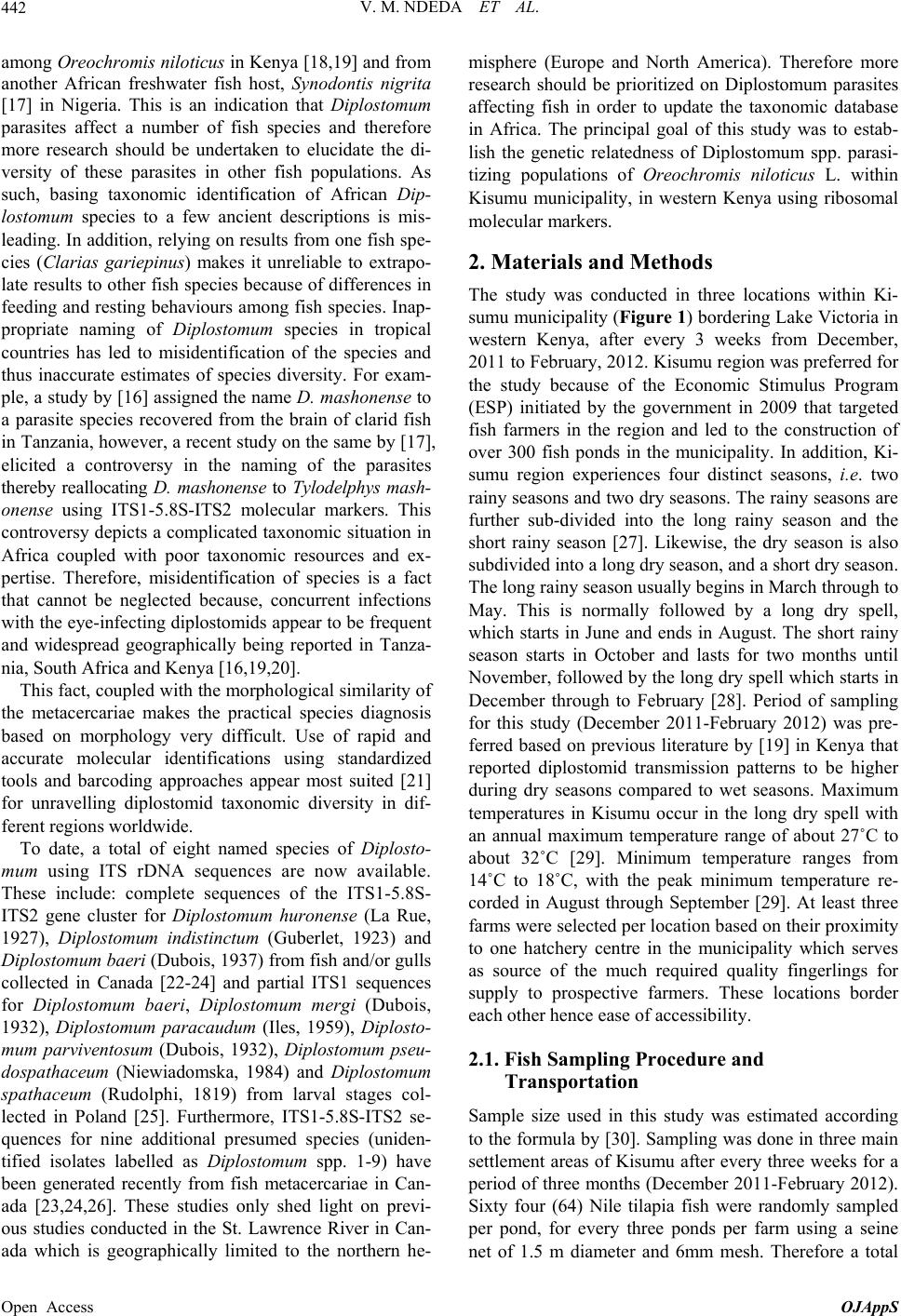 V. M. NDEDA ET AL. 442 among Oreochromis niloticus in Kenya [18,19] and from another African freshwater fish host, Synodontis nigrita [17] in Nigeria. This is an indication that Diplostomum parasites affect a number of fish species and therefore more research should be undertaken to elucidate the di- versity of these parasites in other fish populations. As such, basing taxonomic identification of African Dip- lostomum species to a few ancient descriptions is mis- leading. In addition, relying on results from one fish spe- cies (Clarias gariepinus) makes it unreliable to extrapo- late results to other fish species because of differences in feeding and resting behaviours among fish species. Inap- propriate naming of Diplostomum species in tropical countries has led to misidentification of the species and thus inaccurate estimates of species diversity. For exam- ple, a study by [16] assigned the name D. mashonen se to a parasite species recovered from the brain of clarid fish in Tanzania, however, a recent study on the same by [17], elicited a controversy in the naming of the parasites thereby reallocating D. mashonense to Tylodelphys mash- onense using ITS1-5.8S-ITS2 molecular markers. This controversy depicts a complicated taxonomic situation in Africa coupled with poor taxonomic resources and ex- pertise. Therefore, misidentification of species is a fact that cannot be neglected because, concurrent infections with the eye-infecting diplostomids appear to be frequent and widespread geographically being reported in Tanza- nia, South Africa and Kenya [16,19,20]. This fact, coupled with the morphological similarity of the metacercariae makes the practical species diagnosis based on morphology very difficult. Use of rapid and accurate molecular identifications using standardized tools and barcoding approaches appear most suited [21] for unravelling diplostomid taxonomic diversity in dif- ferent regions worldwide. To date, a total of eight named species of Diplosto- mum using ITS rDNA sequences are now available. These include: complete sequences of the ITS1-5.8S- ITS2 gene cluster for Diplostomum huronense (La Rue, 1927), Diplostomum indistinctum (Guberlet, 1923) and Diplostomum baeri (Dubois, 1937) from fish and/or gulls collected in Canada [22-24] and partial ITS1 sequences for Diplostomum baeri, Diplostomum mergi (Dubois, 1932), Diplostomum paracaudum (Iles, 1959), Diplosto- mum parviventosum (Dubois, 1932), Diplostomum pseu- dospathaceum (Niewiadomska, 1984) and Diplostomum spathaceum (Rudolphi, 1819) from larval stages col- lected in Poland [25]. Furthermore, ITS1-5.8S-ITS2 se- quences for nine additional presumed species (uniden- tified isolates labelled as Diplostomum spp. 1-9) have been generated recently from fish metacercariae in Can- ada [23,24,26]. These studies only shed light on previ- ous studies conducted in the St. Lawrence River in Can- ada which is geographically limited to the northern he- misphere (Europe and North America). Therefore more research should be prioritized on Diplostomum parasites affecting fish in order to update the taxonomic database in Africa. The principal goal of this study was to estab- lish the genetic relatedness of Diplostomum spp. parasi- tizing populations of Oreochromis niloticus L. within Kisumu municipality, in western Kenya using ribosomal molecular markers. 2. Materials and Methods The study was conducted in three locations within Ki- sumu municipality (Figure 1) bordering Lake Victoria in western Kenya, after every 3 weeks from December, 2011 to February, 2012. Kisumu region was preferred for the study because of the Economic Stimulus Program (ESP) initiated by the government in 2009 that targeted fish farmers in the region and led to the construction of over 300 fish ponds in the municipality. In addition, Ki- sumu region experiences four distinct seasons, i.e. two rainy seasons and two dry seasons. The rainy seasons are further sub-divided into the long rainy season and the short rainy season [27]. Likewise, the dry season is also subdivided into a long dry season, and a short dry season. The long rainy season usually begins in March through to May. This is normally followed by a long dry spell, which starts in June and ends in August. The short rainy season starts in October and lasts for two months until November, followed by the long dry spell which starts in December through to February [28]. Period of sampling for this study (December 2011-February 2012) was pre- ferred based on previous literature by [19] in Kenya that reported diplostomid transmission patterns to be higher during dry seasons compared to wet seasons. Maximum temperatures in Kisumu occur in the long dry spell with an annual maximum temperature range of about 27˚C to about 32˚C [29]. Minimum temperature ranges from 14˚C to 18˚C, with the peak minimum temperature re- corded in August through September [29]. At least three farms were selected per location based on their proximity to one hatchery centre in the municipality which serves as source of the much required quality fingerlings for supply to prospective farmers. These locations border each other hence ease of accessibility. 2.1. Fish Sampling Procedure and Transportation Sample size used in this study was estimated according to the formula by [30]. Sampling was done in three main settlement areas of Kisumu after every three weeks for a period of three months (December 2011-February 2012). Sixty four (64) Nile tilapia fish were randomly sampled per pond, for every three ponds per farm using a seine et of 1.5 m diameter and 6mm mesh. Therefore a total n Open Access OJAppS 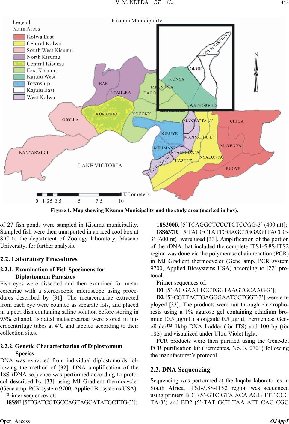 V. M. NDEDA ET AL. Open Access OJAppS 443 Figure 1. Map showing Kisumu Municipality and the study area (marked in box). 18S300R [5’TCAGGCTCCCTCTCCGG-3’ (400 nt)]; of 27 fish ponds were sampled in Kisumu municipality. Sampled fish were then transported in an iced cool box at 8˚C to the department of Zoology laboratory, Maseno University, for further analysis. 18S637R [5’TACGCTATTGGAGCTGGAGTTACCG- 3’ (600 nt)] were used [33]. Amplification of the portion of the rDNA that included the complete ITS1-5.8S-ITS2 region was done via the polymerase chain reaction (PCR) in MJ Gradient thermocycler (Gene amp. PCR system 9700, Applied Biosystems USA) according to [22] pro- tocol. 2.2. Laboratory Procedures 2.2.1. Examina tion of Fish Specimens f or Diplostomum Parasites Primer sequences of: Fish eyes were dissected and then examined for meta- cercariae with a stereoscopic microscope using proce- dures described by [31]. The metacercariae extracted from each eye were counted as separate lots, and placed in a petri dish containing saline solution before storing in 95% ethanol. Isolated metacercariae were stored in mi- crocentrifuge tubes at 4˚C and labeled according to their collection sites. D1 [5’-AGGAATTCCTGGTAAGTGCAAG-3’]; D2 [5’-CGTTACTGAGGGAATCCTGGT-3’] were em- ployed [33]. The products were run through electropho- resis using a 1% agarose gel containing ethidium bro- mide (0.5 μg/mL) alongside 0.5 μg/μl; Fermentas: Gen- eRuler™ 1kbp DNA Ladder (for ITS) and 100 bp (for 18S) and visualized under Ultra Violet light. PCR products were then purified using the Gene-Jet PCR purification kit (Fermentas, No. K 0701) following the manufacturer’s protocol. 2.2.2. Ge netic Cha ra cterization of Diplostomum Species DNA was extracted from individual diplostomoids fol- lowing the method of [32]. DNA amplification of the 18S rDNA sequence was performed according to proto- col described by [33] using MJ Gradient thermocycler (Gene amp. PCR system 9700, Applied Biosystems USA). 2.3. DNA Sequencing Sequencing was performed at the Inqaba laboratories in South Africa. ITS1-5.8S-ITS2 region was sequenced using primers BD1 (5’-GTC GTA ACA AGG TTT CCG TA-3’) and BD2 (5’-TAT GCT TAA ATT CAG CGG Primer sequences of: 18S9F [5’TGATCCTGCCAGTAGCATATGCTTG-3’]; 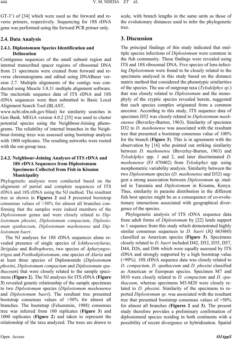 V. M. NDEDA ET AL. 444 GT-3’) of [34] which were used as the forward and re- verse primers, respectively. Sequencing for 18S rDNA gene was performed using the forward PCR primer only. 2.4. Data Analysis 2.4.1. Diplostomum Species Identification and Delineation Contiguous sequences of the small subunit region and internal transcribed spacer regions of ribosomal DNA from 21 specimens were created from forward and re- verse chromatograms and edited using DNABaser ver- sion 2.7. Multiple alignments of the contigs was con- ducted using Muscle 3.8.31 multiple alignment software. The nucleotide sequence data of ITS rDNA and 18S rDNA sequences were then submitted to Basic Local Alignment Search Tool (BLAST, www.ncbi.nlm.nih.gov/blast) for similarity searches in Gen Bank. MEGA version 4.0.2 [35] was used to cluster potential species using the Neighbour-Joining pheno- grams. The reliability of internal branches in the Neigh- bour-Joining trees was assessed using bootstrap analysis with 1000 replicates. The resulting networks were rooted with the out-group taxa. 2.4.2. Neighbour-Joining Analyses of ITS rDNA and 18S rDNA Sequences from Diplostomum Specimens Collected from Fish in Kisumu Municipality Phylogenetic analyses were conducted based on the alignment of partial and complete sequences of ITS rDNA and 18S rDNA using the NJ method. The resultant tree as shown in Figures 2 and 3 presented bootstrap consensus values of >50% for almost all branches con- firming that the samples were indeed members of the Diplostomum genus and were closely related to Dip- lostomum phoxini, Diplostomum compactum, Diplosto- mum spathaceum, Diplostomum mashonense and Dip- lostomum baeri. The NJ analyses for 18S rDNA sequences alone re- vealed presence of single species of Ichthyocotylurus, Strigidae and Bolbophorus, two species of Apharyngos- trigea and Posthodiplostomum, one species of Alaria and at least three species of Diplostomids (Diplostomum phoxini, Diplostomum compactum and Diplostomum spa- thaceum) that were closely related to the sample speci- mens (Figure 2). The NJ analyses for ITS rDNA (Figure 3) revealed genetic relationship of the sample specimens to two Diplostomum species (Diplostomum mashonense and Diplostomum baeri). The resultant tree presented bootstrap consensus values of >50% for almost all branches. The bootstrap (Felsenstein, 1988) consensus tree was inferred from 100 replicates (Figure 3) and 1000 replicates (Figure 2) and taken to represent the relationship of the taxa analyzed. The trees are drawn to scale, with branch lengths in the same units as those of the evolutionary distances used to infer the phylogenetic tree. 3. Discussion The principal findings of this study indicated that mul- tiple species infections of Diplostomum were common in the fish community. These findings were revealed using ITS and 18S ribosomal DNA. Five species of lens-infect- ing Diplostomum were found to be closely related to the specimens analysed in this study based on the distance matrix method that considered the phenotypic similarities of the species. The use of outgroup taxa (Tylodelphys sp.) that was closely related to Diplostomum and the mono- phyly of the cryptic species revealed herein, suggested that each species complex originated from a common ancestor. According to this study, ITS sequence data of specimen D32 was closely related to Diplostomum mash- onense (Beverley-Burton, 1963). Similarity of specimen D32 to D. mashonense was associated with the resultant tree that presented a bootstrap consensus value of 100% for the branch (Figure 3). This is in agreement with the observation by [16] who pointed out striking similarity between D. mashonense (Beverley-Burton, 1963) and Tylodelphys spp. 1 and 2, and later discriminated D. mashonense (FJ 470402) from Tylodephys spp. using morphometric variability analysis. Similarity between the two Diplostomum species (D. mashonense and D32) sug- gest a strong association between Diplostomum sp. stud- ied in Tanzania and Diplostomum in Kisumu, Kenya. Thus, similarity in parasite distribution in the different fish host species might be as a consequence of co-evolu- tionary interactions associated with geographical diver- gence of the species. Phylogenetic analysis of ITS rDNA sequence data from adult forms of Diplostomum by [22] lends support to 1 sequence from this study which demonstrated highly similar consensus sequences to D. baeri (JQ 665460) classified as American species (Figure 3). Specimens closely related to D. baeri included D42, D52, D35, D57, D44, D26, and D46 which were equally assessed by ITS rDNA and strongly supported by a high bootstrap value (>99%). 18S rDNA sequence data was closely related to D. compactum, D. spathaceum and D. phoxini classified as American or European species. Specimen M7 and M10 were closely related to D. compactum and D. spa- thaceum, whereas specimens M5-M20 were closely re- lated to D. phoxini. Similarity of the specimens to re- ported Diplostomum sp. was associated with the resultant tree that presented bootstrap consensus values of >50% for almost all branches (Figures 2 and 3). The present study therefore provides a preliminary confirmation of diplostomoid species residing in both continents with a possibility of recent divergence or hybridization. Spatial Open Access OJAppS 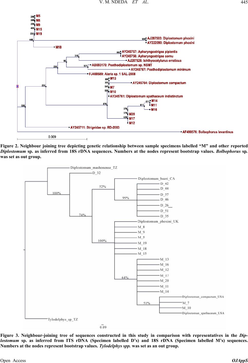 V. M. NDEDA ET AL. Open Access OJAppS 445 Figure 2. Neighbour joining tree depicting genetic relationship between sample specimens labelled “M” and other reported Diplostomum sp. as inferred from 18S rDNA sequences. Numbers at the nodes represent bootstrap values. Bolbophorus sp. was set as out group. Figure 3. Neighbour-joining tree of sequences constructed in this study in comparison with representatives in the Dip- lostomum sp. as inferred from ITS rDNA (Specimen labelled D’s) and 18S rDNA (Specimen labelled M’s) sequences. Numbers at the nodes represe nt bootstr a p value s. Tylodelphys spp. was set as an out group. 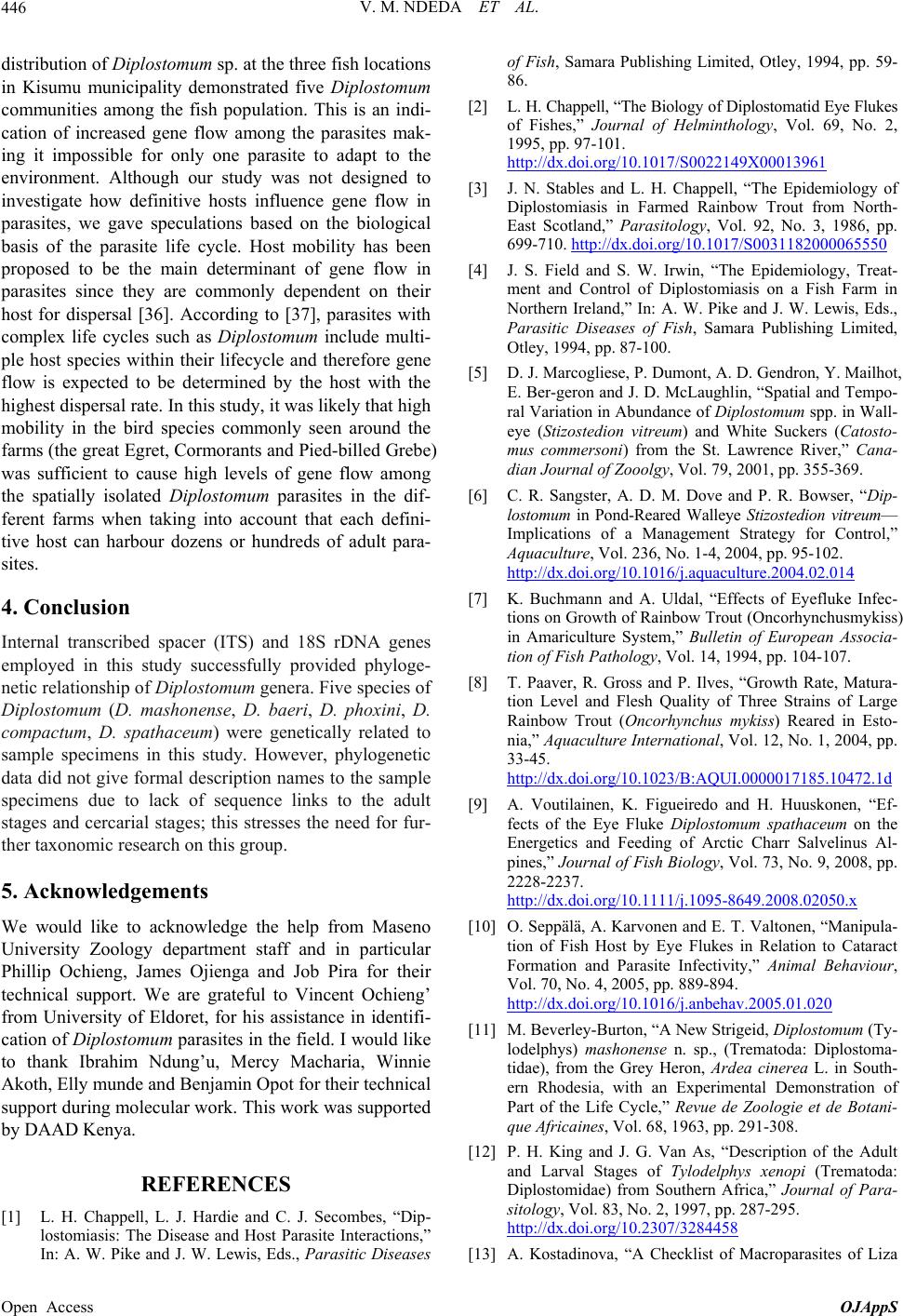 V. M. NDEDA ET AL. 446 istribution of Diplostomum sp. at the three fish locations of Fish, Samara Publi d in Kisumu municipality demonstrated five Diplostomum communities among the fish population. This is an indi- cation of increased gene flow among the parasites mak- ing it impossible for only one parasite to adapt to the environment. Although our study was not designed to investigate how definitive hosts influence gene flow in parasites, we gave speculations based on the biological basis of the parasite life cycle. Host mobility has been proposed to be the main determinant of gene flow in parasites since they are commonly dependent on their host for dispersal [36]. According to [37], parasites with complex life cycles such as Diplostomum include multi- ple host species within their lifecycle and therefore gene flow is expected to be determined by the host with the highest dispersal rate. In this study, it was likely that high mobility in the bird species commonly seen around the farms (the great Egret, Cormorants and Pied-billed Grebe) was sufficient to cause high levels of gene flow among the spatially isolated Diplostomum parasites in the dif- ferent farms when taking into account that each defini- tive host can harbour dozens or hundreds of adult para- sites. 4. Conclusion Internal transcribed spacer (ITS) and 18S rDNA dge the help from Maseno REFERENCES [1] L. H. Chappe. Secombes, “Dip- shing Limited, Otley, 1994, pp. 59- ishes,” Journal of Helminthology, Vol. 69, No. 2, genes employed in this study successfully provided phyloge- netic relationship of Diplostomum genera. Five species of Diplostomum (D. mashonense, D. baeri, D. phoxini, D. compactum, D. spathaceum) were genetically related to sample specimens in this study. However, phylogenetic data did not give formal description names to the sample specimens due to lack of sequence links to the adult stages and cercarial stages; this stresses the need for fur- ther taxonomic research on this group. 5. Acknowledgements We would like to acknowle University Zoology department staff and in particular Phillip Ochieng, James Ojienga and Job Pira for their technical support. We are grateful to Vincent Ochieng’ from University of Eldoret, for his assistance in identifi- cation of Diplostomum parasites in the field. I would like to thank Ibrahim Ndung’u, Mercy Macharia, Winnie Akoth, Elly munde and Benjamin Opot for their technical support during molecular work. This work was supported by DAAD Kenya. ll, L. J. Hardie and C. J lostomiasis: The Disease and Host Parasite Interactions,” In: A. W. Pike and J. W. Lewis, Eds., Parasitic Diseases 86. [2] L. H. Chappell, “The Biology of Diplostomatid Eye Flukes of F 1995, pp. 97-101. http://dx.doi.org/10.1017/S0022149X00013961 [3] J. N. Stables and L. H. Chappell, “The Epidemiolog Diplostomiasis in Farmed Rainbow Trout from North- y of East Scotland,” Parasitology, Vol. 92, No. 3, 1986, pp. 699-710. http://dx.doi.org/10.1017/S0031182000065550 [4] J. S. Field and S. W. Irwin, “The Epidemiology, Treat- ment and Control of Diplostomiasis on a Fish Farm in Laughlin, “Spatial and Tempo- — Northern Ireland,” In: A. W. Pike and J. W. Lewis, Eds., Parasitic Diseases of Fish, Samara Publishing Limited, Otley, 1994, pp. 87-100. [5] D. J. Marcogliese, P. Dumont, A. D. Gendron, Y. Mailhot, E. Ber-geron and J. D. Mc ral Variation in Abundance of Diplostomum spp. in Wall- eye (Stizostedion vitreum) and White Suckers (Catosto- mus commersoni) from the St. Lawrence River,” Cana- dian Journal of Zooolgy, Vol. 79, 2001, pp. 355-369. [6] C. R. Sangster, A. D. M. Dove and P. R. Bowser, “Dip- lostomum in Pond-Reared Walleye Stizostedion vitreum Implications of a Management Strategy for Control,” Aquaculture, Vol. 236, No. 1-4, 2004, pp. 95-102. http://dx.doi.org/10.1016/j.aquaculture.2004.02.014 [7] K. Buchmann and A. Uldal, “Effects of Eyefluke I tions on Growth of Rainbow Trout (Oncorhynchusm nfec- ykiss) arge in Amariculture System,” Bulletin of European Associa- tion of Fish Pathology, Vol. 14, 1994, pp. 104-107. [8] T. Paaver, R. Gross and P. Ilves, “Growth Rate, Matura- tion Level and Flesh Quality of Three Strains of L Rainbow Trout (Oncorhynchus mykiss) Reared in Esto- nia,” Aquaculture International, Vol. 12, No. 1, 2004, pp. 33-45. http://dx.doi.org/10.1023/B:AQUI.0000017185.10472.1d [9] A. Voutilainen, K. Figueiredo and H. Huuskonen, “Ef- fects of the Eye Fluke Diplostomum spathaceum on the Energetics and Feeding of Arctic Charr Salvelinus Al- pines,” Journal of Fish Biology, Vol. 73, No. 9, 2008, pp. 2228-2237. http://dx.doi.org/10.1111/j.1095-8649.2008.02050.x [10] O. Seppälä, A. Karvonen and E. T. Valtonen, “Manipula- tion of Fish Host by Eye Flukes in Relation to Cat aract Formation and Parasite Infectivity,” Animal Behaviour, Vol. 70, No. 4, 2005, pp. 889-894. http://dx.doi.org/10.1016/j.anbehav.2005.01.020 [11] M. Beverley-Burton, “A New Strigeid, Diplostom lodelphys) mashonense n. sp., (Trematoda: Dip um (Ty- lostoma- (Trematoda: tidae), from the Grey Heron, Ardea cinerea L. in South- ern Rhodesia, with an Experimental Demonstration of Part of the Life Cycle,” Revue de Zoologie et de Botani- que Africaines, Vol. 68, 1963, pp. 291-308. [12] P. H. King and J. G. Van As, “Description of the Adult and Larval Stages of Tylodelphys xenopi Diplostomidae) from Southern Africa,” Journal of Para- sitology, Vol. 83, No. 2, 1997, pp. 287-295. http://dx.doi.org/10.2307/3284458 [13] A. Kostadinova, “A Checklist of Macroparasites of Liza Open Access OJAppS  V. M. NDEDA ET AL. 447 Haematocheila (Temminck & Schlegel 1845) (Mugili- dae),” Parasites and Vectors, Vol. 1, 2008, p. 48. http://dx.doi.org/10.1186/1756-3305-1-48 [14] C. J. Mwita and G. Nkwengulila, “Determinants of th Parasite Community of Clariid Fishes from e Lake Victoria, Tanzania,” Journal of Helminthology, Vol. 82, No. 1, 2008, pp. 7-16. http://dx.doi.org/10.1017/S0022149X07839745 [15] C. J. Mwita and tionships of the Metazoan Parasites of the Clari G. Nkwengulila, “Phylogenetic Rela- id Fishes s of Diplostomid Metacercariae (Digenea: of Lake Victoria Inferred from Partial 18S rDNA Se- quences,” Tanzanian Journal of Sciences, Vol. 37, 2011, pp. 179-185. [16] F. D. Chibwana and G. Nkwengulila, “Variation in the Morphometric Trematoda) Infecting the Catfish, Clarias gariepinus in Tanzania,” Journal of Helminthology, Vol. 84, No. 1, 2010, pp. 61-70. http://dx.doi.org/10.1017/S0022149X09990083 [17] F. Chibwana, I. Bla G. Nkwengulila, T. Tomaš Scholz and A. Ko sco-Costa, S. Georgieva, M. K Hosea, stadinov , “First Insight into the Barcodes for African Diplostomids (Digenea: Diplostomidae): Brain Parasites in Clarias gariepinus (Siluriformes: Clariidae),” Infection Genetics and Evolution, Vol. 17, 2013, pp. 62-70. http://dx.doi.org/10.1016/j.meegid.2013.03.037 [18] G. K. Matolla, “Parasites of Tilapia (Oreo cus) in Chepkoilel-Fish Farm of Moi University chromis niloti- , Eldore t, Kenya,” In: S. S. Drexler and H. Waidbacher, Eds., Pro- ceedings of the 6th Framework Programme on BOMOSA Projects: Integrating BOMOSA Cage Fish Farming Sys- tem in Reservoirs, Ponds and Temporary Water Bodies in Eastern Africa, Machakos, 2-4 September 2009, p. 28. [19] E. M. Kembenya, G. K. Matolla, V. Ochieng’ and M. Ngaira, “Diplostomum Parasites Affecting Oreochromis niloticus in Chepkoilel Fish Farm and Two Dams in El- doret-Kenya,” International Journal of Scientific & En- gineering Research, Vol. 7, No. 3, 2012, pp. 1-7. [20] G. N. Madanire-Moyo, W. J. Luus-Powell and P. A. S. Olivier, “Ecology of Metazoan Parasites of Clariasgarie- pinus (Osteichthyes: Clariidae) from the Nwanedi-Lu- phephe Dams of the Limpopo River System, South Af- rica,” African Zoology, Vol. 45, No. 2, 2010, pp. 233-243. http://dx.doi.org/10.3377/004.045.0202 [21] J. April, R. L. Mayden, R. H. Hanner and L. Bernatcheza, “Genetic Calibration of Species Diversity among North America’s Freshwater Fishes,” PNAS, Vol. 108, No. 26, 2011, pp. 10602-10607. http://dx.doi.org/10.1073/pnas.1016437108 [22] D. Gallazzo, E. S. Dayana D. McLaughlin, “Molecular Systematics of ndan, D. J. Marcogliese and J. Some North y and Specificity in Diplostomum American Species of Diplostomum (Digenea) Based on rDNA-Sequence Data and Comparisons with European Congeners,” Canadian Journal of Zoology, Vol. 30, 2002, pp. 2207-2217. [23] S. Locke, J. D. Mclaughlin, S. Dayanandan and D. J. Marcogliese, “Diversit spp. Metacercariae in Freshwater Fishes Revealed by Cytochrome c Oxidase I and Internal Transcribed Spacer Sequences,” International Journal for Parasitology, Vol. 40, No. 3, 2010, pp. 333-343. http://dx.doi.org/10.1016/j.ijpara.2009.08.012 [24] S. Locke, J. D. Mclaughlin and D. J. Marcogliese, “DNA Barcodes Show Cryptic Diversity and a Potent logical Basis for Host Specificity among Diplo ial Physio- stomoidea (Platyhelminthes: Digenea) Parasitizing Freshwater Fishes in the St. Lawrence River, Canada,” Molecular Ecology, Vol. 19, No. 13, 2010, pp. 2813-2827. http://dx.doi.org/10.1111/j.1365-294X.2010.04713.x [25] K. Niewiadomska and Z. Laskowski, “Systematic Rela- tionships among Six Species of Diplostomum nordma 1832 (Digenea) Based on Morphological and Molec nn, ular Data,” Acta Parasitologica, Vol. 47, No. 1, 2002, pp. 20-28. [26] C. Rellstab, K. Louhi, A. Karvonen and J. Jokela, “Ana- lysis of Trematode Parasite Communities in Fish Eye Lenses by Pyrosequencing of Naturally Pooled DNA,” Infection, Genetics and Evolution, Vol. 11, No. 6, 2011, pp. 1276-1286. http://dx.doi.org/10.1016/j.meegid.2011.04.018 [27] Republic of Kenya, “Nyando District Development Plan, Nairobi; Kenya,” 2002. [28] National Environmental Management Authority [NEMA], “Strategy for Flood Management in Lake Victoria Basin, Kenya,” 2004. [29] Lake Basin Development Authority, “Feasibility Study on Kano Plain Irrigation Project,” Vol. 1, No. 71, 1992. [30] W. W. Daniel, “Biostatistics: A Foundation for Analysis in the Health Sciences,” John Wiley & Sons, New York, 1999. [31] S. Yamaguti, “Synopsis of Digenetic Trematodes of Ver- tebrates,” Keigaku Publishing, Tokyo, 1971. [32] G. E. Truett, R. L. Heeger, A. A. Mynatt, J. A. Truett and M. L. Warman, “Preparation of PCR-Quality Mouse Ge- nomic DNA with Hot Sodium Hydroxide and Tris (Hot- ne in atoda: Dige- SHOT),” BioTechniques, Vol. 29, 2000, pp. 52-54. [33] A. Moszczynska, S. A. Locke, J. D. McLaughlin, D. J. Marcogliese and T. J. Crease, “Development of Primers for the Mitochondrial Cytochrome c Oxidase I Ge Digenetic Trematodes Illustrates the Challenge of Bar- coding Parasitic Helminthes,” Molecular Ecology Re- sources, Vol. 9, No. S1, 2009, pp. 75-82. [34] K. Luton, D. Walker and D. Blair, “Comparison of Ribo- somal Internal Transcribed Spacers from Two Congeneric Species of Flukes (Platyhelminthes: Trem nea),” Molecular Biochemistry of Parasitology, Vol. 56, No. 2, 1992, pp. 323-328. http://dx.doi.org/10.1016/0166-6851(92)90181-I [35] K. Tamura, J. Dudley, M. Nei and S. Kumar, “MEGA 4: Molecular Evolutionary Genetics Analysis (MEGA) Soft- ware Version 4.0,” Molecular Biology and Evolution, Vol. 24, No. 8, 2007, pp. 1596-1599. http://dx.doi.org/10.1093/molbev/msm092 [36] M. S. Blouin, C. A. Yowell, C. H. Courtney and J. B. Dame, “Host Movement and the Genetic Populations of Parasitic Nematodes,” Gene Structure of tics, Vol. 141, No. 3, 1995, pp. 1007-1014. [37] F. Prugnolle, A. Theron, J. P. Pointier, R. Jabbour-Zahab, P. Jarne, P. Durand and T. de Meeus, “Dispersal in a Open Access OJAppS  V. M. NDEDA ET AL. Open Access OJAppS 448 o Hosts: Consequence for bbreviations and Acronyms Parasitic Worm and Its Tw Local Adaptation,” Evolution, Vol. 59, No. 2, 2005, pp. 296-303. A ITS: Internal Transcribed Spacer rDNA: Ribosomal DNA |