

Paper Menu >>
Journal Menu >>
 J. Biomedical Science and Engineering, 2013, 6, 38-46 JBiSE http://dx.doi.org/10.4236/jbise.2013.612A006 Published Online December 2013 (http://www.scirp.org/journal/jbise/) A parameterized analysis of the mechanical stress for coronary plaque fibrous caps Ramses Galaz, Catherine Pagiatakis, Emmanuel Gaillard, Rosaire Mongrain* Department of Mechanical Engineering, McGill University, Montreal, Canada Email: *[email protected] Received 31 October 2013; revised 28 November 2013; accepted 8 December 2013 Copyright © 2013 Ramses Galaz et al. This is an open access article distributed under the Creative Commons Attribution License, which permits unrestricted use, distribution, and reproduction in any medium, provided the original work is properly cited. ABSTRACT The fibrous cap is a protective layer of connective tissue that covers the core of an atherosclerotic plaque. The rupture of this layer has been commonly associated with acute myocardial infarctions. The thickness of the fibrous cap, the percentage of stenos- ed area, and the stiffness of the core were studied (commonly associated with vulnerable plaque char- acteristics) to quantify their effects on the cap’s me- chanical stress state by performing analyses using computational fluid-structure interaction (FSI) me- thods. The mechanical stress levels are significantly increased within the fibrous cap structure at the up- stream side of the plaque. As expected, the highest stresses occurred for a severe stenosis and a thin fi- brous cap. Interestingly, a weak structural support such as a soft lipid pool beneath the fibrous cap al- lowed for the hemodynamic pressure gradient forces to displace the fibrous cap in the direction of the flow, resulting in higher strains and thus higher mechani- cal stresses in the upstream portion of the plaque cap, potentially increasing the risk of cap rupture. The peak stress behavior of the most critical cases (thin fibrous cap and soft lipid core) at various degrees of stenosis was analyzed. For mid-range stenosis from 43% to 75%, there was a plateau region revealing that mild and moderate plaques were quickly exposed to the high stress condition of severe plaques. In con- clusion, the particular combi nation of a mild to severe stenosis, a thin fibrous cap and a soft lipid core re- sulted in the highest mechanical stresses calculated at the proximal side of the plaque. Mild and moderate plaques can be subjected to stresses similar to severe plaques, possibly contributing to their rupture. Keywords: Coronary Atherosclerotic Plaque; Fibrous Cap; Stenosis Severity; Lipid Core; Fluid-Structure Interaction 1. INTRODUCTION Atherosclerosis is a chronic inflammatory response in the arterial walls that narrows the lumen of vessels by the gradual deposition of fatty substances, cholesterol crys- tals, cellular waste products and calcium minerals, and the growth of connective tissue [1,2]. These localized pathological lesions are known as atheromatous plaques. Covering the plaque’s core is the fibrous cap, which is a layer of fibrous connective tissue and smooth muscle cells that prevents the core’s highly thrombogenic con- tents from coming into contact with blood. The rupture of the fibrous cap, either by mechanical or by biological factors, may trigger a biological response of platelet ac- tivation and subsequently thrombus formation. It is esti- mated that two thirds to three quarters of all arterial thrombi are caused by plaque rupture [3]. This event could potentially occlude the vessel and prevent blood perfusion downstream, resulting in an acute myocardial infarction. Clinicians have established from histopatho- logical studies that ruptured plaques share certain mor- phological and biological features that would make them more vulnerable to rupture. Unstable lesions with a large and soft lipid core, many inflammatory cells, and a thin fibrous cap are typically associated with acute coronary syndromes [4-6]. It is now recognized that plaque vul- nerability leading to rupture is an important cause of myocardial infarction, and sudden cardiac death. The characteristics of culprit lesions suggest the con- tribution of mechanical factors in plaque vulnerability for rupture. From a mechanical point of view, plaque rupture would occur when the stresses within the fibrous cap tissue surpass the ultimate strength of the material. Therefore, identifying the critical stress state within the diseased tissue could potentially elucidate plaque me- chanical failure mechanisms. Numerous studies have demonstrated that plaque component material properties *Corresponding author. OPEN ACCESS 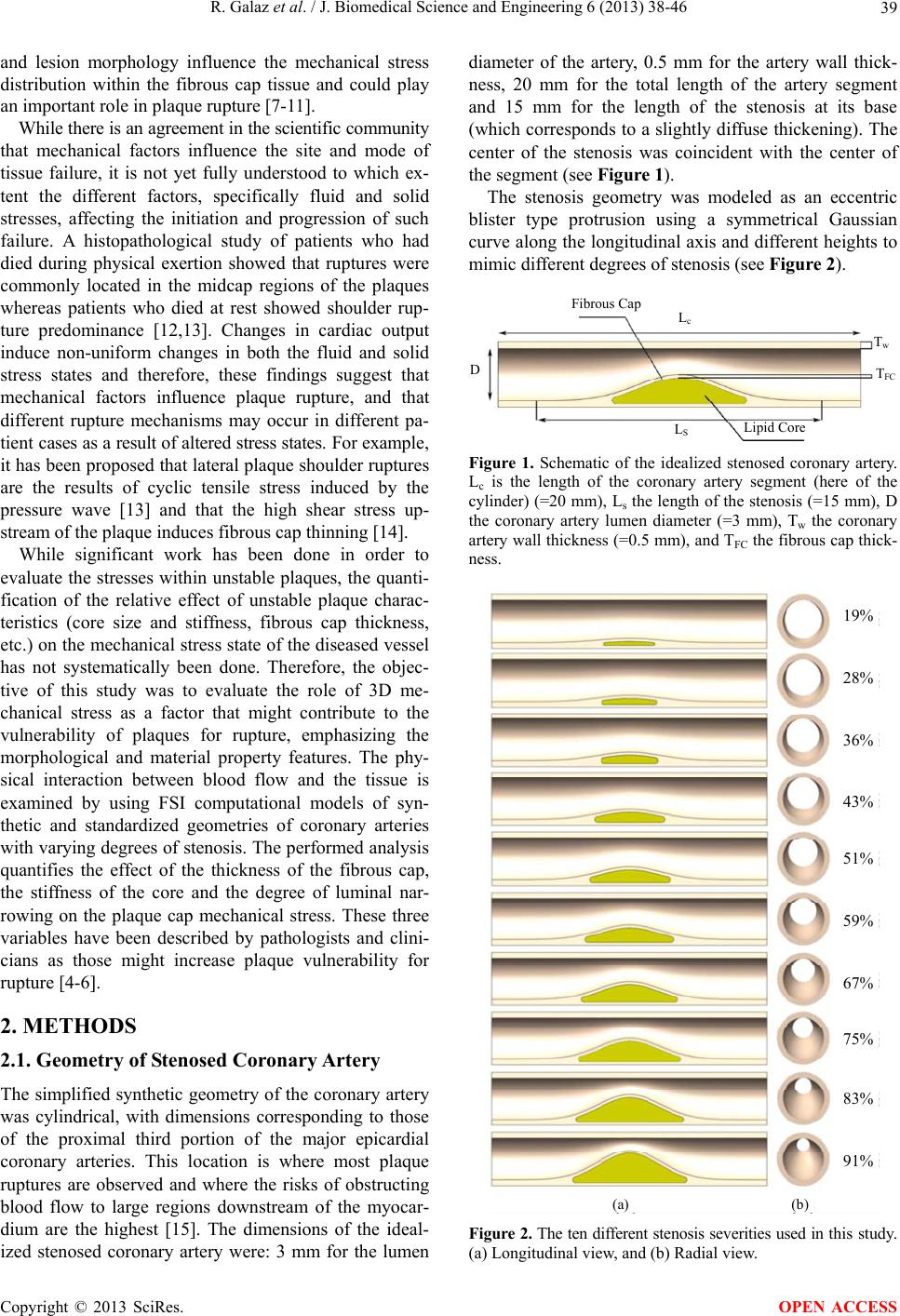 R. Galaz et al. / J. Biomedical Science and Engineering 6 (2013) 38-46 39 and lesion morphology influence the mechanical stress distribution within the fibrous cap tissue and could play an important role in plaque rupture [7-11]. While there is an agreement in the scientific community that mechanical factors influence the site and mode of tissue failure, it is not yet fully understood to which ex- tent the different factors, specifically fluid and solid stresses, affecting the initiation and progression of such failure. A histopathological study of patients who had died during physical exertion showed that ruptures were commonly located in the midcap regions of the plaques whereas patients who died at rest showed shoulder rup- ture predominance [12,13]. Changes in cardiac output induce non-uniform changes in both the fluid and solid stress states and therefore, these findings suggest that mechanical factors influence plaque rupture, and that different rupture mechanisms may occur in different pa- tient cases as a result of altered stress states. For example, it has been proposed that lateral plaque shoulder ruptures are the results of cyclic tensile stress induced by the pressure wave [13] and that the high shear stress up- stream of the plaque induces fibrous cap thinning [14]. While significant work has been done in order to evaluate the stresses within unstable plaques, the quanti- fication of the relative effect of unstable plaque charac- teristics (core size and stiffness, fibrous cap thickness, etc.) on the mechanical stress state of the diseased vessel has not systematically been done. Therefore, the objec- tive of this study was to evaluate the role of 3D me- chanical stress as a factor that might contribute to the vulnerability of plaques for rupture, emphasizing the morphological and material property features. The phy- sical interaction between blood flow and the tissue is examined by using FSI computational models of syn- thetic and standardized geometries of coronary arteries with varying degrees of stenosis. The performed analysis quantifies the effect of the thickness of the fibrous cap, the stiffness of the core and the degree of luminal nar- rowing on the plaque cap mechanical stress. These three variables have been described by pathologists and clini- cians as those might increase plaque vulnerability for rupture [4-6]. 2. METHODS 2.1. Geometry of Stenosed Coronary Artery The simplified synthetic geometry of the coronary artery was cylindrical, with dimensions corresponding to those of the proximal third portion of the major epicardial coronary arteries. This location is where most plaque ruptures are observed and where the risks of obstructing blood flow to large regions downstream of the myocar- dium are the highest [15]. The dimensions of the ideal- ized stenosed coronary artery were: 3 mm for the lumen diameter of the artery, 0.5 mm for the artery wall thick- ness, 20 mm for the total length of the artery segment and 15 mm for the length of the stenosis at its base (which corresponds to a slightly diffuse thickening). The center of the stenosis was coincident with the center of the segment (see Figure 1). The stenosis geometry was modeled as an eccentric blister type protrusion using a symmetrical Gaussian curve along the longitudinal axis and different heights to mimic different degrees of stenosis (see Figure 2). Fibrous Cap L c L S D T w T FC Lipid Core Figure 1. Schematic of the idealized stenosed coronary artery. Lc is the length of the coronary artery segment (here of the cylinder) (=20 mm), Ls the length of the stenosis (=15 mm), D the coronary artery lumen diameter (=3 mm), Tw the coronary artery wall thickness (=0.5 mm), and TFC the fibrous cap thick- ness. (a) (b) 19% 28% 36% 43% 51% 59% 67% 75% 83% 91% Fi gure 2. The ten different stenosis severities used in this study. (a) Longitudinal view, and (b) Radial view. Copyright © 2013 SciRes. OPEN ACCESS 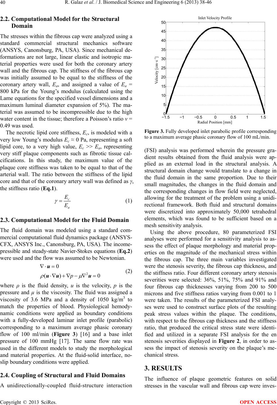 R. Galaz et al. / J. Biomedical Science and Engineering 6 (2013) 38-46 40 2.2. Computational Model for the Structural Domain The stresses within the fibrous cap were analyzed using a standard commercial structural mechanics software (ANSYS, Canonsburg, PA, USA). Since mechanical de- formations are not large, linear elastic and isotropic ma- terial properties were used for both the coronary artery wall and the fibrous cap. The stiffness of the fibrous cap was initially assumed to be equal to the stiffness of the coronary artery wall, Ea, and assigned a value of Ea = 800 kPa for the Young’s modulus (calculated using the Lame equations for the specified vessel dimensions and a maximum luminal diameter expansion of 5%). The ma- terial was assumed to be incompressible due to the high water content in the tissue; therefore a Poisson’s ratio ν = 0.49 was used. The necrotic lipid core stiffness, Ec, is modeled with a very low Young’s modulus Ec ≈ 0 Pa, representing a soft lipid core, to a very high value, Ec >> Ea, representing very stiff plaque components such as fibrotic tissue cal- cifications. In this study, the maximum value of the plaque core stiffness was taken to be equal to that of the arterial wall. The ratio between the stiffness of the lipid core and that of the coronary artery wall was defined as γ, the stiffness ratio (Eq.1). c a E E (1) 2.3. Computational Model for the Fluid Domain The fluid domain was modeled using a standard com- mercial computational fluid dynamics package (ANSYS- CFX, ANSYS Inc., Canonsburg, PA, USA). The income- pressible and steady-state Navier-Stokes equations (Eq.2) were used and the flow was assumed to be Newtonian. 2 0 ()p u uu u0 (2) where ρ is the fluid density, u is the velocity, p is the pressure and µ is the viscosity. The fluid was assigned a viscosity of 3.6 MPa and a density of 1050 kg/m3 to match the properties of blood. Physiological hemody- namic conditions were applied as boundary conditions with a fully-developed laminar inlet profile (parabolic) corresponding to a maximum average phasic coronary flow of 100 ml/min (Figure 3) [16] and a base inlet pressure of 100 mmHg [17]. The same flow rate was used in the different models to study the morphological and material properties. At the fluid-solid interface, no- slip boundary conditions were applied. 2.4. Coupling of Structural and Fluid Domains A unidirectionally-coupled fluid-structure interaction Inlet Velocity Profile Radial Position [mm] −1.5 −1 −0.5 0 0.5 1 1.5 Velocity [cm·s −1 ] 50 45 40 35 30 25 20 15 10 5 0 Figure 3. Fully developed inlet parabolic profile corresponding to a maximum average phasic coronary flow of 100 mL/min. (FSI) analysis was performed wherein the pressure gra- dient results obtained from the fluid analysis were ap- plied as an external load in the structural analysis. A structural domain change would translate to a change in the fluid domain in the same proportion. Due to their small magnitudes, the changes in the fluid domain and the corresponding changes in flow field were neglected, allowing for the treatment of the problem using a unidi- rectional framework. Both fluid and structural domains were discretized into approximately 50,000 tetrahedral elements, which was found to be sufficient based on a mesh sensitivity analysis. Using the above procedure, 80 parameterized FSI analyses were performed for a sensitivity analysis to as- sess the effect of plaque morphology and material prop- erties on the magnitude of the mechanical stress within the fibrous cap. The three main variables investigated were the stenosis severity, the fibrous cap thickness, and the stiffness ratio. Four different coronary artery stenosis severities were selected: 36%, 51%, 75% and 91% and four fibrous cap thicknesses varying from 200 to 500 microns and five stiffness ratios varying from 0.001 to 1 were taken. The results of the parameterized FSI analy- ses were used to construct surface plots of the resulting peak stress values within the plaque. The conditions, with respect to the fibrous cap thickness and the stiffness ratio, that produced the critical stress state were identi- fied and utilized in a separate FSI analysis for the en stenosis severities displayed in Figure 2, in order to as- sess the impact of stenosis severity on the plaque’s me- chanical stress. 3. RESULTS The influence of plaque geometric features on solid stresses in the vascular wall and fibrous cap were inves- Copyright © 2013 SciRes. OPEN ACCESS 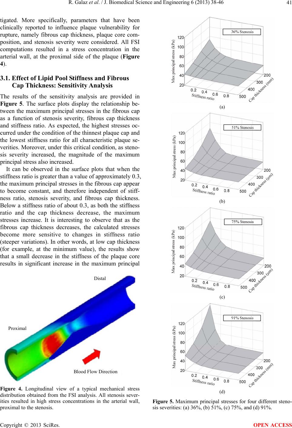 R. Galaz et al. / J. Biomedical Science and Engineering 6 (2013) 38-46 41 tigated. More specifically, parameters that have been clinically reported to influence plaque vulnerability for rupture, namely fibrous cap thickness, plaque core com- position, and stenosis severity were considered. All FSI computations resulted in a stress concentration in the arterial wall, at the proximal side of the plaque (Figure 4). 3.1. Effect of Lipid Pool Stiffness and Fibrous Cap Thickness: Sensitivity Analysis The results of the sensitivity analysis are provided in Figure 5. The surface plots display the relationship be- tween the maximum principal stresses in the fibrous cap as a function of stenosis severity, fibrous cap thickness and stiffness ratio. As expected, the highest stresses oc- curred under the condition of the thinnest plaque cap and the lowest stiffness ratio for all characteristic plaque se- verities. Moreover, under this critical condition, as steno- sis severity increased, the magnitude of the maximum principal stress also increased. It can be observed in the surface plots that when the stiffness ratio is greater than a value of approximately 0.3, the maximum principal stresses in the fibrous cap appear to become constant, and therefore independent of stiff- ness ratio, stenosis severity, and fibrous cap thickness. Below a stiffness ratio of about 0.3, as both the stiffness ratio and the cap thickness decrease, the maximum stresses increase. It is interesting to observe that as the fibrous cap thickness decreases, the calculated stresses become more sensitive to changes in stiffness ratio (steeper variations). In other words, at low cap thickness (for example, at the minimum value), the results show that a small decrease in the stiffness of the plaque core results in significant increase in the maximum principal Distal Proximal Blood Flow Direction Figure 4. Longitudinal view of a typical mechanical stress distribution obtained from the FSI analysis. All stenosis sever- ities resulted in high stress concentrations in the arterial wall, proximal to the stenosis. (a) (b) (c) (d) Figure 5. Maximum principal stresses for four different steno- sis severities: (a) 36%, (b) 51%, (c) 75%, and (d) 91%. Copyright © 2013 SciRes. OPEN ACCESS  R. Galaz et al. / J. Biomedical Science and Engineering 6 (2013) 38-46 42 stresses present in the fibrous cap. When the stiffness ratio is taken to be constant, a decrease in the plaque cap thickness results in a more direct increase in stresses to- wards the maximum value at the minimum plaque thick- ness. It is important to note that the fibrous cap thick- nesses considered in this study are above the value for which plaques have been clinically observed to rupture (approximately 65 microns) [18]. It can be observed that the magnitude of the stresses in the fibrous cap appear to be independent of the stenosis severity, even for severe area reductions, when the stiff- ness ratio is in the range of 0.1 to 0.3. Moreover, when the stiffness ratio decreases to a low level such that the core is essentially a soft lipid pool, the magnitude of the principal cap stresses still appears to be relatively con- stant for intermediate stenoses, yet increases significantly when the area reduction becomes severe. 3.2. Effect of Stenosis Severity The effect of stenosis severity on the stress state of the fibrous cap was investigated by performing FSI analyses for ten different area reductions (ranging from 19% to 91%, Figure 2) under the critical stress condition of small fibrous cap thickness and low lipid core stiffness ratio. In Figure 6, the normalized principal stress in the fibrous cap is plotted as a function of the percent area reduction for the minimal cap thickness (200 microns) and the minimal lipid core stiffness ratio (0.001), which appeared from the sensitivity analysis to be the condition for which the stresses were most sensitive to percent stenosis. The results displayed in Figure 6 show three distinct regions where stresses increase with different rates as a function of the area stenosis, which correspond to mild, Percentage of stenosed area [%] 0 10 20 30 40 50 60 70 80 90 100 1 0.8 0.6 0.4 0.2 0 Normalized principal stress Figure 6. Normalized principal stress for different stenosis severities in the case of a fibrous cap thickness of 200 microns and a stiffness ratio between the lipid core and the coronary artery wall, γ, of 0.001. intermediate and severe stenoses, based on the percent area reduction. For both mild (19% to 43% area reduc- tion) and severe (greater than 75% area reduction) stenoses, the rate of increase of the peak stress with in- creasing area reduction is significant, particularly for severe stenoses which display a steep slope. However, in the case of intermediate stenoses (43% to 75% area re- duction), the plot displays a plateau region where there is a relatively small increase in peak stress over the range of lesion severity. 4. DISCUSSION In previous investigations of the role of mechanical fac- tors on plaque vulnerability for rupture, much focus has been put on 2D/3D structural finite element analyses separate from fluid dynamic analyses. For example, stu- dies have shown through 2D (cross-sectional views) and 3D finite element models that high stress regions corre- spond to the culprit (rupture) zones in histopathological studies and that the magnitudes of these stresses are af- fected by plaque composition and morphology [19,20]. On the other hand, computational fluid dynamics analy- ses have also correlated the location of high wall shear stress to that of plaque ulceration [21]. However, when structural and fluid analyses are con- sidered separately (and particularly in a 2D model), the true state of the stress tensor is not fully represented for either domains and the complex interactions between plaque morphology, plaque composition and fluid and solid forces on the global stress state cannot be evaluated. Therefore, in order to obtain a better understanding of the true physical events taking place in the diseased ves- sel, it becomes necessary to use 3D models that incorpo- rate the interactions between the fluid and structural do- mains. These models would provide more accurate nu- merical evidence on the spatial stress distribution and the longitudinal component of the shear stress caused by blood flow. Moreover, when combined with localized anisotropic material properties and a mechanical failure criterion, the analyses could define the proper orientation and the extent of rupture propagation. The 3D numerical results could serve as a basis to compare the spatial loca- tion of the rupture with clinical evidence that often re- ports plaque rupture locations, for example, at the up- stream regions of the stenosis [22,23]. Tang and col- leagues have developed 3D fluid-structure interaction (FSI) models based on MRI scans and have displayed that regions of localized maximum stresses, particularly solid stresses, correspond well with the location of plaque ulceration identified from histopathological eva- luations. The magnitude of the stresses in ulcerated plaques was also found to be higher than that of stable plaques, and also displayed a general increasing trend in unstable plaques with larger lipid pools and thinner Copyright © 2013 SciRes. OPEN ACCESS  R. Galaz et al. / J. Biomedical Science and Engineering 6 (2013) 38-46 43 plaque caps [17,24,25]. The idea that mechanical factors play a role in the rupture of plaques has been agreed upon in the scientific community although the exact mechanisms by which these factors, specifically solid and fluid stresses, initiate plaque damage, are unclear. From a mechanical perspec- tive, it can be expected that the fibrous cap will rupture when the solid stresses therein surpass the material’s ultimate strength (Lee and Loree [26]). Consequently, the global solid stress state within the fibrous cap is a central factor in the study of plaque fracture mechanics; it is affected by the fluid forces (cyclic pressure, pressure gradients) exerted on and the morphology and material properties of the tissue. Fluid shear stresses could affect the surface of the plaque as well as activate endothelial cells to produce and secrete degrading enzymes that weaken the structure of the fibrous cap. A stenosis impedes the flow of blood and induces an energy loss which is reflected as a pressure drop across the stenosis. This loss in energy is a result of inertial pressure losses from geometrical changes, and viscous effect. It is dissipated from the system in the form of heat (carried away by flow advection), and in the form of ir- reversible mechanical deformation, specifically of the viscoelastic fibrous cap which is the most mechanically solicited component of the plaque. The hemodynamic pressure gradient exerts a force on and causes deforma- tion of the plaque in the direction of the bulk flow, which results in higher solid stresses at the proximal side of the fibrous cap, compared to the distal side. This effect is amplified when the fibrous cap of the plaque is thin and has a weak structural support underneath it, as was shown in our study for the case of a soft lipid core which acts as a weak elastic foundation. This effect has also been proposed by Doriot [27] and Li [11] using theoreti- cal and numerical calculations with only 2D longitudinal models of stenosed arteries. Our numerical results were consistent with clinical observations. Fujii et al. [22] have shown that most of the ulcerated ruptured plaques in culprit lesions of acute coronary syndrome patients were proximal to site of minimal lumen dimension based on intravascular ultra- sound imaging studies. Hiro et al. [23] have also reported ultrasonographic longitudinal views of ruptured coronary plaques, and most of them were also located in the proximal region of the plaque. 4.1. Plaque Cap Principal Stresses Thin fibrous caps and soft lipid cores have been clini- cally associated with higher rupture risk. The values ob- tained in this study for the maximum principal stresses are consistent with similar studies. In a 2D (cross-sec- tional) structural analysis, Finet et al. [8] obtained maxi- mum principal stresses in coronary plaques between 100 kPa and 200 kPa (at a pressure of 110 mmHg, a cap thickness of 200 microns and a core stiffness of 1 kPa) for stenoses of approximately 50 - 60 percent area reduc- tion. Our results gave maximum principal stresses of 85 kPa for similar conditions (baseline pressure of 100 mmHg, a cap thickness of 200 microns and a stiffness ratio of 0.001). Arterial dimensions in Finet et al. were greater than those in the present study and can account in part for the differences in stress magnitude. Tang et al. [24] performed FSI analyses on coronary plaques for which the morphology was obtained from ex vivo MRI of cadaveric samples. For an approximate area reduction of 80%, a pressure of 100 mmHg the maxi- mum principal stresses were found to range from 100 - 200 kPa at the interface of the lipid core and the lumen, which again corresponds well to the 90 - 100 kPa stress range found in the current study for the thin plaque cap and the soft lipid core. A similar study by the same au- thors [28], which also addressed the issue of cyclic bend- ing, found principal stress values within the plaque cap within the 50 - 80 kPa range. 4.2. Plaque Cap Thickness and Core Stiffness Our results showed that plaque cap stresses were found to be very sensitive to small changes in the core stiffness at low stiffness ratios. These findings are similar to those of [8] who showed that for a critical cap thickness of 50 microns, a small change from a core stiffness of 25 kPa to 1 kPa could destabilize the plaque (specifically, in- crease the circumferential cap stresses over a predefined threshold corresponding to rupture initiation). Virmani et al. [18,29] first introduced a critical cap thickness below which coronary plaques are at high risk for rupture. This value was found to be approximately 65 microns and has since been adopted by the American Heart Association. Through computational studies, a cir- cumferential fibrous cap stress of 300 kPa has also been found to correspond to the threshold for coronary plaque rupture [19] and is consistent with the critical cap thick- ness. Previous studies have found a curvilinear/exponen- tial relationship between the cap thickness and the circumferential stresses [8,30], and that significant in- crease in principal stress with a small decrease in cap thickness occurs near this critical value. The results of this study displayed a more direct relationship between the stress and the cap thickness which seems to contra- dict previous findings. It is important to note however, that the minimum cap thickness considered in this study is well above the critical value and the corresponding principal stresses are also below the 300 kPa threshold. The relative contribution/effect of plaque core compo- sition and fibrous cap thickness has not been elucidated thus far yet could have significant clinical implications. The results of our study suggest that one of these vari- Copyright © 2013 SciRes. OPEN ACCESS  R. Galaz et al. / J. Biomedical Science and Engineering 6 (2013) 38-46 44 ables may play a more central role in the initiation of plaque ulceration. The relatively constant principle stresses when the stiffness ratio is above 0.3, independent of both fibrous cap thickness and percent stenosis, sug- gests that there may be a threshold for plaque stiffness above the risk of rupture is reduced. Furthermore, the fact that the maximum principal stresses at the minimum core stiffness were well below the critical plaque rupture stress, suggests that the thickness of the plaque cap may have greater influence on plaque vulnerability for rupture compared to core stiffness. 4.3. Stenosis Severity One of the most interesting results of this study was a quasi-constant region (plateau) of peak principal stress with increasing stenosis severity for intermediate lesions (the maximum principal stress was relatively insensitive to percentage of stenosis). A similar observation was reported in a clinical investigation by Li et al. [11] in the carotid artery; they found that the peak principal stress remained relatively constant for a degree of luminal stenosis comprised between 40% and 60%, and for a fibrous cap thickness ranging from 0.2 mm to 1 mm. The clinical implications of these findings are worthy of note. For intermediate stenoses, the results suggest that the vulnerability to rupture may be insensitive to the severity of the lumen reduction (above and below certain thresholds) in terms of stress loading and the size of the lipid pool. 4.4. Limitations The minimum thickness of the fibrous cap in our model was 200 microns although pathologists have reported that the typical thickness at which fibrous caps fail is around 65 microns [29]. This value facilitated the mesh- ing of the fibrous cap and allowed to avoid being in a situation of rupture. However, the computed stress could be underestimated. Another limitation of this model was that the me- chanical properties of the fibrous cap were the same as those of the artery wall. The mechanical properties of the fibrous cap are still unknown, and they can vary from patient to patient. The fact that the fibrous cap is mostly composed of collagen fibers and some smooth muscle cells is hint that it is reasonable to assume that its me- chanical properties are similar to the artery wall. How- ever, it should be somewhat stiffer since it has a higher ratio of collagen although it was found in [17] that in- creasing the stiffness of the fibrous cap by 100% has minor effects (less than 2%) on the stress state within the cap. Finally, in order to obtain a more accurate model in terms of anatomical structure and physiological condi- tions, it becomes necessary to analyze realistic geome- tries of the coronary arteries and include more realistic material properties such as viscoelasticity and anisotropy of the connective tissue. Furthermore, it has been shown that cyclic bending of the arteries can have a significant effect on the magnitude of the peak principal stresses [28]. This idea is of particular importance for coronary arteries which are subjected to such a loading due to the cyclic contraction of the heart, and must be considered in order to better understand the relative importance of plaque morphology (size and cap thickness) and plaque composition on its vulnerability to rupture. 5. CONCLUSION Using FSI simulations, the 3D mechanical stress in dis- eased coronary arteries was calculated for varying plaque cap thickness, lipid core stiffness and stenosis severity and evaluated as a factor that might contribute to a plaque’s vulnerability to rupture. The peak principal stresses were observed at the proximal side of the plaque in all FSI analyses which was consistent with pathologi- cal observations for plaque ulceration locations. The im- portant findings of this investigation included the fact that 3D mechanical stresses were observed to be more sensitive to changes in fibrous cap thickness compared to changes in core thickness which suggested that cap thickness could play a more central role in plaque rupture vulnerability compared to other morphological plaque characteristics. It was also found that at a critical cap thickness, a small change in plaque composition could significantly change the peak principal stresses and thus the plaque’s vulnerability to rupture. Furthermore, the peak principal stresses of moderate stenoses (ranging from 43% to 75% area reductions) were found to be generally unaffected by stenosis severity (at a particular cap thickness and core stiffness), displayed by a plateau in the stresses as a function of severity. This observation not only suggests that moderate and even mild plaques could have the same vulnerability to mechanical failure but that they can be subjected to stresses similar to se- vere plaques that could contribute to their rupture. REFERENCES [1] Kolodgie, F., Virmani, R., Burke, A., Farb, A., Weber, D., Kutys, R., Finn, A. and Gold, H. (2004) Pathologic as- sess-ment of the vulnerable human coronary plaque. Heart, 90, 1385-1391. http://dx.doi.org/10.1136/hrt.2004.041798 [2] Burke, A., Farb, A., Malcolm, G., Liang, Y., Smialek, J. and Virmani, R. (1997) Coronary risk factors and plaque morphology in patients with coronary disease dying sud- denly. New England Journal of Medicine, 336, 1276- 1282. http://dx.doi.org/10.1056/NEJM199705013361802 [3] Libby, P. and Theroux P. (2005) Pathophysiology of Copyright © 2013 SciRes. OPEN ACCESS  R. Galaz et al. / J. Biomedical Science and Engineering 6 (2013) 38-46 45 coro-nary artery disease. Circulation, 111, 3481-3488. http://dx.doi.org/10.1161/CIRCULATIONAHA.105.5378 78 [4] Falk, E. (1989) Morphologic features of unstable athero- thrombotic plaques underlying acute coronary syndromes. American Journal of Cardiology, 63, 114E-120E. http://dx.doi.org/10.1016/0002-9149(89)90242-7 [5] Davies, M. (1992) Anatomic features in victims of sud- den coronary death. Coronary artery pathology. Circula- tion, 85, 119-124. [6] Richardson, P., Davies, M. and Born, G. (1989) Influence of plaque configuration and stress distribution of coro- nary atherosclerotic plaques. Lancet, 2, 941-944. http://dx.doi.org/10.1016/S0140-6736(89)90953-7 [7] Lee, R., Schoen, F., Loree, H., Lark, M. and Libby, P. (1996) Circumferential stress and matrix metalloprotei- nase-1 in human coronary atherosclerosis: Implications for plaque rupture. Arteriosclerosis, Thrombosis, and Vas- cular Biology, 16, 1070-1073. http://dx.doi.org/10.1161/01.ATV.16.8.1070 [8] Finet, G., Ohayon, J. and Rioufol, G. (2004) Biomecha- nical interaction between cap thickness, lipid core com- position and blood pressure in vulnerable coronary plaque: Impact on stability or instability. Coronary Artery Dis- ease, 15, 13-20. http://dx.doi.org/10.1097/00019501-200402000-00003 [9] Williamson, S., Lam, Y., Younis, H., Patel, S., Kaazem- pur-Mofrad, M. and Kamm, R. (2003) On the sensitivity of wall stresses in diseased arteries to variable material properties. Journal of Biomechanical Engineering, 125, 147-155. http://dx.doi.org/10.1115/1.1537736 [10] Lovett, J. and Rothwell, P. (2003) Site of carotid plaque ulceration in relation to direction of blood flow: An an- giographic and pathological study. Cerebrovascular Dis- eases, 16, 369-375. http://dx.doi.org/10.1159/000072559 [11] Li, Z., Howarth, S., Tang, T. and Gillard, J. (2006) How critical is fibrous cap thickness to carotid plaque stability? A flow-plaque interaction model. Stroke, 37, 1195-1199. http://dx.doi.org/10.1161/01.STR.0000217331.61083.3b [12] Burke, A., Farb, A., Malcom, G.T., Liang, Y.-h, Smialek, J.E. and Virmani, R. (1999) Plaque rupture and sudden death related to exertion in men with coronary artery dis- ease. Journal of the American Medical Association, 281, 921- 926. http://dx.doi.org/10.1001/jama.281.10.921 [13] Falk, E., Shah, P. and Fuster, V. (1995) Coronary plaque disruption. Circulation, 92, 657-671. http://dx.doi.org/10.1161/01.CIR.92.3.657 [14] Mattsson, E., Kohler, T., Vergel, S. and Clowes, A. (1997) Increased blood flow induces regression of intimal hyper- plasia. Arteriosclerosis, Thrombosis, and Vascular Biol- ogy, 17, 2245-2249. http://dx.doi.org/10.1161/01.ATV.17.10.2245 [15] Wang, J., Normand, S., Mauri, L. and Kuntz, R. (2004) Coronary artery spatial distribution of acute myocardial infarction occlusions. Circulation, 110, 278-284. http://dx.doi.org/10.1161/01.CIR.0000135468.67850.F4 [16] Berne, R.M. and Levy, M.N. (1967) Cardiovascular phy- siology. Mosby, St. Louis. [17] Tang, D., Yang, C., Zheng, J., Woodard, P.K., Sicard, G.A., Saffitz, J.E. and Yuan, C. (2004) 3D MRI-based multicomponent FSI models for atherosclerotic plaques. Annals of Biomedical Engineering, 32, 947-960. http://dx.doi.org/10.1023/B:ABME.0000032457.10191.e 0 [18] Virmani, R., Kolodgie, F.D., Burke, A.P., Farb, A. and Schwartz, S.M. (2000) Lessons from sudden coronary death: A comprehensive morphological classification scheme for atherosclerotic lesions. Arteriosclerosis, Thrombosis, and Vascular Biology, 20, 1262-1275. http://dx.doi.org/10.1161/01.ATV.20.5.1262 [19] Cheng, G.C., Loree, H.M., Kamm, R.D., Fishbein, M.C. and Lee, R.T. (1993) Distribution of circumferential stress in ruptured and stable atherosclerotic lesions. A structural analysis with histopathological correlation. Cir- culation, 87, 1179-1187. http://dx.doi.org/10.1161/01.CIR.87.4.1179 [20] Imoto, K., Hiro, T., Fujii, T., Murashige, A., Fukumoto, Y., Hashimoto, G., Okamura, T., Yamada, J., Mori, K. and Matsuzaki, M. (2005) Longitudinal structural deter- minants of atherosclerotic plaque vulnerability. A com- putational analysis of stress distribution using vessel mo- dels and three dimensional intravascular ultrasound im- aging. Journal of the American College of Cardiology, 46, 1507-1515. http://dx.doi.org/10.1016/j.jacc.2005.06.069 [21] Groen, H.C., Gijsen, F.J.H., van der Lugt, A., Ferguson, M.S., Hatsukami, T.S., van der Steen, A.F., Yuan, C. and Wentzel, J.J. (2007) Plaque rupture in the carotid artery is located at the high shear stress region. A case report. Stroke, 38, 2379-2381. http://dx.doi.org/10.1161/STROKEAHA.107.484766 [22] Fujii, K., Kobayashi, Y., Mintz, G., Takebayashi, H., Dangas, G., Moussa, I., Mehran, R., Lansky, A.J., Kreps, E., Collins, M., Colombo, A., Stone, G.W., Leon, M.B. and Moses, J.W. (2003) Intravascular ultrasound assess- ment of ulcerated ruptured plaques: A comparison of cul- prit and nonculprit lesions of 15 patients with acute coro- nary syndromes and lesions in patients without acute coronary syndromes. Circulation, 108, 2473-2478. http://dx.doi.org/10.1161/01.CIR.0000097121.95451.39 [23] Hiro, T., Fujii, T., Yoshitake, S., Kawabata, T., Yasu- moto, K. and Matsuzaki, M. (2000) Longitudinal visuali- zation of spontaneous coronary plaque rupture by 3D in- travascular ultrasound. Circulation, 101, e114. http://dx.doi.org/10.1161/01.CIR.101.12.e114 [24] Tang, D., Yang, C., Zheng, J., Woodard, P.K., Saffitz, J.E., Petruccelli, J.D., Sicard, G.A. and Yuan, C. (2005) Local maximal stress hypothesis and computational pla- que vulnerability index for atherosclerotic plaque assess- ment. Annals of Biomedical Engineering, 33, 1789-1801. http://dx.doi.org/10.1007/s10439-005-8267-1 [25] Tang, D., Teng, Z., Canton, G., Yang, C., Ferguson, M., Huang, X., Zheng, J., Woodard, P.K. and Yuan, C. (2009) Sites of rupture in human atherosclerotic carotid plaques are associated with high structural stresses: An in vivo MRI-based 3D fluid-structure interaction study. Stroke, 40, 3258-3263. http://dx.doi.org/10.1161/STROKEAHA.109.558676 [26] Lee, R.T., Loree, H.M., Cheng, G.C., Lieberman, E.H., Copyright © 2013 SciRes. OPEN ACCESS  R. Galaz et al. / J. Biomedical Science and Engineering 6 (2013) 38-46 Copyright © 2013 SciRes. 46 OPEN ACCESS Jaramillo, N. and Schoen, F.J. (1993) Computational structural analysis based on intravascular ultrasound im- aging before in vitro angioplasty: Prediction of plaque fracture locations. Journal of the American College of Cardiology, 21, 777-782. http://dx.doi.org/10.1016/0735-1097(93)90112-E [27] Doriot, P. (2003) Estimation of the supplementary axial wall stress generated at peak flow by an arterial stenosis. Physics in Medicine and Biology, 48, 127-138. http://dx.doi.org/10.1088/0031-9155/48/1/309 [28] Yang, C., Tang, D., Kobayashi, S., Zheng, J., Woodard, P.K., Teng, Z., Back, R. and Ku, D.N. (2008) Cyclic bending contributes to high stress in human coronary atherosclerotic plaque and rupture risk. Molecular & Cel- lular Biomechanics, 5, 259-274. [29] Virmani, R., Burke, A., Farb, A. and Kolodgie, F. (2002) Cardiovascular plaque rupture. Marcel Dekker Publishers Inc., New York. [30] Loree, H.M., Kamm, R.D., Stringfellow, R.G. and Lee, R.T. (1992) Effects of fibrous cap thickness on peak circumferential stress in model atherosclerotic vessels. Circulation Research, 71, 850-858. http://dx.doi.org/10.1161/01.RES.71.4.850 |